Abstract
Background:
Anterior cruciate ligament (ACL) reconstruction is uncommonly complicated by postoperative infections, the causes of which are rarely identified.
Hypothesis/Purpose:
The goal of this study was to characterize the relationship between methodological sterilization failure and ACL reconstruction infection at an army medical center.
Study Design:
Case series.
Methods:
Demographic, clinical, and laboratory data were collected on 5 postoperative infections during a 14-week period in 2003. All ACL reconstructions completed within the past 6 years at the institution were reviewed to establish a baseline infection rate.
Results:
There was a 14-week period in which 5 cases of infection occurred postoperatively, an infection rate of 12.2%. Previous and subsequent to the identified period, the established rate of infection after ACL reconstruction was 0.3%. There were no violations of sterile technique noted in any of the identified cases. All cases utilized hamstring autograft. All cases also used the DePuy Mitek Intrafix system for tibial fixation of the graft. Two of these cases had positive cultures.
Conclusions:
An isolated series of increased infection rate led to an investigation into the sterile technique. This revealed gross biomaterial remaining inside instrumentation common to all the cases, the DePuy Mitek Intrafix system. The modular cannulated hex driver, made to fit over a small caliber wire, had no wire brushes of a small-enough diameter for the cleaning and sterilization procedure. After recognition of infection, all patients were treated with surgical irrigation and debridement of the affected knee, as well as individualized antibiotic therapy. Patients were followed postoperatively and no patients required revision ACL reconstruction or radical debridement of the graft.
Keywords: anterior cruciate ligament, postoperative infection, surgical site infections
Infection rarely occurs in arthroscopic anterior cruciate ligament (ACL) reconstruction, but it can have serious ramifications.3,7,10 There is controversy surrounding different treatment modalities and their implications on management and outcome of postoperative ACL infection.5 To our knowledge, there has not been a study that has reported a series of infections that had a common cause. A systematic failure of the sterilization procedure could cause a series of infections. However, to our knowledge, this has never been reported in the literature. The purpose of this study was to review our experience with ACL infection after introduction of new instrumentation, describe our management and results, and determine the cause of the outbreak of infections. A series of postopoperative infections after ACL reconstructions that were associated with a specific recognized source are reported.
Methods
The institution in which the cases were performed is a 205-bed army medical center—specifically, a Level II trauma center with an orthopaedic residency program. There are 14 operating rooms. The orthopaedic service consists of 10 attending surgeons, 12 residents, and 3 interns. An average of 90 ACL reconstructions were performed each year during the period reviewed.
A retrospective review of all ACL infections from June 2002 until June 2008 was performed. A chart review of operative notes, in-patient records, and postopoperative clinical visits was performed. Operative reports as well as nursing records were reviewed to determine if any violations in sterile technique were noted. Specifically, serum white blood cell count, C-reactive protein, erythrocyte sedimentation rate, and white blood cell count in aspirated synovial fluid were reviewed. Gram stain and cultures of the synovial fluid were also evaluated. Cases were defined as a positive infection based on a positive culture or clinical picture consistent with infection (fever, elevated white blood cell count, C-reactive protein, erythrocyte sedimentation rate, and white blood cell count in synovial fluid).
Results
Demographics and Clinical Presentations
In October 2003, an increased rate of postoperative ACL reconstruction infections was noted. In the period of June through October 2003, 5 infections were identified out of 41 cases (12.2%). Outside the 14-week period, the infection rate for the remainder of the 6-year review was less than 0.3%. Demographic variables—including sex, age, active-duty military status, American Society of Anesthesiologists class, and length of surgery—showed no significant differences between infection and noninfection groups (Table 1). The study group was found to be similar to the control group with regard to all variables. The majority of patients were active-duty military service members.
Table 1.
Demographics of control and study groups.
| Control n (%) | Study n (%) | P | |
|---|---|---|---|
| Cases | 653 | 5 | |
| Age, y | 28 | 26 | .51 |
| Men | 535 (82) | 3 (60) | .22 |
| Women | 118 (18) | 2 (40) | |
| Active duty military | 562 (86) | 3 (60) | .15 |
| Other | 91 (14) | 2 (40) | |
| American Society of Anesthesiologists | |||
| Class 1 | 398 (61) | 4 (80) | .65 |
| Class 2 | 255 (39) | 1 (20) | |
| Length of surgery (min) | 169.8 | 185.0 | .50 |
There were no violations of sterile technique noted in the operative logs or nursing records in any of the identified cases. All cases had intravenous first-generation cephalosporin antibiotics administered before the start of the procedure as per department routine. All 5 cases used hamstring autograft with the same tibial fixation, the DePuy Mitek Intrafix system (Mitek, Westwood, Massachusetts). Four of these cases used the DePuy Mitek Slingshot system for femoral fixation of the graft. The Arthrex Transfix system was used for the femoral fixation in the remaining case. Surgical data and clinical presentation were noted for each member of this group (Table 2).
Table 2.
Demographics of patients and procedures.a.
| Case | Sex | Age, y | Diagnosis | Other Procedures | Femoral Fixation | Tibial Fixation |
|---|---|---|---|---|---|---|
| 1 | M | 47 | Right ACL acute rupture | None | DMS | DMI |
| 2 | M | 21 | Left ACL acute rupture, medial meniscal tear, lateral meniscal tear | Medial meniscus all-inside repair, lateral meniscus debridement | DMS | DMI |
| 3 | F | 23 | Left ACL acute rupture | None | DMS | DMI |
| 4 | F | 16 | Left ACL acute rupture | None | Arthrex Transfix | DMI |
| 5 | M | 24 | Right ACL acute rupture, medial menisal tear | Medial meniscus debridement | DMS | DMI |
ACL, anterior cruciate ligament; DMS, DePuy Mitek Slingshot; DMI, DePuy Mitek Intrafix. All cases were performed with hamstring autograft.
All patients were seen for evaluation at postopoperative day 7 to 12. One patient presented with symptoms consistent with an infection in this period. In the remaining patients, there was no clinical suspicion for infection. All patients returned to the clinic for symptoms of increased pain, effusion, or erythema about the operative knee (Table 3). This occurred on average at postopoperative day 27 (range, 7-42 days). All patients had laboratory studies performed, which revealed elevated serum white blood cell, C-reactive protein, and erythrocyte sedimentation rate. All patients also had sterile aspiration of the operative knee effusion, which revealed elevated synovial fluid white blood cell. Aspirated synovial fluid was also sent for culture, which yielded positive results for 2 patients (40%), with the isolate being coagulase-negative Staphylococcus (unable to further identify) in 1 patient and coagulase-negative Staphylococcus (further identified as S epidermidis) in the other.
Table 3.
Infection presentation and treatment.
| Case | Sex | Age, y | Culture Results | Days to Encounter | Initial WBC | Initial CRP | Initial ESR | Aspiration WBCa | Surgical I&Ds | Days to Initial I&D | Antibiotic Type | Antibiotic Duration (Weeks) |
|---|---|---|---|---|---|---|---|---|---|---|---|---|
| 1 | M | 47 | Neg | 42 | 10.3 | 10.2 | 82 | 62 | 1 | 53 | Gatifloxacin | 4 |
| 2 | M | 21 | Neg | 42 | 11.6 | 18.2 | 83 | 235 | 3 | 43 | Augmentin | 2 |
| 3 | F | 23 | Staph epi | 19 | 5.7 | 9.23 | 86 | 10 | 2 | 22 | Gatifloxacin | 6 |
| 4 | F | 16 | Neg | 22 | 8.7 | 23 | 97 | 54 | 4 | 22 | Vancomycin / Linezolid | 4 |
| 5 | M | 24 | CNS | 10 | 11.1 | 23 | 34 | 27 | 2 | 10 | Gatifloxacin | 8 |
| Average | 26.0 | 27.0 | 9.5 | 16.7 | 76.4 | 77.5 | 2.4 | 30.0 | 4.8 | |||
| Range | 16-47 | 7-42 | 5.7-11.6 | 9.23-23.0 | 34-97 | 10-235 | 1-4 | 7-53 | 2-8 | |||
WBC, white blood cell; CRP, C-reactive protein; ESR, erythrocyte sedimentation rate; I&D, irrigation and debridement; staph epi, Staphylococcus epidermidis; CNS, coagulase-negative staphylococci (unable to further identify).
In thousands.
Three patients were taken for immediate arthroscopic irrigation and debridement (I&D) with graft retention. Two patients presented with pain only and were initially followed. At 3 and 11 days after their initial presentation, their symptoms had progressed. At that point, they underwent laboratory analysis and were taken to the operating room for I&D.
All patients underwent at least 1 I&D and were placed on individual antibiotic coverage of gram-positive cocci on the basis of patient allergies and available culture susceptibilities and in consultation with infectious disease specialists. Antibiotics were initially given intravenously in the hospital and then transitioned to oral administration for a duration tailored to the clinical picture by the treating surgeon. Patients returned to the operating room for subsequent I&Ds based on response to treatment, with pain and improvement in motion as primary criteria for repeat I&Ds. One patient had 1 I&D performed; 2 patients had 2; 1 patient had 3; and 1 patient had 4. No patients underwent radical graft debridement or removal of their graft during these I&Ds. All patients were uniformly healthy without any existing comorbidities; 3 were active-duty service members; and 2 were dependent family members of active-duty personnel.
Patients were followed at 2, 6, and 12 weeks and 6 and 12 months postopoperatively. None of these patients returned with symptoms of continued pain, erythema, or effusion of the operative knee. These patients did not complain of knee instability during these follow-ups, and none required revision surgery for an incompetent ACL graft.
Based on the increased infection rate, an investigation into the possible violation of sterile technique began. An investigative team was composed of operating surgeons, nurses, central medical supply personnel and hospital quality assurance personnel, and infectious disease service and infection control personnel, and ACL reconstructions were temporarily halted. Patient characteristics, compliance with postopoperative instructions, and operative notes were reviewed. No obvious patterns were noted in any of the patient characteristics or postopoperative course. All instrumentation before each case had undergone steam sterilization in prevacuum autoclaves, which is the institution’s standard.
The investigative team compiled a report documenting the medical supply personnel who assembled each instrument tray used in the involved cases. Multiple medical supply personnel had been responsible for assembling the instrument trays; no one person was identified who was involved in every case. The investigative committee then performed spot reviews on all similar types of cases being performed to evaluate any gross deviation from practice. These reviews revealed no blatant errors in sterile technique, but they did report deviations from standard hospital protocol, such as excessive movement around sterile fields. Also noted were frequent movement of personnel in and out of the operating room and excessive movement of sterile-attired surgeons and nursing staff. Educational sessions on the importance of maintaining sterile techniques and limiting operating room traffic were offered to operating room personnel; however, the infection team deemed it unlikely that these few findings were responsible for the cases of infection.
Review of the operative notes revealed that all 5 cases utilized hamstring autograft reconstruction with the DePuy Mitek Intrafix system. The investigative team carefully examined all instruments common to the Intrafix system as well as the instruments used for harvesting of hamstring grafts. The examination revealed biomaterial remaining inside the cannulated portion of the Intrafix tibial fixation hex driver. Cultures of this material were taken, which grew out separate species of rare coagulase-negative Staphylococcus, rare S epidermidis, and very rare Streptococcus mitis, which is a human flora organism.
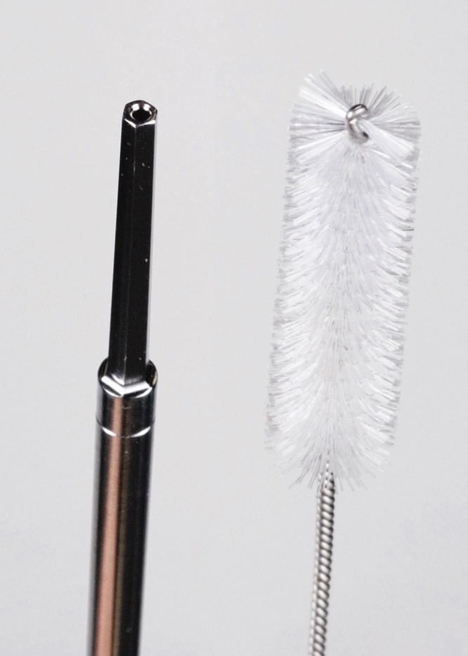
The DePuy Mitek Intrafix cannulated hex driver is made to fit over a small caliber wire. The institution had recently started using this system for tibial fixation. This new system had a specific modular hex driver that was different from ones previously used. At the inception of its use, the institution had no wire brushes of a small-enough diameter for the cleaning and sterilization procedure (Figure 1). As a result, biomaterial was discovered that had remained in the equipment between usage, leading to a breakdown in sterility.
Figure 1.

Original wire brush used for cleaning cannulated devices; this brush is clearly larger in diameter than the center of the hex driver.
The investigative team enacted other processes and changes following their review. All central medical supply personnel went through mandatory reeducation on proper cleaning processes and techniques. The instrumentation involved with the Depuy Mitek Intrafix system was covered in detail, with specific instruction on the cleaning of cannulated instruments with appropriate wire brushes as well as the sonic irrigator. A department-wide operating-room in-service on ACL equipment was given as well. An additional staff member was added to the decontamination station to ensure that proper time was spent on all instrumentation. There were spot checks performed on all ACL instrumentation after all cases for the 4 weeks following this investigation, and weekly cultures were taken for the same period. No biomaterial was found during these subsequent reviews, and all cultures were negative during the same period.
Conclusions
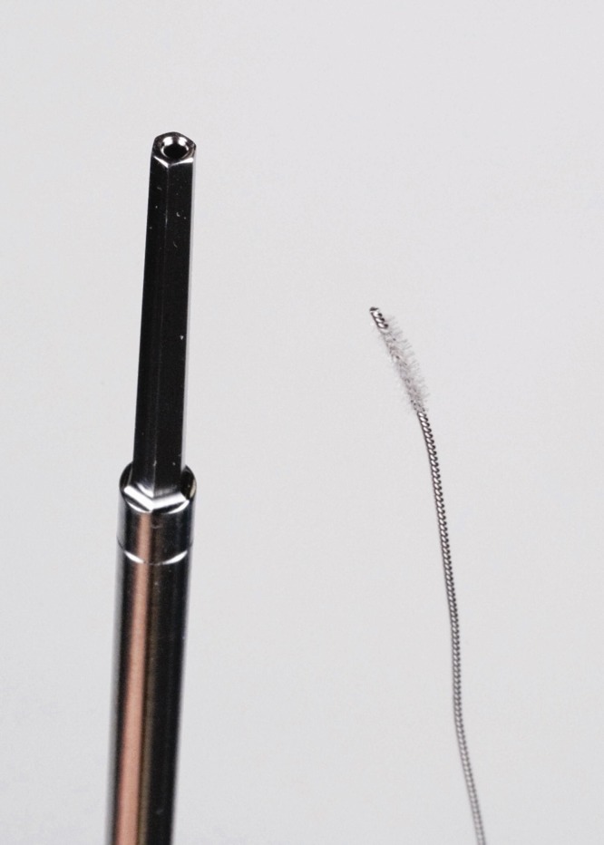
In this case series, a breach in sterile technique was discovered. It was corrected through the use of new wire brushes that were of a smaller diameter, which allowed cleaning of the cannulated portion of the contaminated screwdriver (Figure 2). Once this issue had been identified and corrected, the institution had a return to the preincident rate of infection. With the advent of a multitude of new products by various vendors, the proper execution of sterilization technique has become an increasingly difficult task for hospitals. Similar outbreaks of this type warrant detailed evaluation of sterilization procedures.
Figure 2.

New brush obtained to clean the hex driver, with a small-enough diameter to pass through it.
To our knowledge, this is the first reported series of infection presumptively caused by system failure in sterilization technique. Viola et al reported a retrospective study of 10 patients who had ACL reconstruction and subsequent infections that were traced back to coagulase-negative Staphylococcus in the supposedly sterile inflow cannula.9 No cause for this finding was identified in the study. Babcock et al conducted an outbreak investigation and identified risk factors for developing postoparthroscopic infections.1 They were not able to identify a cause in their outbreak period, but they did identify intra-articular corticosteroid joint injection as an independent risk factor. Blevins et al reported a case series of postopoperative infections seen after arthroscopic meniscus repairs.3 They found a cluster of 3 cases in which they were able to isolate coagulase-negative Staphylococcus. Organic material present in some of the cannulas was also found upon investigation of materials common to all cases. They performed an experiment in which they contaminated the cannulas with coagulase-negative Staphylococcus and then attempted to culture the bacteria after sterilization. Cultures were negative after either flash sterilization or standard sterilization. They concluded that S aureus and coagulase-negative Staphylococcus are the most common cause of septic arthritis following arthroscopy; but owing to the ubiquitous nature of these bacteria, it is often impossible to identify a source of contamination.
Kurzweil presented the data for and against the use of antibiotic prophylaxis for arthroscopic surgery.6 The author agreed with the use of universal prophylaxis before arthroscopic surgery but validated the opposite viewpoint: antibiotic use owing to the multifactorial nature of arthroscopic infections. Bert et al reported that there is no confirmed value in administering antibiotics before routine arthroscopic meniscectomy to prevent joint sepsis.2 All patients in this series, both those who developed infection and those who did not, received preoperative antibiotics. It is impossible to determine whether outcomes were affected.
All cases were managed with antibiotic therapy and serial I&D procedures. The need for radical graft debridement has been reported in previous studies but was not required in this series.4 We did not experience graft rupture in our patients, which has been a complication of postopoperative infection following ACL reconstruction. Schollin-Borg et al reported 10 postopoperative infections following ACL reconstruction.8 They found positive bacteria cultures in 6 patients. They also reported a range of presentation, as this series showed. The authors made note of the delay in treatment after presentation of the infection (average, 5 days; range, 0-34 days). In our experience, all but 2 patients were taken immediately to the operating room for an irrigation and intra-articular debridement, with graft retention. This may have limited the impact of the infection and helped to avoid the need for radical debridement.
There were no complaints of instability after treatment for infection. As such, this series suggests that graft salvage after infection is possible if prompt recognition and treatment of ACL infections is undertaken with aggressive I&D and proper antibiotic therapy.
Footnotes
No potential conflict of interest declared.
References
- 1. Babcock HM, Carroll C, Matava M, L’ecuyer P, Fraser V. Surgical site infections after arthroscopy: outbreak investigation and case control study. Arthroscopy.2003;19:172-181 [DOI] [PubMed] [Google Scholar]
- 2. Bert JM, Giannini D, Nace L. Antibiotic prophylaxis for arthroscopy of the knee: is it necessary? Arthroscopy.2007;23:4-6 [DOI] [PubMed] [Google Scholar]
- 3. Blevins FT, Salgado J, Wascher DC, Koster F. Septic arthritis following arthroscopic meniscus repair: a cluster of three cases. Arthroscopy.1999;15:35-40 [DOI] [PubMed] [Google Scholar]
- 4. Burks RT, Friederichs MG, Fink B, Luker MG, West HS, Greis PE. Treatment of postoperative anterior cruciate ligament infections with graft removal and early reimplantation. Am J Sports Med.2003;31:414-418 [DOI] [PubMed] [Google Scholar]
- 5. Judd D, Bottoni C, Kim D, Burke M, Hooker S. Infections following arthroscopic anterior cruciate ligament reconstruction. Arthroscopy.2006;22:375-384 [DOI] [PubMed] [Google Scholar]
- 6. Kurzweil PR. Antibiotic prophylaxis for arthroscopic surgery. Arthroscopy.2006;22:452-454 [DOI] [PubMed] [Google Scholar]
- 7. Matava MJ, Evans TA, Wright RW, Shively RA. Septic arthritis of the knee following anterior cruciate ligament reconstruction: results of a survey of sports medicine fellowship directors. Arthroscopy.1998;14:717-725 [DOI] [PubMed] [Google Scholar]
- 8. Schollin-Borg M, Michaëlsson K, Rahme H. Presentation, outcome, and cause of septic arthritis after anterior cruciate ligament reconstruction: a case control study. Arthroscopy.2003;19:941-947 [DOI] [PubMed] [Google Scholar]
- 9. Viola R, Marzano N, Vianello R. An unusual epidemic of Staphylococcus-negative infections involving anterior cruciate ligament reconstruction with salvage of the graft and function. Arthroscopy.2000;16:173-177 [DOI] [PubMed] [Google Scholar]
- 10. Williams RJ, Laurencin CT, Warren RF, Speciale AC, Brause BD, O’Brien S. Septic arthritis after arthroscopic anterior cruciate ligament reconstruction: Diagnosis and management. Am J Sports Med.1997;25:261-267 [DOI] [PubMed] [Google Scholar]


