Abstract
Background:
The tennis serve is a complex stroke characterized by a series of segmental rotations involving the entire kinetic chain. Many overhead athletes use a basic 6-stage throwing model; however, the tennis serve does provide some differences.
Evidence Acquisition:
To support the present 8-stage descriptive model, data were gathered from PubMed and SPORTDiscus databases using keywords tennis and serve for publications between 1980 and 2010.
Results:
An 8-stage model of analysis for the tennis serve that includes 3 distinct phases—preparation, acceleration, and follow-through—provides a more tennis-specific analysis than that previously presented in the clinical tennis literature. When a serve is evaluated, the total body perspective is just as important as the individual segments alone.
Conclusion:
The 8-stage model provides a more in-depth analysis that should be utilized in all tennis players to help better understand areas of weakness, potential areas of injury, as well as components that can be improved for greater performance.
Keywords: biomechanics, serve, tennis, kinetic chain
The tennis serve is the most complex stroke in competitive tennis.32 The complexity of the movement results from the combination of limb and joint movements required to summate and transfer forces from the ground up through the kinetic chain and out into the ball. Effective servers maximally utilize their entire kinetic chain via the synchronous use of selective muscle groups, segmental rotations, and coordinated lower extremity muscle activation (quadriceps, hamstrings, and hip rotators, internal and external). This lower body–core force production is then transferred up into the upper body and out through the racket into the ball. If any of the links in the chain are not synchronized effectively, the outcome of the serve will not be optimal.38
The serve has been studied in a similar manner to the throwing motion in baseball, although some significant differences do exist between the serving motion and the throwing motion. These differences include planes of motion, the nondominant arm tossing the tennis ball, the trajectory of forces produced and released, the tennis racket (which alters the lever arm), the technical components of the serve, and the variety of placements and goals of the motion (spin, speed, angle, direction, etc).
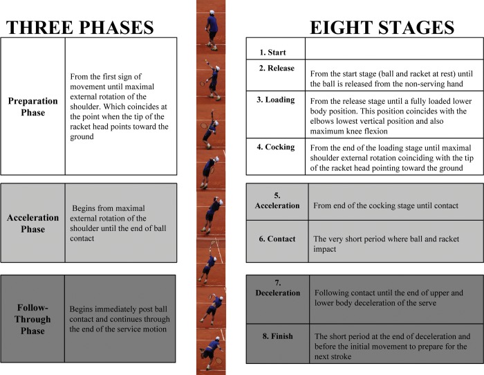
The components usually seen in the traditional throwing analysis30,35 have been altered in this proposed 8-stage tennis-specific serve model (Figure 1). The 8-stage model has 3 distinct phases: preparation, acceleration, and follow-through. Each stage is a direct result of muscle activation and technical adjustments made in the previous stage. When a serve is evaluated, the total body perspective is just as important as the individual segments alone.
Figure 1.
The 3 phases and 8 stages of the tennis serve.
The Kinetic Chain And The Tennis Service Motion
Over a quarter century ago, the kinetic chain was first studied in nationally ranked tennis players.25 Players increase the maximum linear velocity from the knee to the racquet.25 The preparation phase (stages 1-4) results in the storing of potential energy that can be utilized as kinetic energy during the acceleration phase. In an efficiently functioning kinetic chain, the legs and trunk segments are the engine for the development of force and the stable proximal base for distal mobility.26,37,58 This link develops 51% to 55% of the kinetic energy and force delivered to the hand.37 This link also creates the back-leg-to-front-leg angular momentum to drive the arm up and forward.33,55 The large cross-sectional area of the legs and trunk, with its large mass and high moment of inertia, creates an anchor that allows for centripetal motion to occur.10,58 An analysis of the kinetic chain using mathematical modeling revealed that a 20% reduction in kinetic energy from the trunk requires a 34% increase in velocity or a 70% increase in mass to achieve the same kinetic energy to the hand.37 These data highlight the importance of developing effective lower body force and efficient energy transfer up through the kinetic chain.
The 3 Major Types Of Serves In Tennis
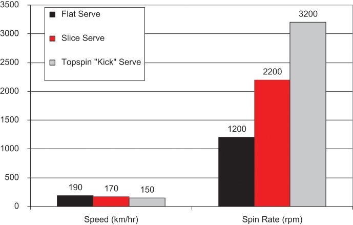
The 3 major types of serves used in tennis are the flat (limited spin), slice (sidespin), and topspin “kick” serves (Figure 2). It is important to understand the differences in these serves and how they may affect the kinetic chain muscle activation patterns and summation of forces. There is an inverse relationship between speed of serves and spin rate (Figure 3). The lower body does not show major differences in the 3 serves. The hypothesis that greater lower trunk muscle activation occurs in topspin (kick) serves has not been supported.7 No major differences were found in 8 lower trunk muscles during flat, topspin, and slice serves. However, bilateral differences in muscle activation were more pronounced in the rectus abdominis and external oblique than in internal oblique and lumbar erector spinae muscles.8
Figure 2.
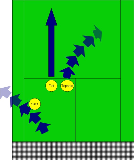
The 3 major types of tennis serves (a right-handed server).
Figure 3.
Speed versus spin comparisons among flat, slice, and topspin (kick) serves. Data adapted from Elliott et al22 and Sakurai et al.53
The major differences seen in serves occur higher in the kinetic chain—namely, at the racket face angle, as determined by forearm pronation and internal shoulder rotation.22
The 8-Stage Model
The 8-stage model has 3 distinct phases: preparation, acceleration, and follow-through. The phases reflect the distinct dynamic functions of the serve: store energy (preparation phase), release energy (acceleration phase), and deceleration (follow-through phase).
Preparation
Phase 1: Start
The start of a player’s serve (Figure 4) reflects style and individual tendency rather than substance. Muscular activation in the shoulder and scapular regions is very low during this phase because demand is low.52 The goal of the start is to align the body to utilize the ground for force/power generation throughout the service motion.
Figure 4.
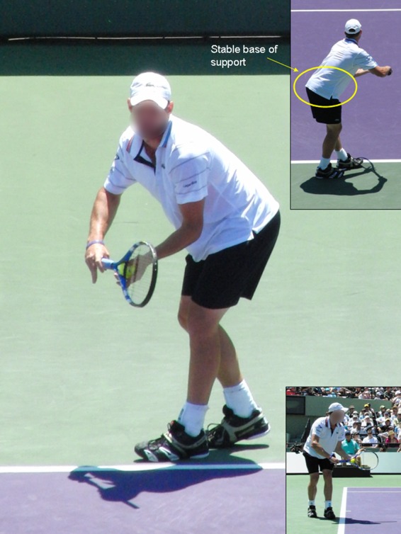
Preparation phase. Stage 1: Start.
Phase 2: Release
This stage occurs in an instant when the ball is released from the nondominant hand (left) (Figure 5). Muscle activation is very limited in the left erector spinae during the start and release stages.7 The activity of the right erector spinae increases steadily from the beginning of the serve through the end.7 The location of the toss relative to the player affects arm abduction and subacromial humeral position. The toss should be out slightly lateral to the overhead position of the server, facilitating ball contact at approximately 100° of arm abduction.29 Improper toss location too close to the head (12 o’clock position) can increase arm abduction and cause subacromial impingement.28 Trunk position and toss location are factors in shoulder pain during the acceleration and contact phases of the tennis serve.12
Figure 5.
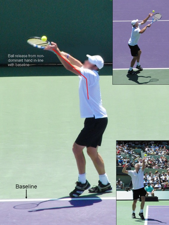
Preparation phase. Stage 2: Release.
Phase 3: Loading
Loading positions the body segments to generate potential energy (Figure 6). There are 2 broad types of lower body loading (foot position) options: the foot-up (Figure 7) or the foot-back (Figure 8) technique.
Figure 6.
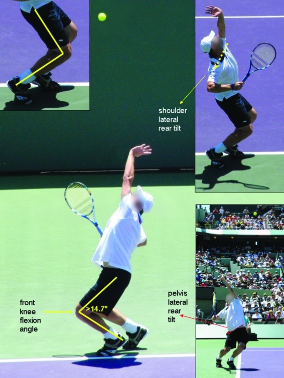
Preparation phase. Stage 3: Loading.
Figure 7.
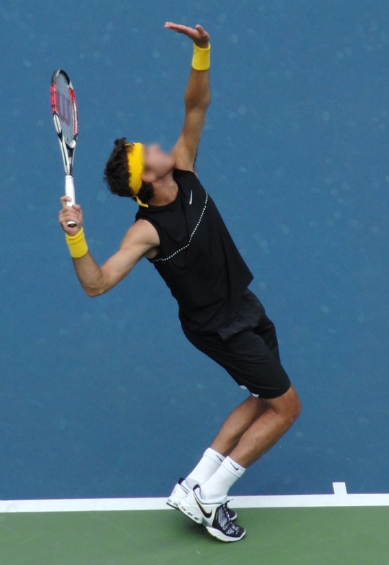
Foot-up serving technique.
Figure 8.
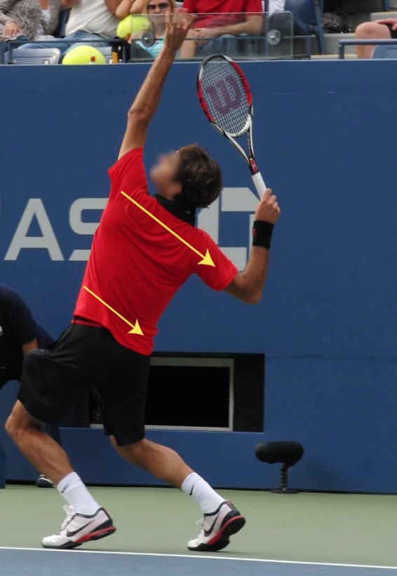
Foot-back serving technique.
Players using a foot-up technique (Figure 7) develop vertical forces, which allow them to reach a greater height than that of the foot-back technique (Figure 8).27 Ball velocities are not different between foot-up and foot-back techniques.27 The back leg provides most of the upward and forward push, whereas the front leg provides a stable post to allow rotational momentum. Large horizontal braking forces are developed with the front foot at landing (stage 8), which is important from a training standpoint. The foot-up serving technique requires eccentric training of the lower body for landing.
A foot-back serving technique requires greater front knee joint extension (65.5° ± 12.6°) compared with the foot-up serving technique (54.1° ± 11.7°, end position).48 This difference is a by-product of the wider base of support, permitting greater squat depth.48 A larger mean range of rear knee joint extension occurs in foot-up serving (59.4° ± 6.6°) compared with the foot-back technique (44.8° ± 8.3°).48 The higher vertical ground reaction forces with a foot-up serving stance4,27 correlate with the peak angular velocities of rear knee joint extension (foot-up, 9.3 ± 1.2 rad·s-1; foot-back, 7.2 ± 0.9 rad·s-1).48
Service velocity correlates with greater muscle force during the loading stage (stage 3),3 while service efficiency is related to internal rotation of the arm.25,26,54 Optimal leg drive mechanics and internal rotation arm flexibility are critical for efficiency and velocity. Maximizing leg motion can produce a consistent leg drive that may enhance shoulder rotation and more efficient serves.32 Compared with beginner servers, elite servers have greater vertical and horizontal force production and earlier activation of the major lower body muscle.32
Shoulder and pelvis lateral rear tilt before the cocking phase (ie, during the loading phase) is a feature of powerful servers (Figures 8 and 9).48 This tilted alignment facilitates the development of angular momentum through lateral trunk flexion during the forward swing: a critical factor in a high-velocity serve.2
Figure 9.
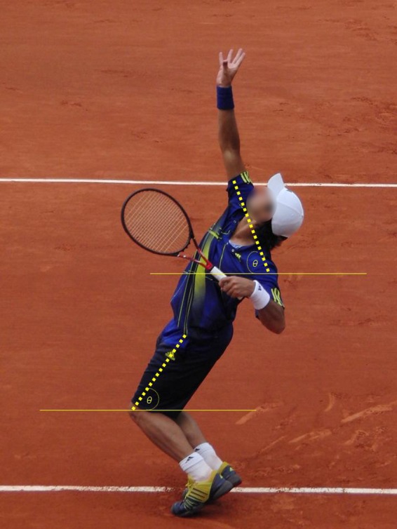
Shoulder and pelvis lateral rear tilt during the loading stage.
The ground reaction forces created in stage 3 (loading) result in an off-center angular impulse, which elevates the racquet side (dominant arm side) of the body and lowers the opposite side (nondominant arm). This produces a shoulder-over-shoulder rotation as the server explosively moves the arm toward the position of ball contact (stage 6) and allows for greater racket height. These movements transfer angular momentum from the lower limbs to the upper limb.2 To achieve this optimum position, lateral trunk flexion (right to left) requires good flexibility and optimal core stability throughout the range of motion.
A front knee flexion angle greater than 15° during the loading stage is recommended for effective front “leg drive.”21 Elite servers with optimal front “leg drive” have lower anterior shoulder and medial elbow loads. The benefits of effective kinetic chain involvement include reducing injury potential in the high-performance tennis serve. The activation patterns of the lower trunk muscles clearly demonstrate a high degree of co-contraction during a tennis serve, especially during stages 3 to 7.7
During right trunk rotation in a right-handed server (Figure 10), the ipsilateral erector spinae is more active than the contralateral.44 The ipsilateral erector spinae increase activity from stage 3 through the end of the deceleration phase of the serve.7 The lateral left erector spinae assist lateral flexion after stage 3 (loading).7
Figure 10.
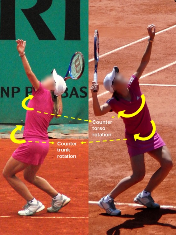
Trunk and torso rotation during the loading stage of the serve.
During the loading and cocking phases, the spine moves into hyperextension, ipsilateral lateral flexion, and ipsilateral rotation. This loads the spinal facets and is a potential factor in the development of spondylolysis in elite developing players.1 Electromyogram (EMG) studies demonstrate high trunk muscle activation in this stage.7 The plyometric stretch-shortening pattern in the tennis serve leads to selective development of the trunk flexors (abdominals).51 Isokinetic testing shows flexion:extension ratio imbalance in elite tennis players.18,51 Symmetric trunk rotation dictates the need for bilateral trunk rotation strength and conditioning exercise (core stabilization) as well as extensor targeting.18,51
Phase 4: Cocking
The cocking position (Figure 11) depends on an efficient loading stage (stage 3). Increasing the efficiency of the dominant arm in driving the racket down and behind the torso lengthens the trajectory of the racket to the ball.24 This position allows for greater potential energy but does require optimal range of motion, positioning, and stabilization throughout the shoulder region.
Figure 11.
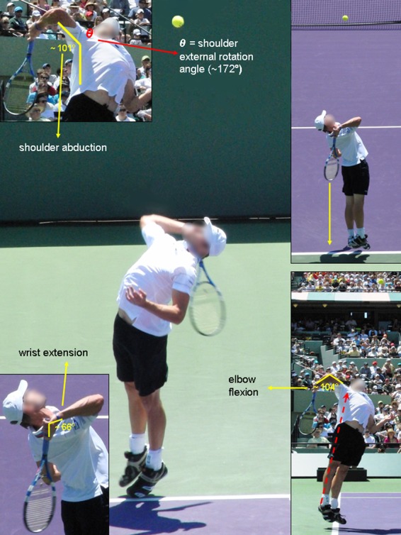
Preparation phase. Stage 4: Cocking.
High internal rotator eccentric loads are applied during the late preparation phase (backswing) (Figure 12), later transitioning into the acceleration phase (stage 5) before impact.3 Effective leg drive forces the racket in a downward motion away from the back. This energy is recovered to assist in generating racket velocity during the acceleration phase of the service motion.21
Figure 12.
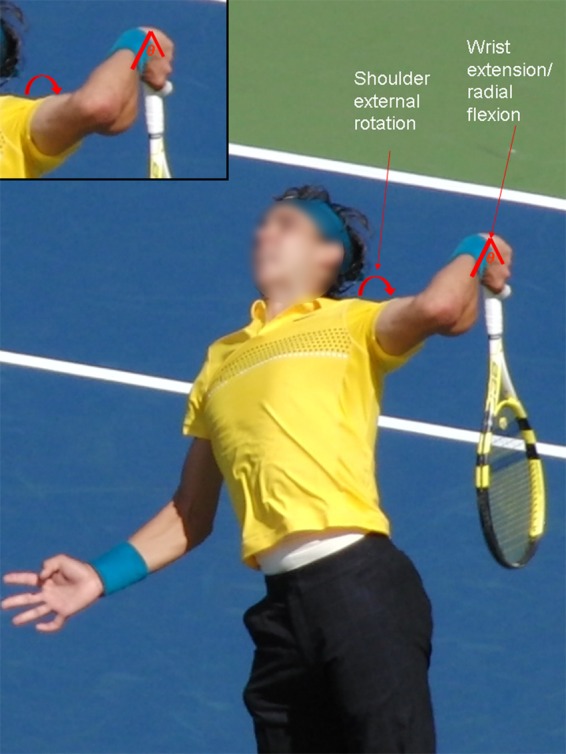
Shoulder and serving arm position during the cocking stage of the serve.
Between the loading stage (position 3) and the beginning of the acceleration stage (position 5), there is an increase in vertical ground reaction forces, while increased muscle activation occurs in the vastus medialis, vastus lateralis, and gastrocnemius.4 Maximal shoulder external rotation is achieved 0.090 ± 0.014 seconds before contact in professional tennis players.29 Leg drive is near completion at this stage.29 At the instant of maximum external rotation, the shoulder is abducted 101° ± 13°, horizontally adducted 7° ± 13°, and externally rotated 172° ± 12°; the elbow is flexed 104° ± 12°; and the wrist is extended 66° ± 19°.29 This resulted in a near parallel position between the racket and the trunk. The magnitude of external rotation is similar to that for elite baseball pitchers, 175° to 185°.11,31 This degree of external rotation is a combination of glenohumeral and scapulothoracic motion, as well as trunk extension motion.11
Repeated external rotation in the tennis serve can lead to increased shoulder external rotation on the dominant arm at the expense of internal rotation.13,19 These increases in external rotation do not match the magnitude of the increases reported in the dominant arm of professional baseball pitchers.19,56 Loss of both internal rotation and total rotation up to 10° to 15° in elite level tennis players occurs at 90° in the abducted shoulder.13,19,41 Stretches of the posterior shoulder (sleeper and cross arm39,41,43) counter internal rotation losses in developing and elite-level competitive tennis players.15,16,34,40,41
During the cocking stage, shoulder loads in the abducted, externally rotated position can lead to injury.5 Muscle activity (percentage maximum voluntary contraction) during the cocking stage is moderately high in the supraspinatus (53%), infraspinatus (41%), subscapularis (25%), biceps brachii (39%), and serratus anterior (70%) to provide stabilization.52 The moderately high activity during this stage demonstrates the importance of anterior and posterior rotator cuff and scapular stabilization for proper execution of the cocking stage.
Tennis serves depend on glenohumeral position during the cocking stage: 7° of horizontal adduction from the coronal plane places the glenohumeral joint anterior to the coronal plane (Figure 12).29 There is increased contact pressure between the supraspinatus/infraspinatus and the posterior glenoid (internal impingement) in cadavers with the abducted, externally rotated shoulder.45 This hyperabduction position is a risk factor in the throwing shoulder.30 Premature dropping of the tossing arm, coupled with early hip and trunk forward rotation, can lead to an exaggerated horizontal abduction, or arm lag, subjecting the shoulder to posterior impingement and loading the anterior capsular structures.5,45
The position of maximal external rotation in the cocking phase shows high abduction angles (83° and 101°),25,29 which risks impingement.57 Lower external:internal rotation ratios on the dominant extremity in elite-level tennis players indicate selective development of the internal rotators relative to the external rotators.13,14,17 Elite junior tennis players show normal external:internal rotation ratios at 90° of abduction with isokinetic testing: 66% to 75% for the dominant arm and 80% to 85% for the nondominant arm.49
Very high activation of the left internal oblique is seen during the preparation phase (stage 4: cocking) and acceleration phase.7 Regardless of the type of serve, the activation patterns of the left and right rectus abdominis are similar. Activation at the end of the preparation and acceleration phases is greater than that at the start of the preparation phase and follow-through.7
Acceleration
Phase 5: Acceleration
The acceleration stage is determined by the previous 4 stages. Elite servers have a quicker acceleration phase (stages 5 and 6) than do beginner servers as a result of a more vigorous knee extension from stages 3 to 6.32 Advanced servers move from maximum glenohumeral joint external rotation to ball contact in less than 1/100 of a second.29
High muscle activity (% maximum voluntary isometric contraction) was found in the pectoralis major (115%), subscapularis (113%), latissimus dorsi (57%), and serratus anterior (74%) during internal rotation of the humerus consistent with EMG recordings during the acceleration phase of throwing.30,52 The pectoralis major, deltoid, trapezius, and triceps are active during the acceleration phase.46,55
Power production during the acceleration stage (concentric action) depends on 2 factors: strength and neuromuscular coordination.47 Vertical force production produced in the serve is approximately 1.68 to 2.12 times one’s body weight.27,32
Peak EMG values for the vastus lateralis, vastus medialis, and gastrocnemius occurs near the end of stage 5 (Figure 13).32 Trunk muscles show their highest EMG values during the acceleration phase.7 The greatest kinetic energy produced during the tennis serve is in the legs/trunk.32,50 Maximum lead knee extension velocity (800° ± 400° per second) occurred 0.180 ± 0.065 seconds before contact29 in professional tennis players competing at the 2000 Olympic Games.
Figure 13.
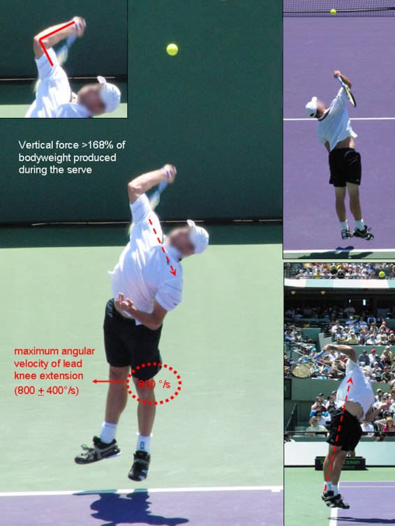
Acceleration phase. Stage 5: Acceleration.
Acceleration of the racquet before ball impact is accompanied by a rapid lumbar spine rotation reversal for a right-handed server from hyperextension and right lateral flexion to flexion and left lateral flexion. This movement (corkscrew) transfers the torque to the spinal segments.23 The serve may place more stress on the lumbar spine because repetitive trunk hyperextensions are a predisposing mechanism for spondylolysis.1,9,36
Both sexes produce segment rotations in the same order; maximum upper torso velocity for females (0.075 seconds before impact) which is earlier than for males (0.058 seconds).29 Females may need a longer phase before maximum torso velocity to increase stored energy and the resultant torso velocity.
Phase 6: Contact
At contact (Figures 14 and 15), the trunk has an average tilt of approximately 48° above horizontal; the arm is abducted 101°; and the elbow, wrist, and lead knee are slightly flexed in Olympic tennis players.29 Elite tennis players generate racket velocities of approximately 38 to 47 m·s-1 (85 to 105 miles·h-1).6,48 The mean shoulder abduction just before contact is approximately 100°,29 which is similar to the 100° ± 10° angle to produce maximal ball velocity and minimal shoulder joint loading in baseball pitching.42,48 This suggests an optimum contact point of 110° ± 15° for the tennis serve.
Figure 14.
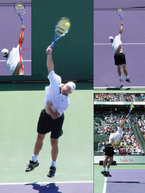
Acceleration phase. Stage 6: Contact.
Figure 15.
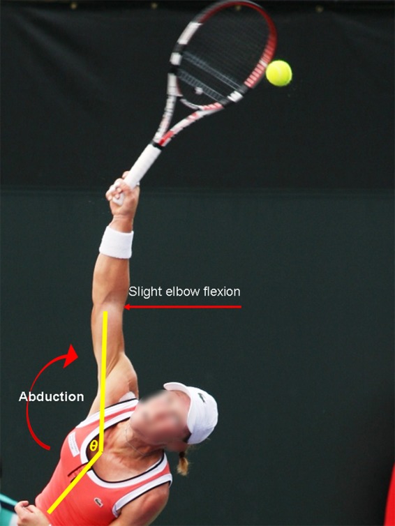
Upper body position during contact.
At ball contact, ball velocity is determined by shoulder internal rotation and wrist flexion.25,26 Elbow flexion (20° ± 4°), wrist extension (15° ± 8°), and front knee flexion (24° ± 14°) are minimal at contact.29 Trunk is tilted 48° ± 7° above horizontal in Olympic professional tennis players.29
Greater maximal left lateral flexion angle indicates greater height during a tennis serve7 and greater left rectus abdominis activity.7 Highly skilled players are subjected to greater asymmetric loads on their lumbar spines due to the greater lateral flexion.
Follow-Through
Phase 7: Deceleration
The follow-through phase (stages 7 and 8) is the most violent of the tennis serve, requiring deceleration eccentric loads in both the upper and lower body (Figure 16). Continued glenohumeral internal rotation and forearm pronation occur during the acceleration stage and continue after ball contact during deceleration. This coupled motion has been termed long axis rotation.21,26
Figure 16.
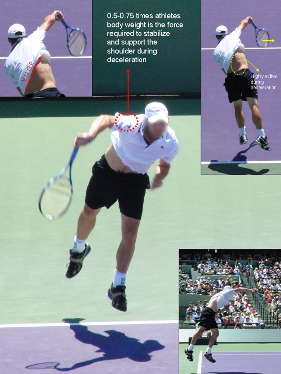
Follow-through phase. Stage 7: Deceleration.
The deceleration force activity between the trunk and the arm during the deceleration stage can be as high as 300 N·m.20 This force stabilizes the shoulder against the distraction forces of 0.5 to 0.75 times one’s body weight.20 There is also moderately high activity in the posterior rotator cuff, serratus anterior, biceps brachii, deltoid, and latissimus dorsi musculature.52 The posterior cuff activation ranges between 30% and 35% as the humerus is decelerated following contact52 to offset the distraction forces and maintain glenohumeral congruity. Serratus anterior maximum voluntary isometric contraction (53%) indicates the continued need for scapular stabilization.52
The left internal oblique is more active than the right internal oblique throughout a serve except during the deceleration stage.7 To stabilize the trunk during an unbalanced posture (ie, the deceleration stage), the right erector spinae become highly active during the deceleration stage.7
Phase 8: Finish
The last stage of the service motion results in lower body landing, which generates eccentric forces (Figure 17). Larger horizontal braking forces are developed with the front foot (at landing, stage 8) using the foot-up technique (vs the foot-back technique) since the center of mass is shifted forward, which may hinder serve and volley players.4
Figure 17.
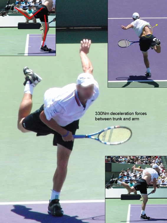
Follow-through phase. Stage 8: Finish.
There are few significant differences in serve kinematics between professional male and female players, thereby eliminating the need to teach different mechanics to them.29
Summary
The 3-phase, 8-stage model of the high-performance serve provides a framework for analysis. There is an inverse relationship between spin and speed; service velocity is correlated with leg drive during the loading stage. There are 2 main foot positions utilized during the loading stage of the serve (foot-up and foot-back). Effective servers utilize rear lateral shoulder and pelvis tilt to store potential energy for speed and spin during the acceleration phase of the serve. Acceleration of the racquet before ball impact is accompanied by a rapid reversal of the rotation of the lumbar spine.
References
- 1. Alyas F, Turner M, Connell D. MRI findings in the lumbar spines of asymptomatic adolescent elite tennis players. Br J Sports Med. 2007;41:836-841 [DOI] [PMC free article] [PubMed] [Google Scholar]
- 2. Bahamonde RE. Changes in angular momentum during the tennis serve. J Sports Sci. 2000;18(8):579-592 [DOI] [PubMed] [Google Scholar]
- 3. Bahamonde RE. Joint power production during flat and slice tennis serves. In: Wilkerson JD, Ludwig KM, Zimmerman WJ, eds. Proceedings on the 15th International Symposium on Biomechanics in Sports Denton, TX; 1997:489-494 [Google Scholar]
- 4. Bahamonde RE, Knudson D. Ground reaction forces of two types of stances and tennis serves. Med Sci Sports Exerc. 2001;33:S102 [Google Scholar]
- 5. Burkhart SS, Morgan CD, Kibler WB. The disabled throwing shoulder: spectrum of pathology. Part 1: pathoanatomy and biomechanics. Arthroscopy. 2003;19:404-420 [DOI] [PubMed] [Google Scholar]
- 6. Chow JW, Carleton LG, Lim YT. Comparing the pre- and post-impact ball and racquet kinematics of elite tennis players’ first and second serves: a preliminary study. J Sports Sci. 2003;21(7):529-537 [DOI] [PubMed] [Google Scholar]
- 7. Chow JW, Park S, Tillman MD. Lower trunk kinematics and muscle activity during different types of tennis serves. Sports Med Arthrosc Rehabil Ther Technol. 2009;1(24). [DOI] [PMC free article] [PubMed] [Google Scholar]
- 8. Chow JW, Shim JH, Lim YT. Lower trunk muscle activity during the tennis serve. J Sci Med Sport. 2003;6:512-518 [DOI] [PubMed] [Google Scholar]
- 9. Congeni J, McCulloch J, Swanson K. Lumbar spondylolysis: a study of natural progression in athletes. Am J Sports Med. 1997;25:248-253 [DOI] [PubMed] [Google Scholar]
- 10. Cordo PJ, Nasher LM. Properties of postural adjustments associated with rapid arm movements. J Neurophysiol. 1982;47:287-308 [DOI] [PubMed] [Google Scholar]
- 11. Dillman CJ, Fleisig GS, Andrews JR. Biomechanics of pitching with emphasis upon shoulder kinematics. J Orthop Sports Phys Ther. 1993;18:402-408 [DOI] [PubMed] [Google Scholar]
- 12. Ellenbecker TS. Rehabilitation of shoulder and elbow injuries in tennis players. Clin Sports Med. 1995;14(1):87-110 [PubMed] [Google Scholar]
- 13. Ellenbecker TS. Shoulder internal and external rotation strength and range of motion in highly skilled tennis players. Isokinet Exerc Sci. 1992;2:1-8 [Google Scholar]
- 14. Ellenbecker TS. A total arm strength isokinetic profile of highly skilled tennis players. Isokinet Exerc Sci. 1991;1:9-21 [Google Scholar]
- 15. Ellenbecker TS, Cools A. Rehabilitation of shoulder impingement syndrome and rotator cuff injuries: an evidence based review. Br J Sports Med. 2010;44:319-327 [DOI] [PubMed] [Google Scholar]
- 16. Ellenbecker TS, Reinold MM, Nelson CO. Clinical concepts for treatment of the elbow in the adolescent overhead athlete. Clin Sports Med. 2010;29(4):705-724 [DOI] [PubMed] [Google Scholar]
- 17. Ellenbecker TS, Roetert EP. Age specific isokinetic glenohumeral internal and external rotation strength in elite junior tennis players. J Sci Med Sport. 2003;6(1):63-70 [DOI] [PubMed] [Google Scholar]
- 18. Ellenbecker TS, Roetert EP. An isokinetic profile of trunk rotation strength in elite tennis players. Med Sci Sports Exerc. 2004;36(11):1959-1963 [DOI] [PubMed] [Google Scholar]
- 19. Ellenbecker TS, Roetert EP, Baillie DS, Davies GJ, Brown SW. Glenohumeral joint total rotation range of motion in elite tennis players and baseball pitchers. Med Sci Sports Exerc. 2002;34(12):2052-2056 [DOI] [PubMed] [Google Scholar]
- 20. Ellenbecker TS, Roetert EP, Kibler WB, Kovacs MS. Applied biomechanics of tennis. In: Magee DJ, Manske RC, Zachazewski JE, Quillen WS, eds. Athletic and Sports Issues in Musculoskeletal Rehabilitation. St Louis, MO: Saunders; 2010:chap 11 [Google Scholar]
- 21. Elliott B, Fleisig GS, Nicholls R, Escamilla R. Technique effects on upper limb loading in the tennis serve. J Sci Med Sport. 2003;6(1):76-87 [DOI] [PubMed] [Google Scholar]
- 22. Elliott B, Reid M, Crespo M. Technique Development in Tennis Stroke Production. London, UK: International Tennis Federation; 2009 [Google Scholar]
- 23. Elliott BC. Biomechanics of the serve in tennis: a biomedical perspective. Sports Med. 1988;6:285-294 [DOI] [PubMed] [Google Scholar]
- 24. Elliott BC. Biomechanics of tennis. In Renstrom P., ed. Tennis. Oxford, UK: Blackwell; 2002:1-28 [Google Scholar]
- 25. Elliott BC, Marhs T, Blanksby B. A three-dimensional cinematographical analysis of the tennis serve. Int J Sport Biomech. 1986;2:260-270 [Google Scholar]
- 26. Elliott BC, Marshall RN, Noffal GJ. Contributions of upper limb segment rotations during the power serve in tennis. J Appl Biomech. 1995;11:433-442 [Google Scholar]
- 27. Elliott BC, Wood GA. The biomechanics of the foot-up and foot-back tennis service techniques. Aust J Sport Sci. 1983;3:3-6 [Google Scholar]
- 28. Flatow EL, Soslowsky LJ, Ticker JB, et al. Excursion of the rotator cuff under the acromion: patterns of subacromial contact. Am J Sports Med. 1994;22(6):779-788 [DOI] [PubMed] [Google Scholar]
- 29. Fleisig G, Nicholls R, Elliott B, Escamilla R. Kinematics used by world class tennis players to produce high-velocity serves. Sports Biomech. 2003;2(1):51-71 [DOI] [PubMed] [Google Scholar]
- 30. Fleisig GS, Andrews JR, Dillman CJ, Escamilla RF. Kinetics of baseball pitching with implications about injury mechanisms. Am J Sports Med. 1995;23:233-239 [DOI] [PubMed] [Google Scholar]
- 31. Fleisig GS, Barrentine SW, Zheng N, Escamilla RF, Andrews J. Kinematic and kinetic comparison of baseball pitching among various levels of development. J Biomech. 1999;32:1371-1375 [DOI] [PubMed] [Google Scholar]
- 32. Girard O, Micallef JP, Millet GP. Lower-limb activity during the power serve in tennis: effects of performance level. Med Sci Sports Exerc. 2005;37(6):1021-1029 [PubMed] [Google Scholar]
- 33. Groppel JL. Tennis for Advanced Players and Those Who Would Like to Be. Champaign, IL: Human Kinetics; 1984 [Google Scholar]
- 34. Harryman DT, Sidles JA, Clark JM, Mcquade KJ, Gibb TD, Matsen FA. Translation of the humeral head on the glenoid with passive glenohumeral joint motion. J Bone Joint Surg Am. 1990;72(9):1334-1343 [PubMed] [Google Scholar]
- 35. Jobe FW, Tibone JE, Perry J, Moynes D. An EMG analysis of the shoulder in throwing and pitching: a preliminary report. Am J Sports Med. 1983;11(1):3-5 [DOI] [PubMed] [Google Scholar]
- 36. Kelsey JL, White AA. Epidemiology and impact of low-back pain. Spine. 1980;5:133-142 [DOI] [PubMed] [Google Scholar]
- 37. Kibler WB. Biomechanical analysis of the shoulder during tennis activities. Clin Sports Med. 1995;14(1):79-85 [PubMed] [Google Scholar]
- 38. Kibler WB. The 4000-watt tennis player: power development for tennis. Med Sci Tennis. 2009;14(1):5-8 [Google Scholar]
- 39. Kibler WB, Chandler TJ. Range of motion in junior tennis players participating in an injury risk modification program. J Sci Med Sport. 2003;6(1):51-62 [DOI] [PubMed] [Google Scholar]
- 40. Kibler WB, Chandler TJ, Livingston BP, Roetert EP. Shoulder range of motion in elite tennis players. Am J Sports Med. 1996;24(3):279-285 [DOI] [PubMed] [Google Scholar]
- 41. Manske RC, Meschke M, Porter A, Smith B, Reiman M. A randomized controlled single-blinded comparison of stretching versus stretching and joint mobilization for posterior shoulder tightness measured by internal rotation loss. Sports Health. 2010;2(2):94-100 [DOI] [PMC free article] [PubMed] [Google Scholar]
- 42. Matsuo T, Matsumoto T, Takada Y, Mochizuki Y. Influence of lateral trunk tilt on throwing arm kinetics during baseball pitching. In: Hong Y, Johns DP, eds. Proceedings of XVIII International Symposium on Biomechanics in Sports. Hong Kong: Chinese University of Hong Kong; 2000:882-886 [Google Scholar]
- 43. McClure P, Balaicuis J, Heiland D, Broersma ME, Thorndike CK, Wood A. A randomized controlled comparison of stretching procedures for posterior shoulder tightness. J Orthop Sports Phys Ther. 2007;37:108-114 [DOI] [PubMed] [Google Scholar]
- 44. McGill SM. The influence of lordosis on axial trunk torque and trunk muscle myoelectric activity. Spine. 1992;17:1187-1193 [DOI] [PubMed] [Google Scholar]
- 45. Mihata T, McGarry MH, Kinoshita M, Lee TQ. Excessive glenohumeral horizontal abduction as occurs during the late cocking phase of the throwing motion can be critical for internal impingement. Am J Sports Med. 2010;38(2):369-374 [DOI] [PubMed] [Google Scholar]
- 46. Miyashita M, Tsundoda T, Sakurai S, Nishizona H, Mizunna T. Muscular activities in the tennis serve and overhead throwing. Scand J Med Sci Sports. 1980;2:52-58 [Google Scholar]
- 47. Nagano A, Gerritsen KGM. Effects of neuromuscular strength training on vertical jumping performance-a computer simulation study. J Appl Biomech. 2001;17:113-128 [Google Scholar]
- 48. Reid M, Elliott B, Alderson J. Lower-limb coordination and shoulder joint mechanics in the tennis serve. Med Sci Sports Exerc. 2008;40(2):308-315 [DOI] [PubMed] [Google Scholar]
- 49. Roetert EP, Ellenbecker TS, Brown SW. Shoulder internal and external rotation range of motion in nationally ranked junior tennis players: a longitudinal analysis. J Strength Cond Res. 2000;14(2):140-143 [Google Scholar]
- 50. Roetert EP, Groppel JL. Mastering the kinetic chain. In: Roetert EP, Groppel JL, eds. World Class Tennis Technique. Champaign, IL: Human Kinetics; 2001:99-113 [Google Scholar]
- 51. Roetert EP, McCormick T, Brown SW, Ellenbecker TS. Relationship between isokinetic and functional trunk strength in elite junior tennis players. Isokinet Exerc Sci. 1996;6:15-30 [Google Scholar]
- 52. Ryu KN, McCormick FW, Jobe FW, Moynes DR, Antonell DJ. An electromyographic analysis of shoulder function in tennis players. Am J Sports Med. 1988;16:481-485 [DOI] [PubMed] [Google Scholar]
- 53. Sakurai S, Jinii T, Reid M, Cuitenho C, Elliott B. Direction of spin axis and spin rate of the ball in tennis service. J Biomech. 2007;40(2):S197 [Google Scholar]
- 54. Sprigings E, Marshall R, Elliott B, Jennings L. A three-dimensional kinematic method for determining the effectiveness of arm segment rotations in producing racket head speed. J Biomech. 1994;27:245-254 [DOI] [PubMed] [Google Scholar]
- 55. Van Gheluwe B, Hebbelinck M. Muscle actions and ground reaction forces in tennis. Int J Sport Biomech. 1986;2:88-99 [Google Scholar]
- 56. Wilk KE, Reinold MM, Macrina LC, et al. Gleohumeral internal rotation measurements differ depending on stabilization techniques. Sports Health. 2009;1(2):131-136 [DOI] [PMC free article] [PubMed] [Google Scholar]
- 57. Wuelker N, Korell M, Thren K. Dynamic glenohumeral joint stability. J Shoulder Elbow Surg. 1998;7:43-52 [DOI] [PubMed] [Google Scholar]
- 58. Zattara M, Bouisset S. Posturo-kinetic organization during the early phase of voluntary upper-limb movement. J Neurol Neurosurg Psychiatry. 1988;51:956-965 [DOI] [PMC free article] [PubMed] [Google Scholar]


