

Abstract
Context:
Various epidemiological studies have estimated that up to 70% of runners sustain an overuse running injury each year. Although few overuse running injuries have an established cause, more than 80% of running-related injuries occur at or below the knee, which suggests that some common mechanisms may be at work. The question then becomes, are there common mechanisms related to overuse running injuries?
Evidence Acquisition:
Research studies were identified via the following electronic databases: MEDLINE, EMBASE PsycInfo, and CINAHL (1980–July 2008). Inclusion was based on evaluation of risk factors for overuse running injuries.
Results:
A majority of the risk factors that have been researched over the past few years can be generally categorized into 2 groups: atypical foot pronation mechanics and inadequate hip muscle stabilization.
Conclusion:
Based on the review of literature, there is no definitive link between atypical foot mechanics and running injury mechanisms. The lack of normative data and a definition of typical foot structure has hampered progress. In contrast, a large and growing body of literature suggests that weakness of hip-stabilizing muscles leads to atypical lower extremity mechanics and increased forces within the lower extremity while running.
Keywords: running, injuries, cause, prevention
Although runners often sustain acute injuries such as ankle sprains and muscle strains, a majority of running injuries can be classified as cumulative micro-trauma injuries (ie, overuse injuries).13,56 Running is one of the most widespread activities during which overuse injuries of the lower extremity occur. Various epidemiological studies have estimated that anywhere from 27% to 70% of recreational and competitive distance runners sustain an overuse running injury during any 1-year period.2,26,32,35,38,53,64 The runners in these studies vary considerably in their running experience and training habits, but they generally run at least 20 to 30 km per week and have been running consistently for at least 1 to 3 years.
The knee is the most common site of overuse running injuries, accounting for close to 50% of all injuries.7,51,54,59 A recent systematic review and meta-analysis reported that the knee was the most common site of musculoskeletal injury for runners.61 According to a clinical study of more than 2000 injured runners, the most common knee condition is patellofemoral pain syndrome (PFPS), followed by iliotibial band syndrome (ITBS), meniscal injuries, and patellar tendinitis.59 Injuries to the foot, ankle, and lower leg—such as plantar fasciitis, Achilles tendinitis, and medial tibial stress syndrome (also known as shin splints)—account for almost 40% of the remaining injuries, whereas less than 20% of the running injuries occur superior to the knee. Although few overuse running injuries have an established cause,5 more than 80% of these injuries occur at or below the knee, thus suggesting that some common mechanisms may be involved.
The cause of these injuries is multifactorial and diverse,38,54,61,62 and several identifiable factors may predict who is at risk.
Evidence Acquisition
For the purpose of this clinical review, research studies were identified via the following electronic databases: MEDLINE, EMBASE, PsycInfo, and CINAHL (1980–July 2008). Included studies were directly related to risk factors for overuse. The following keywords were used: running, injury, mechanics, and knee (resulting in 283 articles). Criteria for screening included (1) running injuries in long-distance runners, (2) a minimum of 20 km per week, (3) recreational or competitive runners but not elite, and (4) epidemiology (prevalence, incidence) or etiology (determinants). Two reviewers categorized the studies to determine whether a majority identified common risk factors. As such, risk factors were generally categorized into 2 groups: atypical foot pronation mechanics and inadequate hip muscle stabilization.
Foot Pronation Mechanics
Pronation is a combination of ankle dorsiflexion, rearfoot eversion, and forefoot abduction, and it occurs during the first half of the stance phase in running. Excessive rearfoot frontal plane motion (eversion) influences lower extremity mechanics via tibial rotation.11,36,39,65 During the first half of the stance phase, the calcaneus everts and the head of the talus internally rotates.24,34 The tibia internally rotates with the talus, owing to the tight articulation of the ankle joint mortise.24,34 In weightbearing activities such as running, there is a direct relationship between degree of pronation and internal tibial rotation (ie, for runners who exhibit a heel-toe footfall pattern).12 Pronation is a necessary and protective mechanism during running; it allows impact forces to be attenuated over a long period. Researchers have suggested that a higher level of pronation is favorable during running, if it falls within normal physiological limits and does not continue beyond midstance.22,58,66 After midstance, it is necessary for the foot to become more rigid and supinate in preparation of toe-off (ie, the tibia and talus externally rotate and the calcaneus inverts). As such, the rearfoot inverts and the tibia externally rotates.24,34 Severe overpronators, or runners who exhibit prolonged pronation, may be at an increased risk of injury because of the potentially large torques generated within the lower extremity and the subsequent increase in internal tibial rotation.3,20,39,42,43 Specifically, the tibialis posterior and soleus muscles function to minimize these torsional forces within the shank and ankle complex.45 If these forces are experienced within the knee or hip joints, then the hamstring and deep external rotator muscles must concomitantly contract to control the subsequent torsional forces, respectively.45
Excessive pronation, pronation velocity, and time to maximum pronation have often been implicated as contributing factors to overuse running injuries.5,27,28,41,42,54 In many studies, a static evaluation of pronation was conducted on injured runners, with the results suggesting that injured runners were more often overpronators when compared to uninjured runners. However, minimal and conflicting experimental evidence supports excessive foot pronation as a contributing factor in the cause of injuries. The majority of these studies were cross-sectional. One study partially supported the speculation regarding a cause-and-effect relationship between foot pronation and injury; it reported that groups of injured runners, when compared to a control group comprising uninjured runners, exhibited greater maximum pronation angles and had greater maximum pronation velocities.43 The results were evident in a group who had medial tibial stress syndrome. Viitasalo and Kvist63 reported similar results when comparing runners with medial tibial stress syndrome to an uninjured control group during barefoot running. However, contradictory results were found in a study in which runners who had never sustained an overuse injury exhibited greater pronation velocity and greater touchdown supination angle when compared to runners who had sustained an overuse injury.23 Messier et al43 compared runners with PFPS to an uninjured control group42 and found no differences in any rearfoot variables. Thus, the relationship between rearfoot position and running injury susceptibility is not clear given these retrospective cross-sectional design studies.
Unfortunately, only 2 prospective studies have been conducted to investigate the link between foot mechanics and overuse injuries. Willems et al67,68 evaluated lower leg pain in a group of 400 physically active young individuals. Plantar pressure measurements and 3-dimensional rearfoot kinematic data were collected, and participants were followed for 1 academic year. Seventy-five injured runners were identified, and their data were compared to those of 167 noninjured runners. The injured runners exhibited significantly prolonged rearfoot pronation, increased medial foot pressure, and accelerated reinversion when compared to controls, thus suggesting that atypical foot pronation is a contributing factor in the cause of running-related injuries. In contrast, Thijs et al,60 using plantar pressure measurements, examined gait-related risk factors for patellofemoral pain in a group of 84 officer cadets over the course of a 6-week basic military training period. Thirty-six cadets developed patellofemoral pain and were therefore compared to the remaining 48. Compared with the control group, the injured group exhibited a supinated heel strike position and reduced pronation (greater lateral contact pressure). Thus, the only 2 prospective studies conducted to date provide conflicting results: one suggests that excessive foot pronation mechanics are related to injury development, whereas the other suggests that reduced foot pronation mechanics are the culprit. Based on these data and the contradictory results derived from the various retrospective cross-sectional studies outlined previously, no definitive answer can be put forth regarding potential running-related injury mechanisms and excessive foot pronation.
Establishing the Typical Foot
The range of physiological foot pronation has not been established. Several investigators have based selection of participants on relatively arbitrary criteria. Mündermann et al46 classified 20 runners as overpronators on the basis of a 2- dimensional standing rearfoot-shank angle greater than 13° (see Figure 1). This value was based on work by Clarke et al,6 who averaged the maximum pronation angle from 9 studies conducted between 1978 and 1983. The average angle when running was 9.4° (± 3.5°), with a maximum pronation of 13° or greater labeled excessive.
Figure 1.
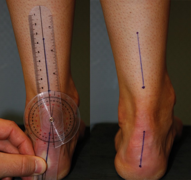
Right, markings to bisect the long axis of the shank and rearfoot; left, goniometric measurement of standing rearfoot-shank angle (left foot shown).
McClay and Manal40 investigated lower extremity mechanics for a group of 20 recreational runners exhibiting normal6 rearfoot mechanics. Participants for this study were selected using a dynamic assessment of 2-dimensional peak rearfoot-shank angle between 8° and 15° while running on a treadmill.
Cheung and Ng3 identified 22 overpronators on the basis of a dynamic 3-dimensional rearfoot angle greater than 6° while running. Genova and Gross19 classified 8 overpronators on the basis of a standing rearfoot-shank angle greater than 10°.29
Cornwall and McPoil8 reported a measure of 6.3° (± 4.0°) for static rearfoot-shank angle from 82 participants. Sobel et al55 reported a similar measure of 6.07° (± 2.71°) for 88 adults, whereas Kendall et al31 reported an average angle of 6.10° (± 2.58°) in 221 runners. Based on these large samples, a static rearfoot-shank angle of 6.00° (± 3.00°) could be considered normal and thus an appropriate measure for screening.
Inadequate Hip Stabilization
The ability to dynamically stabilize the lower extremity during running may play a role in the cause of running-related injuries. For example, the gluteus medius muscle eccentrically controls hip adduction during the stance phase of gait, and the posterolateral fibers assist in eccentric control of hip internal rotation.45 The deep external rotators of the hip (piriformis, quadratus femoris, etc) play a critical role in hip stabilization and primarily function to eccentrically control internal rotation of the hip during the stance phase of gait.10
Ireland et al25 investigated the hypothesis of reduced hip muscle strength as a contributor to injuries and reported that, when compared to matched controls, female PFPS patients demonstrated 26% less hip abductor and 36% less hip external rotator strength. In addition, runners with ITBS exhibited significantly weaker hip abductor muscle strength in the affected limb, when compared to the unaffected limb and to healthy controls.18 Ten patients with PFPS exhibited 27% less hip abduction and 30% less hip external rotation strength on the injured limb, when compared to the contralateral limbs and to controls.52 Injured runners also demonstrated significantly weaker hip abductor and hip flexor muscles, as compared to the noninjured limb and to the control group.47 Cichanowski et al4 reported significantly reduced hip abductor and external rotator muscle strength for a group of 13 PFPS patients, compared to the noninjured limbs and to controls. Finally, Kendall et al30 investigated the influence of proximal and distal clinical measures between 60 runners with PFPS and 52 who served as noninjured controls. As such, 90% of the patients in the PFPS group exhibited significantly reduced hip external rotator, abductor, and flexor strength. These studies suggest a relationship among hip muscle weakness, side-to-side strength imbalances, and running-related overuse injuries.
Unfortunately, the relationship between hip mechanics and running-related injuries is not well understood. Noehren et al49 examined differences in hip mechanics between runners who had sustained ITBS and those who had no knee-related running injuries. Compared to the control group, the ITBS group exhibited a significantly greater peak hip adduction angle and significantly greater frontal plane knee joint moments. Weakness of the hip abductor muscles may result in greater hip adduction, which may necessitate greater passive restraint from the iliotibial band and so result in the greater frontal plane knee joint moments while running. In support of Noehren et al,49 Ferber et al15 retrospectively evaluated 35 runners with a history of ITBS, who demonstrated significantly greater peak knee internal rotation angle and peak hip adduction angle when compared to 35 controls.
Several studies link common clinical variables, such as muscle strength, anatomical alignment, and the development of running-related injuries. Ferber et al17 compared differences in kinematic and kinetic patterns of the hip and knee in 20 male and female recreational runners. Compared to men, women exhibited significantly greater peak hip adduction angle and hip frontal plane negative work, which may be the result of a greater pelvis width–femoral length ratio in women.21 Female runners also demonstrated a significantly greater peak knee abduction angle and were in a more abducted knee position throughout stance. Malinzak et al37 showed that female runners exhibit a significantly greater knee abduction angle throughout the stance phase of running, greater peak hip adduction, and hip internal rotation angle. The combination of greater hip adduction and knee abduction may be related to greater genu valgum1 and increased Q angle21,33,44,50,70 in women.
Fredericson et al18 reported on the importance of hip abductor strengthening for participants experiencing ITBS. After participating in a 6-week intervention, 22 of 24 runners experienced a significant decrease in pain and a 35% to 51% increase in hip abductor strength. At a 6-month follow-up, there were no reports of ITBS recurrence. Ferber and Kendall16 evaluated 284 consecutive patients with various musculoskeletal running injuries. Patients were asked to report the average amount of pain they were experiencing while running, using a 10-cm visual analogue scale. A rehabilitation program was prescribed to improve hip abductor, flexor, and external rotator muscle strength. After 4 to 6 weeks of rehabilitation, 165 patients (58%) returned for follow-up assessment, among whom 89% reported at least a 50% improvement in pain. These results suggest that a hip strengthening rehabilitation program can be effective.
The current treatment of PFPS is usually not effective, and research has revealed that patients remain at risk for recurring bouts of pain.9,14,48,57,69 Nimon et al48 reported that 25% of PFPS patients continued to have significant knee pain over a 20-year period. Features were not identified that predicted which patients would not improve. Stathopulu and Baildam57 found that 91% of PFPS patients continued to exhibit varying intensity of daily symptoms, 45% experienced pain at 4- and 18-year follow-up, and 36% stated that the pain restricted their physical activities. A study of 250 PFPS patients, who were surveyed an average 5.7 years after initial treatment, showed that 73% still experienced knee pain, 35% saw no change in symptoms, and 13% experienced increased pain.69
Conclusion
Various epidemiological studies have estimated that up to 70% of runners sustain an overuse running injury each year. The knee is the most common site, accounting for approximately 50% of all running injuries. Risk factors can be categorized into 2 groups: atypical foot pronation and inadequate hip muscle stabilization.
Acknowledgments
Dr Ferber and his research laboratory is funded, in part, by the Alberta Heritage Foundation for Medical Research. Karen Kendall is funded, in part, by the Workers Compensation Board of Alberta Meredith Graduate Fellowship. We thank these organizations for their support.
Footnotes
No potential conflict of interest declared.
NATA Members: Receive 3 free CEUs each year when you subscribe to Sports Health and take and pass the related online quizzes! Not a subscriber? Not a member? The Sports Health–related CEU quizzes are also available for purchase. For more information and to take the quiz for this article, visit www.nata.org/sportshealthquizzes.
References
- 1. Benas D. Special considerations in women’s rehabilitation programs. In: Hunter LY, Funk FJ, eds. Rehabilitation of the Injured Knee. Princeton, NJ: Mosby Co; 1984:393-405 [Google Scholar]
- 2. Caspersen CJ, Powell KE, Koplan JP, Shirley RW, Campbell CC, Sikes RK. The incidence of injuries and hazards in recreational and fitness runners. Med Sci Sports Exerc. 1984;16:113-114 [Google Scholar]
- 3. Cheung RT, Ng GY. Efficacy of motion control shoes for reducing excessive rearfoot motion in fatigued runners. Phys Ther Sport. 2007;8:75-81 [Google Scholar]
- 4. Cichanowski HR, Schmitt JS, Johnson RJ, Niemuth PE. Hip strength in collegiate female athletes with patellofemoral pain. Med Sci Sports Exerc. 2007;39:1227-1232 [DOI] [PubMed] [Google Scholar]
- 5. Clarke TE, Frederick EC, Hamill CL. The effects of shoe design parameters on rearfoot control in running. Med Sci Sports Exerc. 1983;15:376-381 [PubMed] [Google Scholar]
- 6. Clarke TE, Frederick EC, Hamill CL. The study of rearfoot movement in running. In: Frederick EC, ed. Sport Shoes and Playing Surfaces. Champaign, IL: Human Kinetics; 1984:166-189 [Google Scholar]
- 7. Clement DB, Taunton JE, Smart GW, McNicol KL. A survey of overuse running injuries. Phys Sportsmed. 1981;9:47-58 [DOI] [PubMed] [Google Scholar]
- 8. Cornwall MW, McPoil TG. Influence of rearfoot postural alignment on rearfoot motion during walking. Foot. 2004;14:133-138 [Google Scholar]
- 9. Crossley K, Bennell K, Green S, McConnell J. A systematic review of physical interventions for patellofemoral pain syndrome. Clin J Sport Med. 2001;11:103-110 [DOI] [PubMed] [Google Scholar]
- 10. Delp SL, Hess WE, Hungerford DS, Jones LC. Variation of rotation moment arms with hip flexion. J Biomech. 1999;32:493-501 [DOI] [PubMed] [Google Scholar]
- 11. Duffey MJ, Martin DF, Cannon DW, Craven T, Messier SP. Etiologic factors associated with anterior knee pain in distance runners. Med Sci Sports Exerc. 2000;11:1825-1832 [DOI] [PubMed] [Google Scholar]
- 12. Dugan SA, Bhat KP. Biomechanics and analysis of running gait. Phys Med Rehabil Clin N Am. 2005;16:603-623 [DOI] [PubMed] [Google Scholar]
- 13. Elliott BC. Adolescent overuse sporting injuries: a biomechanical review. Aust Sports Commission Program. 1990;23:1-9 [Google Scholar]
- 14. Fagan V, Delahunt E. Patellofemoral pain syndrome: a review on the associated neuromuscular deficits and current treatment options [published online ahead of print July 14, 2008]. Br J Sports Med. PMID:18424487 [DOI] [PubMed] [Google Scholar]
- 15. Ferber R, Davis IS, Noehren B, Hamill J. Retrospective biomechanical investigation of iliotibial band syndrome in competitive female runners. J Orthop Sports Phys Ther. In press [DOI] [PubMed] [Google Scholar]
- 16. Ferber R, Kendall KD. Biomechanical approach to rehabilitation of lower extremity musculoskeletal injuries in runners. J Athl Train. 2007;42:S114 [Google Scholar]
- 17. Ferber R, McClay-Davis I, Williams DS., III Gender differences in lower extremity mechanics during running. Clin Biomech (Bristol, Avon). 2003;18:350-357 [DOI] [PubMed] [Google Scholar]
- 18. Fredericson M, Cookingham CL, Chaudhari AM, Dowdell BC, Oestreicher N, Sahrmann SA. Hip abductor weakness in distance runners with iliotibial band syndrome. Clin J Sport Med. 2000;10:169-175 [DOI] [PubMed] [Google Scholar]
- 19. Genova JM, Gross MT. Effect of foot orthotics on calcaneal eversion during standing and treadmill walking for subjects with abnormal pronation. J Orthop Sports Phys Ther. 2000;30:664-675 [DOI] [PubMed] [Google Scholar]
- 20. Hamill J, van Emmerik RE, Heiderscheit BC, Li L. A dynamical systems approach to lower extremity running injuries. Clin Biomech (Bristol, Avon). 1999;14:297-308 [DOI] [PubMed] [Google Scholar]
- 21. Horton MG, Hall TL. Quadriceps femoris muscle angle: normal values and relationships with gender and selected skeletal measures. Phys Ther. 1989;69:897-901 [DOI] [PubMed] [Google Scholar]
- 22. Hreljac A. Impact and overuse injuries in runners. Med Sci Sports Exerc. 2004;36:845-849 [DOI] [PubMed] [Google Scholar]
- 23. Hreljac A, Marshall RN, Hume PA. Evaluation of lower extremity overuse injury potential in runners. Med Sci Sports Exerc. 2000;32:1635-1641 [DOI] [PubMed] [Google Scholar]
- 24. Inman VT. The Joints of the Ankle. Baltimore, MD: Williams and Wilkins; 1976 [Google Scholar]
- 25. Ireland ML, Willson JD, Ballantyne BT, Davis IM. Hip strength in females with and without patellofemoral pain. J Orthop Sports Phys Ther. 2003;33:671-676 [DOI] [PubMed] [Google Scholar]
- 26. Jacobs SJ, Berson BL. Injuries to runners: a study of entrants to a 10,000 meter race. Am J Sports Med. 1986;14:151-155 [DOI] [PubMed] [Google Scholar]
- 27. James SL. Running injuries of the knee. Instr Course Lect. 1998;47:407-417 [PubMed] [Google Scholar]
- 28. James SL, Jones DC. Biomechanical aspects of distance running injuries. In: Cavanagh PR, ed. Biomechanics of Distance Running. Champaign, IL: Human Kinetics; 1990:249-269 [Google Scholar]
- 29. Jonson SR, Gross MT. Intraexaminer reliability, interexaminer reliability, and mean values for nine lower extremity skeletal measures in healthy naval midshipmen. J Orthop Sports Phys Ther. 1997;25:253-263 [DOI] [PubMed] [Google Scholar]
- 30. Kendall KD, Ferber R, Louro M. Proximal and distal clinical measures related to patellofemoral pain syndrome in runners. J Athl Train. 2007;42:S114 [Google Scholar]
- 31. Kendall KD, Sheerin K, Keshmiri E, Ferber R. Normative database of common anatomical measures related to running injuries. J Athl Train. 2008;43:S123 [Google Scholar]
- 32. Koplan JP, Powell KE, Sikes RK. The risk of exercise: an epidemiological study of the benefits and risks of running. JAMA. 1982;248:3118-3121 [PubMed] [Google Scholar]
- 33. Livingston LA. The quadriceps angle: a review of the literature. J Orthop Sports Phys Ther. 1998;28:105-109 [DOI] [PubMed] [Google Scholar]
- 34. Lundberg A, Goldie I, Kalin B, Selvik G. Kinematics of the ankle/foot complex: plantarflexion and dorsiflexion. Foot Ankle. 1989;9:194-200 [DOI] [PubMed] [Google Scholar]
- 35. Lysholm J, Wiklander J. Injuries in runners. Am J Sports Med. 1987;15:168-171 [DOI] [PubMed] [Google Scholar]
- 36. Macera CA, Pate RR, Powell KE. Predicting lower-extremity injuries among habitual runners. Arch Intern Med. 1989;149:2565-2568 [PubMed] [Google Scholar]
- 37. Malinzak RA, Colby SM, Kirkendall DT, et al. A comparison of knee joint motion patterns between men and women in selected athletic tasks. Clin Biomech (Bristol, Avon). 2001;16:438-445 [DOI] [PubMed] [Google Scholar]
- 38. Marti B, Vader JP, Minder CE. On the epidemiology of running injuries: the 1984 Bern Grand-Prix study. Am J Sports Med. 1988;16:285-293 [DOI] [PubMed] [Google Scholar]
- 39. McClay I, Manal K. A comparison of three-dimensional lower extremity kinematics during running between excessive pronators and normals. Clin Biomech (Bristol, Avon). 1997;13:195-203 [DOI] [PubMed] [Google Scholar]
- 40. McClay I, Manal K. Three dimensional kinetic analysis of running: significance of secondary planes of motion. Med Sci Sports Exerc. 1999;31:1629-1637 [DOI] [PubMed] [Google Scholar]
- 41. McKenzie DC, Clement DB, Taunton JE. Running shoes, orthotics, and injuries. Sports Med. 1985;2:334-347 [DOI] [PubMed] [Google Scholar]
- 42. Messier SP, Davis SE, Curl WW. Etiologic factors associated with patellofemoral pain in runners. Med Sci Sports Exerc. 1991;23:1008-1015 [PubMed] [Google Scholar]
- 43. Messier SP, Pittala KA. Etiologic factors associated with selected running injuries. Med Sci Sports Exerc. 1988;20:501-505 [PubMed] [Google Scholar]
- 44. Mizuno Y, Kumagai M, Mattessich SM. Q-angle influences tibiofemoral and patellofemoral kinematics. J Orthop Res. 2001;19:834-840 [DOI] [PubMed] [Google Scholar]
- 45. Moore KL, Dalley AF. Clinically Oriented Anatomy. 5th ed. Philadelphia, PA: Lippincott, Williams & Wilkins; 2005 [Google Scholar]
- 46. Mündermann A, Nigg BM, Humble RN, Stefanyshyn DJ. Foot orthotics affect lower extremity kinematics and kinetics during running. Clin Biomech (Bristol, Avon). 2003;18:254-262 [DOI] [PubMed] [Google Scholar]
- 47. Niemuth PE, Johnson RJ, Myers MJ, et al. Hip muscle weakness and overuse injuries in recreational runners. Clin J Sport Med. 2005;15:14-21 [DOI] [PubMed] [Google Scholar]
- 48. Nimon G, Murray D, Sandow M, Goodfellow J. Natural history of anterior knee pain: a 14- to 20-year follow-up of nonoperative management. J Pediatr Orthop. 1998;18:118-122 [PubMed] [Google Scholar]
- 49. Noehren B, Davis I, Hamill J. ASB clinical biomechanics award winner 2006 prospective study of the biomechanical factors associated with iliotibial band syndrome. Clin Biomech (Bristol, Avon). 2007;22:951-956 [DOI] [PubMed] [Google Scholar]
- 50. Pettitt R, Dolski A. Corrective neuromuscular approach to the treatment of iliotibial band friction syndrome: a case report. J Athl Train. 2000;35:96-99 [PMC free article] [PubMed] [Google Scholar]
- 51. Pinshaw R, Atlas V, Noakes T. The nature and response to therapy of 196 consecutive injuries seen at a runners’ clinic. S Afr Med J. 1984;65:291-298 [PubMed] [Google Scholar]
- 52. Robinson RL, Nee RJ. Analysis of hip strength in females seeking physical therapy treatment for unilateral patellofemoral pain syndrome. J Orthop Sports Phys Ther. 2007;37:232-238 [DOI] [PubMed] [Google Scholar]
- 53. Rochcongar P, Pernes J, Carre F. Occurrence of running injuries: a survey among 1153 runners. Sci Sports. 1995;10:15-19 [Google Scholar]
- 54. Rolf C. Overuse injuries of the lower extremity in runners. Scand J Med Sci Sports. 1995;5:181-190 [DOI] [PubMed] [Google Scholar]
- 55. Sobel E, Levitz S, Caselli M, Brentnall Z, Tran MQ. Natural history of the rearfoot angle: preliminary values in 150 children. Foot Ankle Int. 1999;20:119-125 [DOI] [PubMed] [Google Scholar]
- 56. Stanish WD. Overuse injuries in athletes: a perspective. Med Sci Sports Exerc. 1984;16:1-7 [PubMed] [Google Scholar]
- 57. Stathopulu E, Baildam E. Anterior knee pain: a long-term follow-up. Rheumatology (Oxford, England). 2003;42:380-382 [DOI] [PubMed] [Google Scholar]
- 58. Subotnick SI. The biomechanics of running: implications for the prevention of foot injuries. Sports Med. 1995;2:144-153 [DOI] [PubMed] [Google Scholar]
- 59. Taunton JE, Ryan MB, Clement DB, McKenzie DC, Lloyd-Smith DR, Zumbo BD. A retrospective case-control analysis of 2002 running injuries. Br J Sports Med. 2002;36:95-101 [DOI] [PMC free article] [PubMed] [Google Scholar]
- 60. Thijs Y, Van Tiggelen D, Roosen P, De Clercq D, Witvrouw E. A prospective study on gait-related intrinsic risk factors for patellofemoral pain. Clin J Sport Med. 2007;17:437-445 [DOI] [PubMed] [Google Scholar]
- 61. van Gent RN, Siem D, Middelkoop M, van Os AG, Bierma-Zienstra SM, Koes BW. Incidence and determinants of lower extremity running injuries in long distance runners: a systematic review. Br J Sports Med. 2007;41:469-480 [DOI] [PMC free article] [PubMed] [Google Scholar]
- 62. van Mechelen W. Can running injuries be effectively prevented? Sports Med. 1995;19:161-165 [DOI] [PubMed] [Google Scholar]
- 63. Viitasalo JT, Kvist M. Some biomechanical aspects of the foot and ankle in athletes with and without shin splints. Am J Sports Med. 1983;11:125-130 [DOI] [PubMed] [Google Scholar]
- 64. Walter SD, Hart LE, McIntosh JM, Sutton JR. The Ontario Cohort Study of running-related injuries. Arch Intern Med. 1989;149:2561-2564 [PubMed] [Google Scholar]
- 65. Waryasz GR, McDermott AY. Patellofemoral pain syndrome (PFPS): a systematic review of anatomy and potential risk factors. Dyn Med. 2008;26:7-9 [DOI] [PMC free article] [PubMed] [Google Scholar]
- 66. Williams DS, III, McClay IS, Hamill J. Arch structure and injury patterns in runners. Clin Biomech (Bristol, Avon). 2001;16:341-347 [DOI] [PubMed] [Google Scholar]
- 67. Willems TM, De Clercq D, Delbaere K, Vanderstraeten G, De Cock A, Witvrouw E. A prospective study of gait related risk factors for exercise- related lower leg pain. Gait Posture. 2006;23:91-98 [DOI] [PubMed] [Google Scholar]
- 68. Willems TM, Witvrouw E, De Cock A, De Clercq D. Gait-related risk factors for exercise-related lower-leg pain during shod running. Med Sci Sports Exerc. 2007;39:330-339 [DOI] [PubMed] [Google Scholar]
- 69. Witvrouw E, Lysens R, Bellemans J, Peers K, Vanderstraeten G. Open versus closed kinetic chain exercises for patellofemoral pain: a prospective, randomized study. Am J Sports Med. 2000;28:687-694 [DOI] [PubMed] [Google Scholar]
- 70. Woodland LH, Francis RS. Parameters and comparisons of the quadriceps angle of college-aged men and women in the supine and standing positions. Am J Sports Med. 1992;20:208-211 [DOI] [PubMed] [Google Scholar]