

Abstract
Gossypiboma or textiloma are two terms used to describe any cotton matrix such as gauze pads left behind during an operation in the body cavities. They may lead to infections or abscess formations, or may mimic malignant tumours. Here, we present a woman with a history of a previous operation on her thorax who became symptomatic 25 years after the operation because of retained surgical gauzes covered by fibrinous materials with adhesions to the left lung. The cotton matrix had developed into a gossypiboma mimicking a mediastinal tumour.
Keywords: Retained foreign body, Gossypiboma, Thoracic surgery, Mediastinum, Malignant tumour
INTRODUCTION
Gossypiboma is a non-absorbable material with a cotton matrix (such as gauze sponges), which is accidentally left inside a patient's body cavity after an operation [1]. Gossypiboma can cause serious morbidities and is associated with medicolegal consequences [2]. Although gossypiboma can be prevented and should never happen, its incidence is estimated to be as high as one in 1000 to one in 10 000 surgeries [3, 4]. Thus, gossypiboma is among the most common safety violations for operated patients. Gossypiboma may become symptomatic and even mimic malignant tumours [5].
We present a woman with a history of thoracic surgery who became symptomatic 25 years after the surgery because of an intrathoracic mass similar to a mediastinal tumour.
CASE PRESENTATION
A 57-year old woman complaining of dyspnoea, chest pain and dysphagia was visited by her family physician. She had a history of urgent closed mitral commissurotomy through the left anterior thoracotomy for mitral stenosis 25 years earlier. Her vital signs were stable, and the only abnormal finding on the physical examination was atrial fibrillation. The chest radiography showed a mass in the anterior mediastinum. Thus, she was referred to a thoracic surgeon who ordered a chest computed tomography (CT). The chest CT showed a large mass in the anterior mediastinum that was suspected to be a malignant tumour (Fig. 1). Thus, a thoracic surgery was planned and a cardiologist was consulted to assess the risk of thoracic surgery in the presence of atrial fibrillation. Then a transthoracic echocardiography was done, which showed a severe mitral stenosis and a tricuspid regurgitation while coronary arteries were normal on coronary angiography. Thus, simultaneous mitral valve replacement, tricuspid repair and excision of the mediastinal mass were planned.
Figure 1:

Chest tomogram showing an intrathoracic mass in favour of a mediastinal tumour.
After general anaesthesia and through a median sternotomy, a large mass was detected in the anterior mediastinum, which extended to the hilum of the left lung and adhered posteriorly to the trachea, laterally to the lung tissue and medially to the pericardium. During a meticulous dissection of the tumour and because of the severe adhesion of the mass to the adjacent area, there was an occurrence of severe bleeding that obliged us to establish a cardiopulmonary bypass and then complete the tumour resection. The tumour was a hard, non-capsulated 10×10×15 cm mass. The mass was cut before sending for histological examination and a pack of sponge gauzes was found, which was covered by fibrinous exudates (Fig. 2). The severe calcified mitral valve was then replaced by a 29-mm St Jude® prosthetic valve and the severe tricuspid regurgitation, which resulted from an annulus dilatation, was repaired using the De Vega technique. Cardiopulmonary bypass was terminated and the chest was closed after complete control of the bleeding. The histological report of the specimen sent to the pathology department was in favour of a fibrinous material without any malignant cells.
Figure 2:
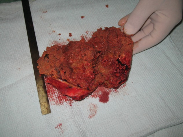
The cut section of the mass showing the gauzes covered by fibrinous exudates simulating a mediastinal tumour.
After surgery, the patient was admitted to the intensive care unit (ICU). The postoperative course was uneventful and after 3 days she was admitted to the post-ICU ward. The patient was discharged from the post-ICU ward after 4 days. The total length of hospital stay was 7 days. The patient was visited in the clinic 1 week later while her condition was good. The patient went back to her hometown; however, 3 months later she came back and was visited again while she was well, with no complaint. She was then referred to her local cardiologist for any possible complaint in future.
DISCUSSION
Gossypibomas are mostly reported in patients undergoing laparotomy [6]. Thus, thoracic gossypiboma is rare [1, 7]. Symptomatic thoracic gossypibomas are mostly seen in the pleural space or in the mediastinum [1], manifesting with chronic chest pain, fever and respiratory symptoms. In our case, the lesion was in the anterior mediastinum and presented with chest pain, a respiratory symptom (dyspnoea) and also dysphagia, probably caused by a compression of the oesophagus.
Gossypibomas are usually detected through radiological studies and radiologists are mostly the first clinicians who face the gossypibomas [8]. CT is the technique of choice to detect a gossypiboma. The tomographic patterns of gossypibomas depend on the time interval between the surgery and the imaging. Early after surgery, in CT scans, a thoracic gossypiboma presents as a heterogeneous hypodense mass with a central spongiform pattern containing air bubbles. If radiopaque strip markers are used, they present as a thin metallic density within the mass. The mass is usually covered with a thin enhancing capsule. However, air bubbles disappear in the late postoperative period and a fibrinous reaction occurs that results in a solid mass, and if radiopaque markers were not used it might mimic malignant tumours [1, 8]. Thus, it will be difficult in the late postoperative period to diagnose a thoracic gossypiboma based on radiological features. In this period, a gossypiboma is usually diagnosed through the histological examination of the resected mass. As patients usually become symptomatic in the late postoperative period [1], radiological studies mostly do not help the diagnosis of gossypiboma if radiopaque markers are not used. Taking biopsy samples, or checking tumour markers, might be indicated in some cases. Non-invasive diagnostic procedures including positron emission tomography scan imaging can be useful; although such a method has false positive or false negative results and is not available in many regions including our country. In our case, the mediastinal gossypiboma was presented 25 years after the first surgery and thus the chest CT showed only a non-specific solid mass and thus a malignant tumour was the most probable diagnosis. However, a specific preoperative diagnosis could not be established because of the non-specific CT findings. The patient was operated on to remove the tumour and the histological study revealed the nature of the mass.
Gossypibomas can be asymptomatic in some cases and can be detected accidentally. As the foreign bodies can cause reactions and may invade the adjacent organs even in such cases, it is wise to operate on the patient to remove the foreign body and to prevent further unpredictable side effects.
As thoracic gossypibomas could cause chronic respiratory symptoms and are hard to diagnose, specially in the late postoperative period, it is important to take preventive strategies during the operation. It could be suggested to use radiopaque gauzes in thoracic surgeries. In this way, radiological studies could be more helpful in the diagnosis of gossypibomas. Also, all sponge gauzes should be counted both preoperatively and postoperatively, which needs effective cooperation between the surgeon and his/her team. If the gauze count is uncertain, a plain chest radiography should be performed before closing the chest [1, 6, 9], as it is suggested by some centres to do a routine plain radiography after each surgery to prevent all cases of retained foreign bodies [10]. The routine protocol in our centre is using the radiopaque gauzes as well as careful counting of the gauzes during surgery. Besides, a chest radiography is requested after all operations in the ICU, and the radiographs will be carefully evaluated by the surgeon to rule out any foreign body deposition.
CONCLUSION
The low incidence, low index of suspicion and non-specific findings on chest CT in the late postoperative period could be the main causes of missed thoracic gossypibomas. Thus, if a thoracic mass is seen in a patient with persistent respiratory symptoms and a history of thoracic surgery, gossypiboma should be included in the differential diagnoses of the mass. Also, definite strategies should be used by all thoracic surgery teams to prevent any case of gossypiboma.
Conflict of interest: none declared.
REFERENCES
- 1.Karabulut N, Herek D, Kiroglu Y. CT features of intrathoracic gossypiboma (textiloma) Diagn Interv Radiol. 2011;17:122–4. doi: 10.4261/1305-3825.DIR.3120-09.0. [DOI] [PubMed] [Google Scholar]
- 2.Whang G, Mogel GT, Tsai J, Palmer SL. Left behind: unintentionally retained surgically placed foreign bodies and how to reduce their incidence—pictorial review. AJR Am J Roentgenol. 2009;193:79–89. doi: 10.2214/AJR.09.7153. [DOI] [PubMed] [Google Scholar]
- 3.Madan R, Trotman-Dickenson B, Hunsaker AR. Intrathoracic gossypiboma. AJR Am J Roentgenol. 2007;189:90–1. doi: 10.2214/AJR.07.2250. [DOI] [PubMed] [Google Scholar]
- 4.Akbulut S, Arikanoglu Z, Yagmur Y, Basbug M. Gossypibomas mimicking a splenic hydatid cyst and ileal tumor: a case report and literature review. J Gastrointest Surg. 2011;15:2101–7. doi: 10.1007/s11605-011-1592-9. [DOI] [PubMed] [Google Scholar]
- 5.Arredondo J, Marti P, Bondia JM, Zozaya G, Cienfuegos JA. Foreign body tumor simulating a gastrointestinal stromal tumor. Rev Esp Enferm Dig. 2010;102:616–7. doi: 10.4321/s1130-01082010001000015. [DOI] [PubMed] [Google Scholar]
- 6.Gumus M, Gumus H, Kapan M, Onder A, Tekbas G, Bac B. A serious medicolegal problem after surgery: gossypiboma. Am J Forensic Med Pathol. 2011;33:54–7. doi: 10.1097/PAF.0b013e31821c09fe. [DOI] [PubMed] [Google Scholar]
- 7.Topal U, Gebitekin C, Tuncel E. Intrathoracic gossypiboma. AJR Am J Roentgenol. 2001;177:1485–6. doi: 10.2214/ajr.177.6.1771485. [DOI] [PubMed] [Google Scholar]
- 8.Szarf G, Mussi de Andrade TC, Nakano E, Szjenfeld D, Costa AS, Jr, Rymkiewicz E, et al. Forty-year-old intrathoracic gossypiboma after cardiac valve surgery. Circulation. 2009;119:3142–3. doi: 10.1161/CIRCULATIONAHA.109.849794. [DOI] [PubMed] [Google Scholar]
- 9.Retnamma RK, Nair SG, Umadethan B, Manoj P. An unusual case of thoracic gossypiboma. Ann Card Anaesth. 2010;13:261–3. doi: 10.4103/0971-9784.69059. [DOI] [PubMed] [Google Scholar]
- 10.McIntyre LK, Jurkovich GJ, Gunn ML, Maier RV. Gossypiboma: tales of lost sponges and lessons learned. Arch Surg. 2010;145:770–5. doi: 10.1001/archsurg.2010.152. [DOI] [PubMed] [Google Scholar]