Abstract
Burkholderia glumae is the primary causal agent of bacterial panicle blight of rice. In this study, 11 naturally avirulent and nine virulent strains of B. glumae native to the southern United States were characterized in terms of virulence in rice and onion, toxofalvin production, antifungal activity, pigmentation and genomic structure. Virulence of B. glumae strains on rice panicles was highly correlated to virulence on onion bulb scales, suggesting that onion bulb can be a convenient alternative host system to efficiently determine the virulence of B. glumae strains. Production of toxoflavin, the phytotoxin that functions as a major virulence factor, was closely associated with the virulence phenotypes of B. glumae strains in rice. Some strains of B. glumae showed various levels of antifungal activity against Rhizoctonia solani, the causal agent of sheath blight, and pigmentation phenotypes on casamino acid-peptone-glucose (CPG) agar plates regardless of their virulence traits. Purple and yellow-green pigments were partially purified from a pigmenting strain of B. glumae, 411gr-6, and the purple pigment fraction showed a strong antifungal activity against Collectotrichum orbiculare. Genetic variations were detected among the B. glumae strains from DNA fingerprinting analyses by repetitive element sequence-based PCR (rep-PCR) for BOX-A1R-based repetitive extragenic palindromic (BOX) or enterobacterial repetitive intergenic consensus (ERIC) sequences of bacteria; and close genetic relatedness among virulent but pigment-deficient strains were revealed by clustering analyses of DNA fingerprints from BOX-and ERIC-PCR.
Introduction
Burkholderia glumae is a seed-born rice pathogen that causes bacterial panicle blight (BPB), which is an emerging major disease problem in many rice-producing areas around the world, including the southeastern United States and South and Central American countries [1]. In particular, significant yield losses from BPB were experienced in rice-producing areas of the southeastern United States, including Louisiana, Texas and Arkansas in 1996, 1997, 2000, and most recently, in 2010 [2]. Burkholderia gladioli also causes BPB but tends to be less virulent and less prevalent than B. glumae [3]. Prolonged high night temperatures and frequent rainfalls at the rice heading stage are thought to be important environmental predispositions for outbreaks of this disease [4], [5].
BPB is a problematic disease not only because it causes severe economic damages, but also because there are few effective methods to control this disease. Oxolinic acid used as a seed treatment or foliar application is the only chemical that can control BPB, but it is not commercially available in the United States [2]. Additionally, the occurrence of oxolinic acid-resistant strains of B. glumae can limit the use of this chemical [1], [6]. Low nitrogen usage, which may reduce disease severity, has not been successful, and early planting, which may avoid high temperatures during the heading stage, can be useless if high temperatures are reached early in the season [2]. Growing disease resistant varieties would be the best option, but only partially resistant varieties that lack desired commercial characteristics are available [1], [7], [8].
Multiple virulence factors are involved in the bacterial pathogenesis of B. glumae. Molecular genetic studies performed by several research groups identified major pathogenic determinants of B. glumae, including the phytotoxin, toxoflavin [9], [10], and lipase [11]. Bacterial motility mediated by flagella is also required for the pathogenicity of B. glumae [12]. Toxoflavin and lipase production as well as the bacterial motility mediated by flagella are controlled by a quorum-sensing system composed of a LuxI-family acyl-homoserine lactone (AHL) synthase, TofI, and a LuxR-family AHL receptor, TofR [9], [11], [12]. Additional virulence factors known to contribute to the full virulence of B. glumae include the PehA and PehB polygalacturonases [13], the KatG catalase [14], and the Hrp type III secretion system (T3SS) [15].
More than 300 B. glumae strains were previously isolated from rice plants with symptoms of BPB growing in rice fields in Louisiana and other states in the southeastern United States, including Texas and Arkansas [3]. In this previous study, some of the isolated strains showed asymptomatic or hypovirulent phenotypes on both rice sheaths and rice panicles in greenhouse tests [3]. In this study, 11of the 19 strains that showed asymptomatic phenotypes in preliminary virulence tests were confirmed to be naturally avirulent after a series of rigorous virulence assays in the greenhouse and in the field. These eleven naturally avirulent strains were tested with nine virulent strains of B. glumae for various phenotypes including virulence on onion bulb scales, toxoflavin production, pigmentation on CPG agar media, and antifungal activity, as well as for genetic variations revealed by BOX- and ERIC-PCR analyses.
Materials and Methods
Bacterial Strains and Culture Conditions
Bacterial strains and plasmids used in this study are listed in Table 1. B. glumae and Escherichia coli strains were routinely grown and maintained in Luria Bertani (LB) agar or broth medium [16] at 30 to 37°C.
Table 1. Bacterial strains used in this study.
| Strain name | Description/Origin | Reference |
| Burkholderia glumae | ||
| 106sh-5 | Avirulent, non-pigmenting/U.S. (Louisiana) | [3] |
| 106sh-9 | Avirulent, non-pigmenting/U.S. (Louisiana) | [3] |
| 11sh2-2-a | Virulent, pigmenting/U.S. (Louisiana) | [3] |
| 117g1-7-a | Virulent, non-pigmenting/U.S. (Louisiana) | [3] |
| 189gr-8 | Virulent, pigmenting/U.S. (Texas) | [3] |
| 191sh-1 | Virulent, pigmenting/U.S. (Texas) | [3] |
| 201sh-1 | Virulent, pigmenting/U.S. (Louisiana) | [3] |
| 237gr-5 | Avirulent, pigmenting/U.S. (Louisiana) | [3] |
| 257sh-1 | Avirulent, pigmenting/U.S. (Louisiana) | [3] |
| 336gr-1 | Virulent, non-pigmenting/U.S. (Louisiana) | [3] |
| 366gr-2 | Avirulent, non-pigmenting/U.S. (Arkansas) | [3] |
| 261gr-9 | Virulent, pigmenting/U.S. (Louisiana) | [3] |
| 961149-4-4 | Avirulent, pigmenting/U.S. (Louisiana) | [3] |
| 395-2 | Avirulent, non-pigmenting/U.S. (Arkansas) | [3] |
| 379gr-1-b | Avirulent, non-pigmenting/U.S. (Arkansas) | [3] |
| 396gr-2 | Avirulent, non-pigmenting/U.S. (Arkansas) | [3] |
| 411gr-6 | Virulent, pigmenting/U.S. (Arkansas) | [34] |
| 957856-41-c | Virulent, pigmenting/U.S. (Louisiana) | [3] |
| 98gr-1 | Avirulent, non-pigmenting/U.S. (Louisiana) | [3] |
| 99sh-7 | Avirulent, non-pigmenting/U.S. (Louisiana) | [3] |
| ATCC33617 | Avirulent, non-pigmenting/Japan (type strain) | [28] |
| AU6208 | Virulent, pigmenting/U.S. (Michigan) | [38] |
| BGR1 | Virulent, non-pigmenting/South Korea | [29] |
| LSUPB223 | A toxA::pKNOCKGm derivative of 336gr-1, NitR, GmR | This study |
| Burkholderia gladioli | ||
| ATCC51989 | An ATCC strain of B. gladioli | [39] |
| Escherichia coli | ||
| HB101 | F– thi-1 hsd20 (r– Bm– B) sup E44 recA13 ara-14 leuB6 proA2 lacY1 rpsL20 (SmR) xyl-5 mtl-1 | [40] |
| S17-1 λpir | recA thi pro hsdR (res– mod+)(RP4::2-Tc::Mu-Km::Tn7) λ pir phage lysogen | [23] |
| Plasmids and Mutants | ||
| pKNOCKGm | A suicide vector, R6K ori, GmR | [22] |
| pKNOCKGm::ToxA-int | A clone of toxA internal region in pKNOCKGm, GmR | This study |
| pRK2013::Tn7 | ColE1 mob + tra RK2 Δrep RK2 repE kan::Tn7 (TpR, SmR, SpR ) | [41] |
| pSC-A-amp/kan | A PCR cloning Vector, ApR, KmR | Agilent Technology (Santa Clara, CA, USA) |
Recombinant DNA Techniques
Procedures for routine DNA cloning and amplification were conducted according to Sambrook et al. (2001).
Confirmation of B. Glumae
Diagnostic PCR for B. glumae was conducted with the species specific primer sets, 5′-ACACGGAACACCTGGGTA-3′ and 5′-TCGCTCTCCCGAAGAGAT-3′ for the 16S-23S rDNA internal transcribed spacer (ITS) region and 5′-GAAGTGTCGCCGATGGAG-3′ and 5′-CCTTCACCGACAGCACGCAT-3′ for gyrB, using the previously described reaction conditions for each primer set [3], [17].
Virulence Assay on Rice Plants
The rice variety Trenasse, which is highly susceptible to BPB, was used for testing the virulence of B. glumae strains on rice panicles. Overnight cultures of B. glumae strains on LB agar plates were resuspended in sterile tap water at a concentration of ca. 1×10⁸ CFU/ml (OD600 = 0.1). Rice plants at the 20 to 30% heading stage were inoculated twice with a two-day interval between sprays (∼ 2 ml/plant). Disease symptoms were evaluated 14 d after the first inoculation. The experiments were conducted in the greenhouse and in the field during the 2009 growing season. For field experiments at the LSU AgCenter Rice Research Station (Crowley, Louisiana, USA), the susceptible variety Trenasse was grown in rows (12 to 15 plants per row) with ca. one-foot intervals between rows. A row of the partially resistant variety Jupiter was grown between every four rows of Trenasse. Virulence was scored using a 0–9 scale in which 0 indicated no symptoms and 9 indicated more than 80% discolored panicles. Overall disease severity of an entire row was scored with four replications for each treatment. For greenhouse experiments, rice plants were grown in plastic pots (15 cm diameter by 20 cm height) containing a soil mixture of clay, Jiffy Mix® (Ferrry-Morse Seed Co, Fulton, KY, USA) and sand in a 3∶1:1 ratio. Rice plants were inoculated with the same method used for field experiments. Greenhouse tests for determining the virulence phenotypes of B. glumae strains were repeated three times. In each test, disease severity of each rice plant was scored for each treatment with four replications. Yield reduction caused by B. glumae infection was determined with the yield data obtained from field experiments. Rice grains from a single row infested with each strain or mock-inoculated with water were collected, dried to 13% moisture, and weighed. Four replications were implemented in the yield data.
Onion Assay
Yellow onions used in this study were purchased from a local market. The virulence of each B. glumae strain on onion bulb scales was tested following a previously developed method with minor modifications [18]. Briefly, 5 µl of bacterial suspension containing ca. 5×10⁵ CFU in 10 mM MgCl2 was applied to a ca. 2 mm-wound on the inner surface of an onion bulb scale made with micropipette tip. Inoculated onion scales were incubated in a wet-chamber at 30°C and virulence was determined by measuring the macerated area on each onion bulb scale after 48 h.
Determination of Toxoflavin Production and Pigmentation
To determine the production of toxoflavin by each B. glumae strain, B. glumae cells were streaked on a King’s B (KB) agar plate [16] and incubated for 24 h at 37°C. Toxoflavin production was determined based on the presence of a yellow pigment diffused from the bacterial colonies into the surrounding agar medium. To determine the pigmentation phenotype of each B. glumae strain, bacterial cells were streaked on a CPG agar plate [16] and incubated for 48 h at 30°C.
Measurement of Antifungal Activity Against Rhizoctonia solani
Antifungal activities of B. glumae strains against R. solani were measured following a previously reported method [19] with some modifications. Briefly, one ml of an overnight culture of each B. glumae strain was centrifuged, washed twice with fresh LB broth, and resuspended in 100 µl of fresh LB broth. Ten-microliter aliquots of each suspension were pipetted onto three locations around the center of a potato dextrose agar (PDA) plate. Inoculated PDA plates were incubated overnight (∼ 16 h) at 37°C. Mycelial plugs 5 mm in diameter were cut from R. solani cultures grown on PDA at 30°C and placed in the center of each PDA plate containing three spots of B. glumae. The length of the inhibition zone between B. glumae and R. solani was measured for each bacterial spot 48 h after incubation at 25°C. Nine replications were performed for each strain of B. glumae.
BOX- and ERIC-PCR and Cluster Analyses of the DNA Fingerprints
BOX- and ERIC-PCR were conducted following a previously established method [20]. PCR products were separated on a 1.8% agarose gel run at 60 V for 18 h and were visualized with a Kodak Gel Logic 1500 imaging system (Rochester, NY, USA). DNA fingerprints of individual strains generated from BOX- and ERIC-PCR were converted into a binary matrix by scoring amenable DNA bands as present or absent. Cluster analyses of the DNA fingerprints were performed with unweighted pair-group method of averages (UPGMA) using MEGA5 [21].
Generation of the toxA Toxoflavin Deficient Mutant, LSUPB223
An internal region of toxA was amplified using the primers, 5′-TTTCGGGCGTGAAATCTATC-3′ and 5′-AGCGGTAGAAGCTGAACTGG-3′. The amplified PCR product was cloned into the PCR cloning vector, pSC-A-amp/kan, using a StrataClone PCR Cloning Kit (Agilent Technologies, Santa Clara, CA, USA) following the manufacturer’s instructions. The insert of the resultant PCR clone, pSC::ToxA-int, was digested with the KpnI and SacII restriction enzymes and then ligated to the KpnI/SacII-cut pKNOCKGm suicide vector [22], to generate pKNOCKGm::ToxA-int (Table 1). Escherichia coli S17-1 λpir [23] was used to maintain the pKNOCK vectors and pKNOCKGm::ToxA-int. E. coli HB101, which carries the helper plasmid, pRK2013 [24], was used in triparental mating to introduce pKNOCKGm::ToxA-int into a virulent strain of B. glumae, 336gr-1. toxA mutants resulting from homologous recombination with the vector containing the internal fragment of toxA were selected on LB agar containing nitrofurantoin and gentamycin. The toxoflavin producing phenotypes of these mutants were verified on KB agar plates.
Results
Determination of Naturally Avirulent Strains of B. glumae Isolated from Rice Fields in the Southeastern United States
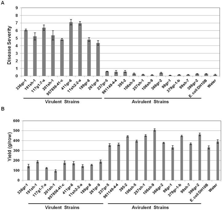
In a previous study, some strains of B. glumae isolated from rice fields in Louisiana and other southeastern states failed to produce symptoms on both rice sheaths and panicles [3]. In this study, 19 out of 24 strains that showed asymptomatic phenotypes in the previous tests were confirmed by diagnostic PCR with species specific primers that anneal to the ITS region or gyrB of B. glumae (data not shown) to be B. glumae. These 19 strains were re-examined for their virulence in rice along with a highly virulent strain of B. glumae, 336gr-1, and Escherichia coli DH10B as a positive and negative control, respectively. From repeated tests in the greenhouse (data not shown) and in the field, 11 of the 19 strains of B. glumae were confirmed to be avirulent, whereas the remaining eight strains were determined to be virulent (Figure 1A). In addition, the 11 avirulent strains did not cause significant yield reductions when compared to the negative control, while virulent strains including the virulent reference strain, 336gr-1, caused 50 to 75% yield reductions (Figure 1B).
Figure 1. Virulence effects of Burkholderia glumae strains on rice panicles.
A) Disease severities of rice panicles inoculated with virulent and avirulent strains of B. glumae. Disease scales: no symptom, 0; 1–10% symptomatic area, 1; 11–20% symptomatic area, 2; 21–30% symptomatic area, 3; 31–40% symptomatic area, 4; 41–50% symptomatic area, 5; 51–60% symptomatic area, 6; 61–70% symptomatic area, 7; 71–80% symptomatic area, 8; more than 80% symptomatic area, 9. B) Rice yields (g/16-foot row) of plants inoculated with virulent and avirulent strains of B. glumae. These data were obtained from the susceptible rice variety, Trenasse, and error bars indicate the standard deviation from four replications.
Test of Onion Bulb Scales as an Alternative for Efficient and High-throughput Virulence Assays for Strains of B. glumae
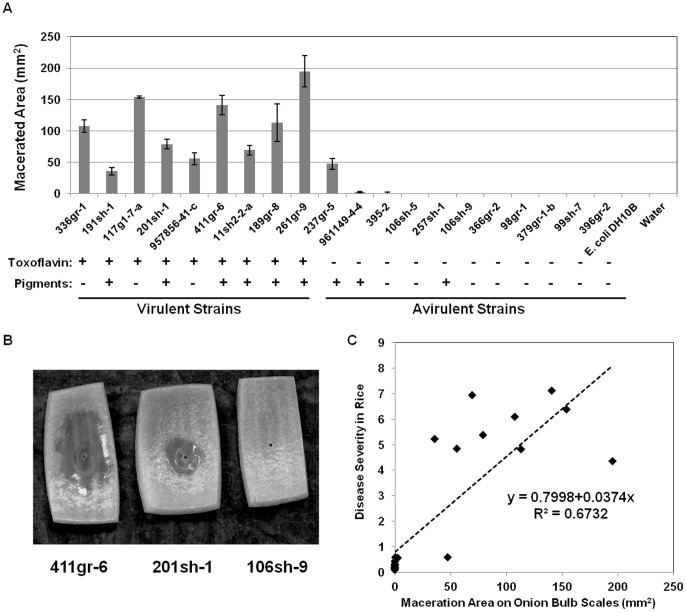
Remarkably, all of the virulent strains of B. glumae that were tested caused maceration of the onion bulb scale tissue, but all of the avirulent strains of B. glumae except 237gr-5 did not cause maceration of the onion bulb scale tissue (Figures 2A and 2B). Moreover, the virulence of B. glumae on onion bulb scales was highly correlated with that on rice panicles (R2 = 0.6732) (Figure 2C).
Figure 2. Virulence of Burkholderia glumae strains on onion bulb scales.
A) Area of macerated tissue of onion bulb scales inoculated with virulent and avirulent strains of B. glumae. Error bars indicate the standard deviation from three replicates. Phenotypes in toxoflavin production and pigmentation are indicated under the name of each strain. B) Onion bulb scales showing various levels of maceration after 48 h incubation at 30°C after inoculation with a highly virulent, a moderately virulent, and an avirulent strain of B. glumae, 411gr-6, 201sh-1, and 106sh-9, respectively. C) Correlation between the abilities of B. glumae strains to cause maceration on onion bulb scales and to produce symptoms on rice panicles: The dotted line represents a linear regression line. All the parameter estimates were statistically significant at α <0.001 indicating strong relationship between the disease severity in rice panicles and the macerated area on onion scales. Statistical analysis was performed by using SAS 9.3 version.
Production of Toxoflavin
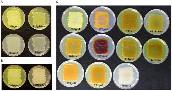
Toxoflavin is a bright yellow pigment that has antibiotic and phytotoxic activity and that is produced by and functions as a major virulence factor of B. glumae [9], [10], [25]. A virulent strain of B. glumae, 336gr-1, produced a yellow pigment in culture media, including LB and KB. In solid media, this yellow pigment is diffused from bacterial colonies into the surrounding agar medium (Figure 3A). Mutation of toxA, a gene required for toxoflavin biosynthesis [9], [10], abolished the production of this yellow pigment (Figure 3B). In addition, the yellow pigment extracted from the culture media with chloroform showed absorbance maxima at 258 and 393 nm (data no shown) like a recent study on toxoflavin [26]. These results together indicate that the yellow pigment is toxoflavin and that the production of toxoflavin can be determined by the presence of the yellow pigment in media with cultures of B. glumae. As shown in Figure 2A, all of the B. glumae strains that were virulent to rice produced toxoflavin, whereas all of the strains that were avirulent to rice did not, indicating that toxoflavin production is closely associated with the virulence of B. glumae to rice. However, one toxoflavin-deficient strain, 237gr-5, produced maceration symptoms on onion bulb scales, indicating that toxoflavin is dispensable for the maceration activity of B. glumae in onion.
Figure 3. Toxoflavin and pigmentation phenotypes of strains of B. glumae.
A) Toxoflavin production on KB agar plates by virulent strains (336gr-1 and 117g1-7-1) and avirulent strains (366gr-2 and 98gr-1) of B. glumae. B) Toxoflavin production by a virulent strain of B. glumae, 336gr-1, and its toxA mutant derivative, LSUPB223. C) Pigmentation of strains of B. glumae on CPG agar plates in comparison with a non-pigmenting virulent strain of B. glumae, 336gr-1, and a non-pigmenting avirulent strain of B. glumae, 396gr-2. The bright yellow pigment of strain 336gr-1 is toxoflavin. Photos were taken after 24 h incubation at 37°C for A) and B) and after 48 h incubation at 30°C for C).
Production of Yellow-green and Purple Pigments in CPG Agar Medium
Nine of the 20 strains tested produced unknown pigments when grown on CPG agar plates (Figures 2A and 3C). These pigments were easily distinguished from toxoflavin, which is bright yellow and diffused into the agar medium from bacterial colonies. Six of the nine virulent strains and three of the 11 avirulent strains produced pigments (Figures 2A and 3C). Partial purification of the pigments produced by the pigmenting strain, 411gr-6, yielded three fractions with different colors; yellow-green, purple, and brown (Figure S1A). The brown fraction (Figure S1A) is likely a mixture of multiple unknown substances, not a single material (data not shown). Thus, the observed pigmentation may be due to at least two different pigments, which were yellow-green and purple (Figures 3C and S1A). The purple pigment was restricted to the bacterial colonies, whereas the yellow-green pigment was diffused into the agar medium (Figure 3C). The yellow-green pigment purified from agar extract exhibited strong green fluorescence when illuminated with UV light with UV absorption maxima at 202, 238, 301, 448 and 464 nm. The purple pigment exhibited a strong antifungal activity (Figure S1B). In terms of color and antifungal activity, the purple pigment resembles the virulence factor of the opportunistic human pathogen Pseudomonas aeruginosa, pyocyanin, which is also known as “blue phenazine” [27]. However UV absorbance spectra and spectroscopic data including 1H-NMR indicated that the purple pigment is different from pyocyanin (data not shown). It is noteworthy that a diverse range of phenotypic variation in pigmentation was observed among the pigment-producing strains (Figure 3C). Especially, 257sh-1 produced excessive amounts of the purple pigment compared to the other pigment-producing strains (Figure 3C). In addition, three virulent strains, 191sh-1, 411gr-6 and 11sh2-2-a, produced larger amounts of the diffusible yellow-green pigment compared to other B. glumae strains (Figure 3C). Unlike toxoflavin, the pigmentation did not appear to be related to bacterial pathogenesis in onion (Figure 2A).
Antifungal Activities of Naturally Avirulent Strains of B. glumae Against Rhizoctonia solani
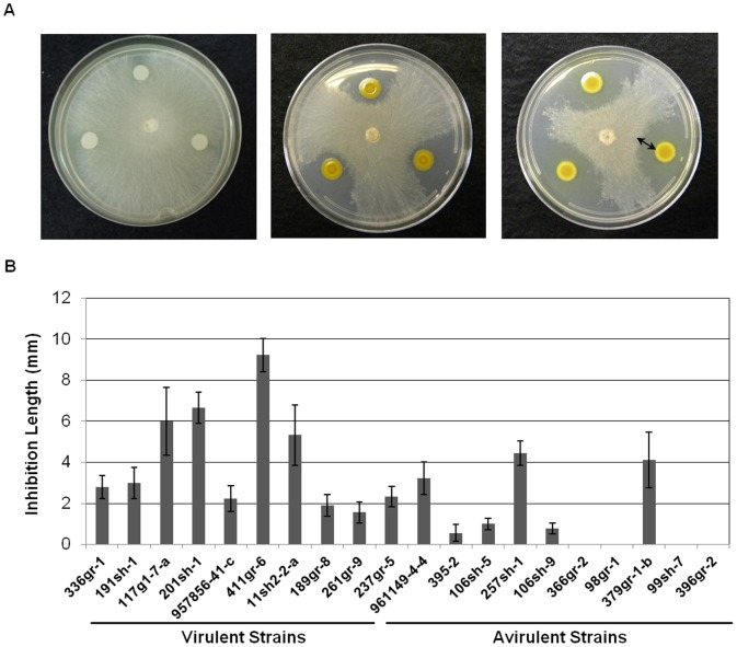
All of the virulent strains of B. glumae showed observable antifungal activities, whereas only seven of the 11 avirulent strains showed observable antifungal activities (Figures 4A and 4B). The highly virulent and toxoflavin- and pigment-producing strain, 411gr-6, showed the highest antifungal activity among all of the strains tested (Figure 4B). An avirulent strain, 379gr-1-b, showed antifungal activity despite its toxofalvin- and pigment-deficient phenotype, indicating that this strain possesses at least one antifungal system that is independent of toxoflavin and pigment production in CPG agar medium (Figure 4B).
Figure 4. Antifungal activities of Burkholderia glumae strains against Rhizoctonia solani.
A) Various levels of antifungal activities against R. solani after 48 h incubation at 25°C. B. glumae in the center of each plate was incubated overnight (∼ 16 h) prior to the inoculation of R. solani. The arrow indicates the measurement used to quantify the antifungal activity. B) Antifungal activities of virulent and avirulent strains of B. glumae determined by the length of the inhibition zone between the edge of the colony of B. glumae and the edge of the mycelial growth of R. solani. Error bars indicate the standard deviation from nine replications.
Comparison of Genomic Structures between Virulent and Avirulent Strains of B. glumae by BOX- and ERIC-PCR
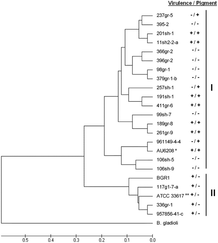
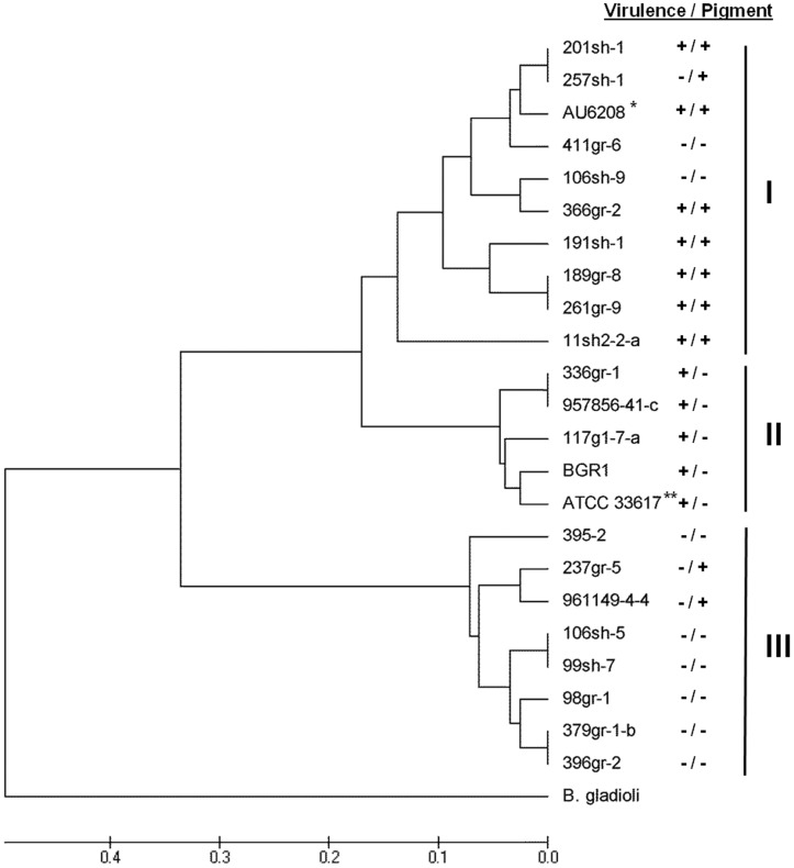
The observed phenotypic variations in virulence-related traits (virulence on rice and onion and toxoflavin production), antifungal activity and pigment production among the strains of B. glumae prompted us to investigate the genetic relatedness among strains. BOX- and ERIC-PCR revealed genomic variation among the 23 tested strains of B. glumae, which included the 20 U.S. strains as well as the type strain ATCC33617T [28], a Korean strain, BGR1 [29], and a clinical strain, AU6208 [11](Figures 5 and 6). Because only DNA samples were available for BGR1 and AU6208, phenotypes of these strains could not be determined in our laboratory. However, the virulent Korean strain, BGR1, did not produce the dark pigments on CPG agar medium [29], whereas AU6208 was shown to be virulent to rice [11] and to produce pigments on CPG medium (J. LiPuma, personal communication). The genomic DNA of the B. gladioli strain ATCC51989 was included as a control and used as an outgroup in phylogenetic analyses.
Figure 5. Phylogenetic tree generated from UPGMA analysis of the BOX-PCR fingerprints of Burkholderia glumae strains.
*: AU6208 was reported to be virulent to rice in a previous study [11] and tested for the pigmentation phenotype by another research group (J. J. LiPuma, personal communication). **: The phenotypes of BGR1 were indicated based on the previous studies reported by Kim et al. (2004) and Jeong et al. (2003). ***: ATCC33617 is the type strain originally isolated as the causal agent of bacterial panicle blight and its lost pathogenicity by a spontaneous mutation of tofR could be restored by the addition of a functional copy of tofR [11].
Figure 6. Phylogenetic tree generated from UPGMA analysis of the ERIC-PCR fingerprints of Burkholderia glumae strains.
*: AU6208 was reported to be virulent to rice in a previous study [11] and tested for the pigmentation phenotype by another research group (J. J. LiPuma, personal communication). **: The phenotypes of BGR1 were indicated based on the previous studies reported by Kim et al. (2004) and Jeong et al. (2003). ***: ATCC33617 is the type strain originally isolated as the causal agent of bacterial panicle blight and its lost pathogenicity by a spontaneous mutation of tofR could be restored by the addition of a functional copy of tofR [11].
Various BOX-PCR fingerprints were observed among strains of B. glumae (data not shown). Twenty-six band classes ranging from 330 bp to 3,500 bp that were reproducible in two independent reactions were scored as previously described and used to generate a dendogram (Figure 5). The dendogram revealed two major phyletic groups. The dendogram indicated that major phyletic groups were not separated on the basis of virulence or pigmentation (Figure 5). Group I was a polyphyletic group that contained the majority of the B. glumae strains, including all the naturally avirulent strains and all the pigment-producing strains. Group II was a polyphyletic group that contained virulent, pigment-deficient strains and the type strain ATCC33617T (Figure 5).
Genetic differences were also observed in ERIC-PCR fingerprints (data not shown). Twenty-one band classes between 130 bp and 3500 bp that were amenable and reproducible in two independent reactions were scored as previously described and used to create a dendogram (Figure 6). This dendogram revealed three major phyletic groups. Group I contained all of the virulent, pigment-producing strains; Group II contained all of the virulent, non-pigmenting strains; and Group III contained only avirulent strains (Figure 6).
Although differences were observed between the BOX and ERIC dendograms, all of the strains that grouped together in Group II in the BOX analysis also grouped together in the ERIC analysis (Figures 5 and 6).
Discussion
In this study, 11 strains of B. glumae indigenous to the southeastern U.S. were confirmed to be naturally avirulent through a series of virulence tests conducted in the field and greenhouse. These naturally avirulent strains did not produce any obvious symptoms of BPB or cause any significant yield reduction compared to the nine virulent strains tested (Figures 1A and B). At least 5% of the B. glumae strains isolated from rice fields in the southeastern U.S. showed avirulent phenotypes (data not shown). Because the avirulent strains studied were isolated from rice plants showing BPB symptoms, they are presumably derivatives of a virulent strain that lost their pathogenicityvia spontaneous mutation after host infection. It might also be possible that populations of B. glumae present in natural conditions typically contain avirulent cells.
Although it is unknown how avirulent strains of B. glumae are generated in nature, this phenomenon may be explained by several possible mechanisms. First, major deletion(s) of genomic DNA could cause a loss of multiple virulence genes. It was recently reported that the cluster of hrp/hrc genes encoding the Hrp T3SS is absent in the genome of a naturally avirulent strain of Pseudomonas syringae, 508, probably due to a deletion event [30]. Second, a point mutation could occur in a core regulatory gene that globally controls multiple virulence factors or in a gene that encodes an important virulence factor. Indeed, the ATCC strain of B. glumae tested in this study, ATCC33617T, was reported to be avirulent because of a frameshift mutation in tofR, which encodes the TofR AHL receptor that functions as a central regulatory element for the production of major virulence factors of B. glumae, including toxoflavin and lipase [11]. In addition, four different natural mutant alleles of avrBs2, the avirulence gene corresponding to Bs2 of pepper in gene-for-gene resistance, were observed in natural strains of Xanthomonas campestris that showed attenuated virulence phenotypes in pepper plants with a bs2/bs2 genetic background [31]. Finally, multiple mutations in a genome could also be responsible for the loss of pathogenicity in nature. To identify the cause(s) of the avirulent phenotypes of B. glumae strains from infected rice plants, the expression of known and potential virulence genes and the presence of the functional quorum-sensing system required for the pathogenicity of B. glumae in the 11 avirulent strains are currently being studied in our laboratory. Whole genome sequencing and comparison with virulent strains would also help to elucidate the genetic basis of naturally-occurring, non-pathogenic strains of B. glumae.
An alternate assay system using onion bulb scales for determining the virulence of B. glumae was also developed in this study. This assay system was previously used by Jacobs et al. (2008) to test strains of B. cepacia and B. cenocepacia pathogenic to onion. In our study, virulent strains of B. glumae were able to cause symptoms on onion bulb scales and virulence on onion bulb scales was highly correlated with virulence on rice panicles (Figure 2C). Strain 237gr-5 was the only strain of the 20 strains tested, that showed different virulence/avirulence features between two different host systems (avirulent to rice but virulent to onion)(Figures 1 and 2A). Virulence tests on rice are time-consuming, labor-intensive, and require a lot of space. The high correlation observed between the virulence of B. glumae in rice and that in onion strongly suggests that onion bulb scales can be used as an excellent alternative assay for convenient and high-throughput virulence tests of B. glumae strains and would serve as a powerful tool for large-scale functional genomic studies of this bacterium. Nevertheless, caution should be made when onion bulb scales are used as a surrogate system for determining the virulence of B. glumae in rice because virulence factors responsible for maceration of onion tissue may be different from those responsible for panicle blighting of rice.
Production of toxoflavin, was closely related to the bacterial virulence phenotypes in rice. All nine virulent strains produced toxoflavin, whereas all 11 avirulent strains did not produce toxoflavin (Figures 1 and 2A). The ability of each bacterial strain to produce toxoflavin was determined by observing the production of the bright yellow pigment released into the culture medium (Figure 3A). Directional mutation of toxA via homologous recombination, which resulted in the loss of ability to produce toxoflavin [10], caused the loss of yellow pigment production (Figure 3B), indicating that the yellow pigment is toxoflavin. These facts strongly suggest that naturally avirulent strains of B. glumae can be readily determined by their disability to produce toxoflavin in the culture media. Even though the pathogenicity of the B. glumae strains in rice was tightly linked to their ability to produce toxoflavin in this study, toxoflavin may not fully account for the pathogenesis of B. glumae because mutation of a toxoflavin synthesis gene could not completely abolish bacterial virulence in previous studies [10](Shrestha and Ham, unpublished). Since all of the avirulent strains produced very weak observable symptoms (Figure 1A), it is likely that these strains are defective in the production of multiple virulence factors that are collectively required for full virulence. In fact, it has been shown that B. glumae produces additional virulence factors, including lipase [11], the Hrp T3SS [15] and polygalacturonases [13], which contribute to the virulence of this pathogen to rice. Production of known and potential virulence factors of B. glumae by individual avirulent strains is currently being analyzed (Karki and Ham, unpublished).
It is intriguing that some strains of B. glumae produce unique pigments in CPG medium (Figure 3C). Diverse variations in the pigmentation phenotype were observed within the pigment-producing strains of B. glumae (Figure 3C). Two different pigments (yellow-green and purple) were partially purified from a pigmenting strain, 411gr-6 (Figure S1), and their chemical and physical properties were also partially determined (data not shown). They were easily distinguished from the bright yellow toxoflavin. To the best of our knowledge, this pigmentation phenomenon of B. glumae has not been reported elsewhere. It was reported that some strains of B. cenocepacia produced melanin-like pigments in tyrosine-enriched media and that those pigments may act as scavengers of reactive oxygen species generated from oxidative burse responses of host cells [32]. Later, it was found that the melanin-like pigment produced by a clinical strain of B. cenocepacia was likely to be a pyomelanin synthesized from a homogentisate (HGA) through the action of 4-hydroxyphenylpyruvic acid dioxygenase (HppD), and that pigment production was abolished by disruption of hppD [33]. However, mutation of hppD in 411gr-6, a pigment-producing strain of B. glumae, did not change the pigmentation phenotype of this strain, suggesting that the pigments produced by B. glumae on CPG agar medium are different from the melanin-like pigments produced by B. cenocepacia (data not shown). We recently found that a two-component regulatory system, composed of the PidS sensor histidine kinase and the PidR response regulator, is essential for the production of all three pigments in B. glumae [34]. In addition, all of the pigment-deficient mutants screened from random Tn5-mutangenesis of the pigment-producing strain 411gr-6 were deficient in the production of all three pigments, implying that they may be synthesized via a common regulatory and biosynthetic pathway [34].
Antifungal activities were detected from most of the strains tested in this study (Figure 4B). In particular, all of the virulent strains showed various antifungal activities (Figure 4B). It is probable that the observed antifungal activities of the virulent strains may be conferred in part, if not fully, by toxoflavin, which is known to have a broad toxic effect on prokaryotes and eukaryotes [35]. However, some avirulent strains that do not produce toxoflavin also showed antifungal activities, indicating that these avirulent strains produce additional antifungal compounds. Interestingly, the purple pigment from 411gr-6 showed a strong antifungal activity against C. orbiculare (Figure S1B) and all the pigmenting avirulent strains also showed high levels of antifungal activities against R. solani (Figure 4B), suggesting that the production of this pigment may contribute to the antifungal activities of the pigmenting B. glumae strains. The chemical structures of the yellow-green and purple pigments are currently being characterized. Meanwhile, the antifungal activities shown by non-pigmenting avirulent strains including 379-gr-1-b (Figure 4B) indicate the presence of additional antifungal compound(s) produced by some B. glumae strains. The avirulent strains with antifungal activities against R. solani may be useful tools for biological control of sheath blight and possibly other fungal rice diseases, including blast caused by Magnaporthe grisea. We are currently testing the antifungal activities of these strains against other fungal pathogens. Antifungal activities of Burkholderia spp. other than B. glumae and their application as biological control agents have previously been reported [36], [37]. Nevertheless, naturally avirulent B. glumae strains could be a more useful tool for the biological control of phylloplane diseases of rice, including sheath blight and blast, because they could inhibit pathogens in the rice phyllosphere better than other Burkholderia spp. that typically persist in the soil and plant rhizophere.
BOX- and ERIC-PCR analyses revealed variations in genome structure among strains of B. glumae showing various phenotypic traits and originating from diverse geographic locations (Figures 5 and 6). All of the virulent strains showing pigment-deficient phenotypes were grouped in a single major polyphyletic group in both BOX-and ERIC-PCR phylograms (Group II in both BOX- and ERIC-PCR phylograms) regardless of geographic origin (Figures 5 and 6). Even though the strain ATCC33617T, previously known to be an avirulent strain, was grouped together with virulent strains in Group II in both rep-PCR analyses, it would not be unacceptable to consider this strain as a virulent one since it was originally isolated as the causal agent of the rice disease [28] and later lost its ability to perceive the quorum-sensing signal due to a spontaneous point mutation in tofR encoding the cognate receptor for the quorum-sensing signal of B. glumae and since the production of virulence factors was restored by the introduction of a functional tofR clone [11]. In our independent study, all of the avirulent U.S. strains with the exception of 237gr-5 that was virulent to onion (Figure 2A), also showed deficiency in quorum-sensing (Karki and Ham, unpublished). However, unlike ATCC33617T, pathogenicity could not be restored by the introduction of a DNA clone carrying both tofI and tofR genes in these strains, suggesting that mutation on the tofI/tofR locus is not the only cause of the avirulent phenotype of the naturally avirulent strains (Karki and Ham, unpublished). It is also noteworthy that all of the virulent pigment-producing strains belonged to Group I in both BOX-PCR and ERIC-PCR analyses, and that Group III from the ERIC-PCR analysis only contained avirulent strains (Figure 6). The clinical strain AU6208, which was previously reported to be virulent to rice [11], was grouped together with the virulent strains producing pigments (Figures 5 and 6). According to the tests by Dr. LiPuma’s research group in University of Michigan, AU6208 also produces pigments on CPG agar plates (J. J. LiPuma, personal communication). These cluster analyses from BOX- and ERIC-PCR data and the phenotypic characteristics associated with major polyphyletic groups suggest that multiple lineages of B. glumae may exist.
Conclusively, significant phenotypic variations were observed among strains of B. glumae, including variation in virulence, pigmentation and antifungal activities. In addition, some phyletic groups based on BOX-and ERIC-PCR fingerprints were associated with virulence and pigmentation phenotypes. Nevertheless, genetic backgrounds of the observed phenotypes, including natural avirulence and pigmentation are still unknown. Comparative genomics approaches with whole genome sequence information, which can now be readily obtained by high-throughput sequencing, would provide important clues to identify the causes of these phenotypic traits. Additional comprehensive population genetics studies should also be conducted to elucidate the genetic lineages of this pathogenic bacterium.
Supporting Information
Partially purified pigments of Burkholderia glumae 411gr-6 (A) and antifungal activity of the partially purified purple pigment (B). Peak 4 from high pressure liquid chromatography showed purple color and an antifungal activity against Collectotrichum orbiculare. The photo of the antifungal activity was taken 48 h after incubation at 28°C.
(TIF)
Acknowledgments
We wish to give special thanks to Dr. J. J. LiPuma for providing the genomic DNA samples of AU6208 and the personal communication for the pigmentation phenotype of AU6208, to Dr. I. Hwang for providing the genomic DNA of BGR1, and to Mr. S. B. Bohora for statistical analyses.
Funding Statement
This work was financially supported by the Louisiana State University Agricultural Center (LAB93918), the Research and Development Program of the Louisiana Board of Regents Support Fund (Grant number: LEQSF(2008-11)-RD-A-02), and the Louisiana Rice Research Board (Grant numbers: 940-38-4158 and 940-38-4173). The funders had no role in study design, data collection and analysis, decision to publish, or preparation of the manuscript.
References
- 1. Ham JH, Melanson RA, Rush MC (2011) Burkholderia glumae: next major pathogen of rice? Mol Plant Pathol 12: 329–339. [DOI] [PMC free article] [PubMed] [Google Scholar]
- 2.Ham JH, Groth D (2011) Bacterial Panicle Blight, an Emerging Rice Disease Louisiana Agriculture. Baton Rouge, Louisiana, USA: Louisiana State University Agricultural Center. 16–17.
- 3. Nandakumar R, Shahjahan AKM, Yuan XL, Dickstein ER, Groth DE, et al. (2009) Burkholderia glumae and B. gladioli cause bacterial panicle blight in rice in the southern United States. Plant Dis 93: 896–905. [DOI] [PubMed] [Google Scholar]
- 4. Cha KH, Lee YH, Ko SJ, Park SK, Park IJ (2001) Influence of weather condition at heading period on the development of rice bacterial grain rot caused by Burkholderia glumae . Res Plant Dis 7: 150–154. [Google Scholar]
- 5. Tsushima S, Naito H, Koitabashi M (1995) Forecast of yield loss suffered from bacterial grain rot of rice in paddy field severely diseased pancles Ann Phytopathol Soc Jpn. 61: 419–424. [Google Scholar]
- 6. Maeda Y, Kiba A, Ohnish K, Hikichi Y (2004) New method to detect oxolinic acid-resistant Burkholderia glumae infecting rice seeds using a mismatch amplification mutation assay polymerase chain reaction J Gen Plant Pathol. 70: 215–217. [Google Scholar]
- 7. Sayler RJ, Cartwright RD, Yang Y (2006) Genetic characterization and real-time PCR detection of Burkholderia glumae, a newly emerging bacterial pathogen of rice in the United States. Plant Dis 90: 603–610. [DOI] [PubMed] [Google Scholar]
- 8. Groth DE, Linscombe SD, Sha X (2007) Registration of two disease-resistant germplasm lines of rice. J Plant Regist 1: 63–64. [Google Scholar]
- 9. Kim J, Kim JG, Kang Y, Jang JY, Jog GJ, et al. (2004) Quorum sensing and the LysR-type transcriptional activator ToxR regulate toxoflavin biosynthesis and transport in Burkholderia glumae. Mol Microbiol 54: 921–934. [DOI] [PubMed] [Google Scholar]
- 10. Suzuki F, Sawada HA, Zegami K, Tsuchiya K (2004) Molecular characterization of the tox operon involved in toxoflavin biosynthesis of Burkholderia glumae . J Gen Plant Pathol 70: 97–107. [Google Scholar]
- 11. Devescovi G, Bigirimana J, Degrassi G, Cabrio L, LiPuma JJ, et al. (2007) Involvement of a quorum-sensing-regulated lipase secreted by a clinical isolate of Burkholderia glumae in severe disease symptoms in rice. Appl Environ Microbiol 73: 4950–4958. [DOI] [PMC free article] [PubMed] [Google Scholar]
- 12. Kim J, Kang Y, Choi O, Jeong Y, Jeong JE, et al. (2007) Regulation of polar flagellum genes is mediated by quorum sensing and FlhDC in Burkholderia glumae. Mol Microbiol 64: 165–179. [DOI] [PubMed] [Google Scholar]
- 13. Degrassi G, Devescovi G, Kim J, Hwang I, Venturi V (2008) Identification, characterization and regulation of two secreted polygalacturonases of the emerging rice pathogen Burkholderia glumae. FEMS Microbiol Ecol 65: 251–262. [DOI] [PubMed] [Google Scholar]
- 14. Chun H, Choi O, Goo E, Kim N, Kim H, et al. (2009) The quorum sensing-dependent gene katG of Burkholderia glumae is important for protection from visible light. J Bacteriol 191: 4152–4157. [DOI] [PMC free article] [PubMed] [Google Scholar]
- 15. Kang Y, Kim J, Kim S, Kim H, Lim JY, et al. (2008) Proteomic analysis of the proteins regulated by HrpB from the plant pathogenic bacterium Burkholderia glumae. Proteomics 8: 106–121. [DOI] [PubMed] [Google Scholar]
- 16.Schaad NW, Jones JB, Chun W (2001) Laboratory guide for identification of plant pathogenic bacteria. 3rd Ed.: The American Society of Phytopathological Society Press.
- 17. Maeda Y, Shinohara H, Kiba A, Ohnishi K, Furuya N, et al. (2006) Phylogenetic study and multiplex PCR-based detection of Burkholderia plantarii, Burkholderia glumae and Burkholderia gladioli using gyrB and rpoD sequences. Int J Syst Evol Microbiol 56: 1031–1038. [DOI] [PubMed] [Google Scholar]
- 18. Jacobs JL, Fasi AC, Ramette A, Smith JJ, Hammerschmidt R, et al. (2008) Identification and onion pathogenicity of Burkholderia cepacia complex isolates from the onion rhizosphere and onion field soil. Appl Environ Microbiol 74: 3121–3129. [DOI] [PMC free article] [PubMed] [Google Scholar]
- 19. O’Grady EP, Nguyen DT, Weisskopf L, Eberl L, Sokol PA (2011) The Burkholderia cenocepacia LysR-type transcriptional regulator ShvR influences expression of quorum-sensing, protease, type II secretion, and afc genes. J Bacteriol 193: 163–176. [DOI] [PMC free article] [PubMed] [Google Scholar]
- 20. Louws FJ, Fulbright DW, Stephens CT, de Bruijn FJ (1994) Specific genomic fingerprints of phytopathogenic Xanthomonas and Pseudomonas pathovars and strains generated with repetitive sequences and PCR. Appl Environ Microbiol 60: 2286–2295. [DOI] [PMC free article] [PubMed] [Google Scholar]
- 21. Tamura K, Peterson D, Peterson N, Stecher G, Nei M, et al. (2011) MEGA5: molecular evolutionary genetics analysis using maximum likelihood, evolutionary distance, and maximum parsimony methods. Mol Biol Evol 28: 2731–2739. [DOI] [PMC free article] [PubMed] [Google Scholar]
- 22. Alexeyev MF (1999) The pKNOCK series of broad-host-range mobilizable suicide vectors for gene knockout and targeted DNA insertion into the chromosome of gram-negative bacteria. Biotechniques 26: 824–828. [DOI] [PubMed] [Google Scholar]
- 23. Simon R, Priefer U, Puhler A (1983) A broad host range mobilization systemfor in vitro genetic engineering: Transposition mutagenesis in gram negative bacteria. Biotechnology 1: 784–791. [Google Scholar]
- 24. Figurski DH, Helinski DR (1979) Replication of an origin-containing derivative of plasmid RK2 dependent on a plasmid function provided in trans. Proc Natl Acad Sci U S A 76: 1648–1652. [DOI] [PMC free article] [PubMed] [Google Scholar]
- 25. Levenberg B, Linton SN (1966) On the biosynthesis of toxoflavin, an azapteridine antibiotic produced by Pseudomonas cocovenenans. J Biol Chem 241: 846–852. [PubMed] [Google Scholar]
- 26. Jung WS, Lee J, Kim MI, Ma J, Nagamatsu T, et al. (2011) Structural and functional analysis of phytotoxin toxoflavin-degrading enzyme. PLoS One 6: e22443. [DOI] [PMC free article] [PubMed] [Google Scholar]
- 27. Lau GW, Hassett DJ, Ran H, Kong F (2004) The role of pyocyanin in Pseudomonas aeruginosa infection. Trends Mol Med 10: 599–606. [DOI] [PubMed] [Google Scholar]
- 28. Urakami T, Itoyoshida C, Araki H, Kijima T, Suzuki KI, et al. (1994) Transfer of Pseudomonas-Plantarii and Pseudomonas-Glumae to Burkholderia as Burkholderia Spp and Description of Burkholderia-Vandii Sp-Nov. International Journal of Systematic Bacteriology 44: 235–245. [Google Scholar]
- 29. Jeong Y, Kim J, Kim S, Kang Y, Nagamatsu T, et al. (2003) Toxoflavin produced by Burkholderia glumae causing rice grain rot is responsible for inducing bacterial wilt in many field crops. Plant Dis 87: 890–895. [DOI] [PubMed] [Google Scholar]
- 30. Mohr TJ, Liu H, Yan S, Morris CE, Castillo JA, et al. (2008) Naturally occurring nonpathogenic isolates of the plant pathogen Pseudomonas syringae lack a type III secretion system and effector gene orthologues. J Bacteriol 190: 2858–2870. [DOI] [PMC free article] [PubMed] [Google Scholar]
- 31. Gassmann W, Dahlbeck D, Chesnokova O, Minsavage GV, Jones JB, et al. (2000) Molecular evolution of virulence in natural field strains of Xanthomonas campestris pv. vesicatoria. J Bacteriol 182: 7053–7059. [DOI] [PMC free article] [PubMed] [Google Scholar]
- 32. Zughaier SM, Ryley HC, Jackson SK (1999) A melanin pigment purified from an epidemic strain of Burkholderia cepacia attenuates monocyte respiratory burst activity by scavenging superoxide anion. Infect Immun 67: 908–913. [DOI] [PMC free article] [PubMed] [Google Scholar]
- 33. Keith KE, Killip L, He P, Moran GR, Valvano MA (2007) Burkholderia cenocepacia C5424 produces a pigment with antioxidant properties using a homogentisate intermediate. J Bacteriol 189: 9057–9065. [DOI] [PMC free article] [PubMed] [Google Scholar]
- 34.Karki HS, Barphagha IK, Ham JH (2012) A conserved two-component regulatory system, PidS/PidR, globally regulates pigmentation and virulence-related phenotypes of Burkholderia glumae. Mol Plant Pathol. [DOI] [PMC free article] [PubMed]
- 35. Wang Z, Ma M, Wang R (1996) Enhanced vasocontraction of rat tail arteries by toxoflavin. Br J Pharmacol 117: 293–298. [DOI] [PMC free article] [PubMed] [Google Scholar]
- 36. Chiarini L, Bevivino A, Dalmastri C, Tabacchioni S, Visca P (2006) Burkholderia cepacia complex species: health hazards and biotechnological potential. Trends Microbiol 14: 277–286. [DOI] [PubMed] [Google Scholar]
- 37. Parke JL, Gurian-Sherman D (2001) Diversity of the Burkholderia cepacia complex and implications for risk assessment of biological control strains. Annu Rev Phytopathol 39: 225–258. [DOI] [PubMed] [Google Scholar]
- 38. Weinberg JB, Alexander BD, Majure JM, Williams LW, Kim JY, et al. (2007) Burkholderia glumae infection in an infant with chronic granulomatous disease. J Clin Microbiol 45: 662–665. [DOI] [PMC free article] [PubMed] [Google Scholar]
- 39. Ross JP, Holland SM, Gill VJ, DeCarlo ES, Gallin JI (1995) Severe Burkholderia (Pseudomonas) gladioli infection in chronic granulomatous disease: report of two successfully treated cases. Clin Infect Dis 21: 1291–1293. [DOI] [PubMed] [Google Scholar]
- 40. Boyer HW, Roulland-Dussoix D (1969) A complementation analysis of the restriction and modification of DNA in Escherichia coli. J Mol Biol 41: 459–472. [DOI] [PubMed] [Google Scholar]
- 41. Ditta G, Stanfield S, Corbin D, Helinski DR (1980) Broad host range DNA cloning system for gram-negative bacteria: construction of a gene bank of Rhizobium meliloti. Proc Natl Acad Sci U S A 77: 7347–7351. [DOI] [PMC free article] [PubMed] [Google Scholar]
Associated Data
This section collects any data citations, data availability statements, or supplementary materials included in this article.
Supplementary Materials
Partially purified pigments of Burkholderia glumae 411gr-6 (A) and antifungal activity of the partially purified purple pigment (B). Peak 4 from high pressure liquid chromatography showed purple color and an antifungal activity against Collectotrichum orbiculare. The photo of the antifungal activity was taken 48 h after incubation at 28°C.
(TIF)


