Abstract
Background
Kawasaki disease is an acute vasculitis that occurs mainly in children. Cervical lymphadenopathy is one of the major presenting manifestations of Kawasaki disease. We report a case of Kawasaki disease with para aortic lymphadenopathy, as an unusual feature in this disease.
Case Presentation
This 2.5 year old girl presented with persistent high grade fever, erythematous rash, bilateral non purulent conjunctivitis, red lips, and edema of extremities. Laboratory results included an elevated erythrocyte sedimentation rate, leukocytosis, anemia, and positive C-reactive protein. On second day after admission she developed abdominal pain. Ultrasonography of abdomen revealed multiple lymph nodes around para aortic area, the largest measuring 12mm×6mm. Treatment consisted of aspirin and high dose intravenous γ-globulin. Ultrasonography and CT scan of abdomen performed one week later showed disappearance of the lymph nodes.
Conclusion
There are few previous reports of lymphadenopathy in unusual sites such as mediastinum in Kawasaki disease. Para aortic lymph nodes enlargement might be an associated finding with acute phase of Kawasaki disease. In these patients a close observation and ultrasonographic follow up will prevent unnecessary further investigation.
Keywords: Kawasaki disease, Lymphadenopathy, Vasculitis, Fever, Children
Introduction
Kawasaki disease (KD) is one of the most common causes of multisystem vasculitis in childhood. Because of its predilection for the coronary arteries, KD is now recognized as the first cause of acquired heart disease in children in the developed world[1]. The Diagnosis of disease requires the presence of fever lasting 5 days or more as well as at least four of the five physical findings including bilateral conjunctival injection, polymorphous rash, cervical lymphadenopathy, mucosal changes, and changes in the extremities[2].
The acute phase of the disease in 50% of cases is associated with anterior cervical lymphadenopathy, which occurs less commonly in the posterior cervical and axillary area[2]. The present report describes a case of KD with para aortic lymphadenopathy which has never been reported.
Case Presentation
This was a 2.5 year-old girl admitted to the hospital with a 5 day history of high grade and persistent fever. The patient received amoxicillin (50mg/kg/day) for 3 days and acetaminophen (15mg/kg/dose every 4-6 h), but she no improvement was noted. After 3 days fever she developed generalized erythematous macular rash. On admission she was febrile (39.5°C) and restless. She had red lips, strawberry tongue, bilateral non suppurative conjunctival injection, and edema of extremities. No rash was noticed.
Laboratory findings: white blood cell (WBC) 14×103/µl, hemoglobin 10.9 gr/dl, platelets 126×103/µl, C-reactive protein 192 mg/dl, erythrocyte sedimentation rate 80 mm/hr, albumin 2.8 gr/dl, Serum glutamic pyruvic transaminase (SGPT) 24 U/L, Serum glutamic oxaloacetic transaminase (SGOT) 25 U/L, alkaline phosphatase 451 U/L, Lactate dehydrogenase (LDH) 430 U/L, Creatine phosphokinase (CPK) 124 and total billirubin 8 mg/dl. Blood, throat, stool, and urine cultures were negative. Electrocardiogram and chest x-ray were normal. Echocardiography showed dilatation of right coronary artery. KD was diagnosed based on the presence of clinical criteria, and lesion of coronary artery. She was treated with aspirin (100mg/kg/day), and intravenous gamma globulin (2 gr/kg). On day 2 from admission she developed abdominal pain.
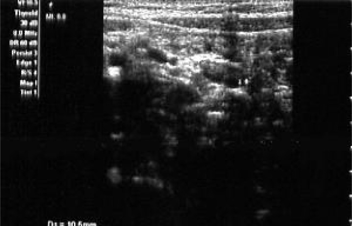
Ultrasound examination of abdomen revealed presence of multiple lymph nodes in paraaortic area distributed from below the pancreas to the bifurcation of the aorta; the largest was 6 mm ×12mm (Fig. 1). After 36 hour from receiving of intravenous gamma globulin, the fever was not subsided, so another dose (2gr/kg) was given. One day later the fever stopped. Four days thereafter we reduced aspirin dose (4mg/kg/day) and discharged the patient in good condition.
Fig. 1.
Ultrasound examination of the abdomen showing multiple lymph nodes in para aortic area.
One week later blood tests revealed platelet count 480×103/µl, WBC count 6.7×10³/µl and plasma albumin 3/5 gr/dl. Ultrasonography was normal without signs of lymphadenopathy. CT scan of abdomen was also normal. A second echocardiography showed ectasia of the right coronary artery. On follow up 8 weeks later, erythrocyte sedimentation rate decreased to 13mm/hr and echocardiography showed disappearance of ectasia of the coronary artery, so aspirin was discontinued. The patient was followed by cardiologist for 1 year and the last echocardiography has been normal.
Discussion
KD is one of the most common vasculitides in children. Owning to lack of diagnostic tests, the diagnosis is based on clinical criteria after the exclusion of other febrile diseases[3].
The present case had high grade, persistent fever, and four characteristic signs and symptoms of KD: erythematous macular rash, bilateral non purulent conjunctival injection, oro pharyngeal changes, and edema of extremities.
In addition to the established signs and symptoms which constituted the basis for diagnosis of the disease, the case had paraaortic lymphadenopathies. The patient had no other laboratory or clinical criteria of macrophage activation syndrome, such as elevated levels of aspartate aminotransferase, decreased WBC, central nervous system dysfunction, hemorrhage or hepatomegaly. The patient had thrombocytopenia though not common in Kawasaki but if present, is associated with an increased risk of coronary artery aneurysm and myocardial infarction[4]. Because of rising platelet count and disappearance of symptoms and signs following treatment, bone marrow aspiration was not performed. As the second ultrasonography of abdomen was normal without a sign of lymphadenopathy, so CT scan of abdomen was done to confirm disappearance of lymphadenopathy that was normal too. Lymphadenopathy is the least occurring (50–75%) diagnostic feature of KD[4]. It occurs most commonly in anterior cervical and less commonly in posterior cervical and axillary areas[2]. It was also reported to occur in mediastinum, with disappearance after 6 weeks[6]. Falcini reported a case of severe KD with multifocal lymphadenopathy mimicking a lymphoproliferative disorder[7].
Conclusion
The present report shows that lymphadenopathy in KD might also occur in paraaortic region. The disappearance of lymphadenopathy upon conventional treatment indicates that lymphadenopathy was associated with the disease. In these patients a close observation and ultrasonographic follow up will prevent unnecessary further investigation.
Acknowledgements
The authors would like to thank Dr Nekuian at Center for Development of Clinical Research, Nemazee Hospital for his assistance.
References
- 1.Taubert KA, Rowley AH, Shulman ST. Nationwide survey of Kawasaki disease and acute rheumatic fever. J Pediatr. 1991;119(2):279–82. doi: 10.1016/s0022-3476(05)80742-5. [DOI] [PubMed] [Google Scholar]
- 2.Sudel RP, Ross E, Cassidy JT. Cassidy Textbook of Pediatric Rheumatology. 5th ed. Philadelphia: Saunders; 2005. Kawasaki disease; pp. 521–36. [Google Scholar]
- 3.Council on Cardiovascular Disease in the Young. Committee on Rheumatic Fever, Endocarditis, and Kawasaki Disease; American Heart Association. Diagnostic guidelines for Kawasaki disease. Circulation. 2001;103(2):335–6. [Google Scholar]
- 4.Nofech MY, Garty BZ. Thrombocytopenia in Kawasaki disease: a risk factor for the development of coronary artery aneurysms. Pediatr Hematol Oncol. 2003;20(8):597–601. [PubMed] [Google Scholar]
- 5.Rowly AH, Gonzalez-Crussi F, Shulman ST. Kawasaki syndrome. Rev Infect Dis. 1988;10(1):1–15. doi: 10.1093/clinids/10.1.1. [DOI] [PubMed] [Google Scholar]
- 6.Bosch Marcet J, Serres Creixams X, Penas Boira M, et al. Mediastinal lymphadenopathy, a variant of incomplete Kawasaki disease. Acta Paediatr. 1998;87(11):1200–2. doi: 10.1080/080352598750031239. [DOI] [PubMed] [Google Scholar]
- 7.Falcini F, Simonini G, Calabri GB, et al. Multifocal lymphadenopathy associated with severe Kawasaki disease. Ann Rheum Dis. 2003;62(7):688–9. doi: 10.1136/ard.62.7.688. [DOI] [PMC free article] [PubMed] [Google Scholar]


