Abstract
Background
Diaphragmatic paralysis in newborns is related to brachial plexus palsy. It can cause respiratory failure necessitating prolonged mechanical ventilation and subsequent extubation failure.
Case Presentation
We present a two-hour-old male newborn with a birth weight of 4500 grams who had a right-sided brachial plexus palsy and right diaphragmatic paralysis due to shoulder dystocia. He developed respiratory distress due to isolated paralysis of the right hemi diaphragm. The clinical course was progressive, his condition worsening despite oxygen application. Physical examination, chest X-rays and M-mode ultrasonography of the diaphragm confirmed the diagnosis diaphragmatic paralysis. Surgical plication of diaphragm was done earlier than the usual time because of recurrent extubation failure. Diaphragmatic plication led to rapid improvement of pulmonary function and allowed discontinuation of mechanical ventilation in less than 3 days.
Conclusion
Early diaphragmatic plication enhances weaning process and may prevent or minimize the morbidity associated with long-term mechanical ventilation in a neonate with diaphragmatic paralysis.
Keywords: Neonate, Diaphragmatic Paralysis, Ventilation, Mechanical, Lower Brachial Plexus Palsy
Introduction
The exact incidence of phrenic nerve injury in infants is not well documented. So, respiratory distress due to diaphragmatic paralysis as a consequence of birth injury in newborn infants seems to be rare[1, 2]. The right side is affected more than the left side and most of it is associated with brachial plexus injury[1, 3]. The most important risk factors are macrosomia, shoulder dystocia and extraction in breech presentation[3]. Iatrogenic injury to the phrenic nerve during the chest tube insertion has been reported[4].
Several cases are also reported after cardiac surgery[5–7]. Blazer and his colleagues described a neonate with severe bilateral diaphragmatic paralysis caused by hemorrhage in the lower brain stem[8]. Other rare causes and associations of diaphragmatic paralysis are procedures like internal jugular vein cut-down[9], venesection at the neck for parenteral feeding[10], followed by neck surgery[11], congenital hypomyelination neuropathy[12] and congenital myotonic dystrophy[13].
Diaphragmatic paralysis usually presents as respiratory distress or difficulty in weaning from the ventilator. With the patient in lateral decubitus position and paralyzed side up, accentuated paradoxical inspiratory inward epigastric motion ipsilateral to the paralyzed hemidiaphragm can be seen[1, 3]. Krabiber et al reported a two-week old baby who had left sided brachial plexus palsy and torticollis with an undiagnosed left diaphragmatic paralysis. Simultaneous occurrence of brachial plexus palsy and diaphragmatic paralysis make an easy clue to early diagnosis and not to overlook diaphragmatic paralysis[14].
Paradoxic cephalic motion of the diaphragm during inspiration prevents air entry on the ipsilateral side and shifts the freely mobile mediastinum away from the affected side, impairing function of the contralateral lung. Infants have a greater reliance on the diaphragm for ventilation and weak intercostal and abdominal accessory muscles, further compromising their respiratory status[15].
Chest radiograph obtained over the first week shows persistent elevation of the hemi diaphragm and real-time ultrasound scans of the diaphragm show decreased and paradoxical excursion of the ipsilateral hemi diaphragm and normal excursion of the contra-lateral side. In suspected cases fluoroscopic examination make the definite diagnosis[3]. Electrodiagnostic studies (EDS) are an adjunctive tool and help to localize and prognosticate the outcome of obstetrical brachial plexus palsy. A poor re-innervation pattern on EMG is correlated with inadequate recovery[16]. Managements are generally supportive and include lying ipsilateral to the involved side, oxygen therapy and in case of respiratory failure mechanical ventilation[3, 9].
Diaphragmatic plication is carried out to manage refractory cases. Generally, it is carried out at the end of one-month via a simple trans-abdominal, transthoracic or trans-laparoscopic operation without great complication[5, 6, 17–21].
The purpose of this case report is to emphasis the benefit of the early operation and diaphragmatic plication.
Case Presentation
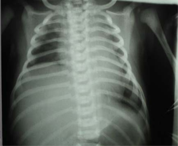
A 38-year-old mother with two previous normal pregnancies delivered vaginally a male neonate weighing 4500 g, at gestation age of 42w+5d with an Apgar score of 8 and 9 at 1st and 5th minute, respectively. The process of delivery was complicated by shoulder dystocia. Shortly after birth, the newborn developed progressively severe respiratory distress, requiring O2 application via head box. He received intravenous fluid and antibiotics. He was subsequently transferred to our hospital where he continued to require O2 therapy. His initial physical examination revealed tachypnea, nasal flaring, chest indrawing, asynchronized chest and abdomen motion and the absence of Moro reflex and absence of any spontaneous movement in the right hand. His right arm remained in adduction and internal rotation position. The grasp reflex was intact. Apart from respiratory distress no other abnormality was detected clinically. In the chest roentgenogram the lung field was normal, no sign of clavicle and homeruns fracture, but the right hemidiaphragm was highly elevated (Fig 1).
Fig. 1.
Chest X-ray before mechanical ventilation reveals the elevation of right hemidiaphragm
According to the history of shoulder dystocia and the findings on physical examination and chest X-ray diagnosis of brachial plexus (Erb) palsy accompanied with diaphragmatic paralysis was made and the neonate transferred to NICU. He was placed on his right side of the body and continued to require increasing concentrations of oxygen. Endotracheal intubation and ventilatory support (Bear cub infant ventilator, Bear Medical System) at 24 h of age was required because of respiratory failure, which was characterized by pH of 7.15 and PCO2 of 68 mmHg on arterial blood gas analysis.
Conventional ventilatory support to maintain oxygenation continued and weaned from ventilator after 6 days, but he started to have increasing respiratory distress and inefficient chest excursions. So he was re-intubated and ventilated for another 5 days because of extubation failure. Chest X-ray was the same as previous one and the inflammatory markers for sepsis like CRP and ESR were normal. The initial and the second blood culture were negative. No other cause of extubation failure, other than the thoracic and inefficient respiration was evident. He was weaned at this stage to the Nasal Continues Positive Pressure (N-CPAP) to manage the extubation failure. The chest X-ray at this stage showed an elevated right hemidiaphragm like the previous one without the radiologic signs of collapse or pneumonia. After a couple of hours he developed respiratory distress and hypoxemia requiring re-intubation and ventilation using the same conventional ventilator for the third time.
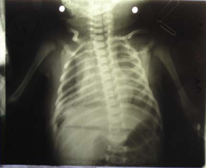
Besides, real-time ultrasonography (Model SSD-620) showed marked decrease in movement of the right hemidiaphragm when compared with the left hemidiaphragm. Cranial ultrasonography was normal. Although most of the authors recommend the plication at the end of the first month, we decided to operate earlier because of multiple extubation failures. Seventy two hours after operation the patient was extubated without extubation failure. Chest X-ray and real time ultra sonography showed normal position and function of the diaphragm (Fig. 2).
Fig. 2.
Chest X-ray after the surgical intervention revealed the right hemidiaphragm in its proper position
The patient was handed over to newborn services on day 24 of life and discharged to home after 7 days. His mother did not show any signs of myotonia in physical examination. On follow-up examinations at 38, 45 and 62 days of life his muscle tone was normal. His growth and development was normal and the Moro reflex of right hand was significantly improved. He demonstrated the normal passive range of motion in his right arm and no respiratory symptom was seen on follow up examination in outpatient clinic at 12 and 18 months of age.
Discussion
Early surgical intervention in our patient resulted in rapid improvement and weaning from ventilator. Extubation failure for two times necessitated diaphragmatic plication.
Management of diaphragmatic paralysis due to phrenic nerve injury is controversial. There are three options: 1) immediate diaphragmatic plication to reduce the need for mechanical ventilation, duration of hospital stay and pulmonary infections, 2) diaphragmatic plication after 2–4 weeks, which is the usual time for spontaneous recovery, and 3) a conservative, non-operative approach using prolonged mechanical ventilation, as improvement can occur up to 2 months, thus avoiding operative/postoperative complications[22, 23].
To avoid the complication associated with prolonged mechanical ventilation, early operative intervention is indicated when extubation to CPAP is unsuccessful over a course of several days[23].
Current evidence indicates that diaphragmatic plication is simple, safe and durable, even in ELBW neonates, and should be undertaken once failure to extubate to CPAP is documented over ‘several’ days[22, 23].
De Vries et al stated that spontaneous im-provement of diaphragmatic paralysis after one month is rare and it would be better to do the operation at the end of one month of age[17].
Langer et al. reported 23 cases of diaphragmatic paralysis due to birth trauma or heart surgery, which were operated 18.5 days after the insult and extubated 2 days after the diaphragmatic plication. They have chosen an arbitrary duration of three weeks of symptomatic phrenic nerve paralysis as the indication for plication [24].
Bowerson et al. reported 4 cases of diaphragmatic paralysis associated with neonatal brachial plexus palsy. Three of these patients required surgical placation of the diaphragm[25].
Escanda et al. managed a neonate with diaphragmatic paralysis with nasal CPAP successfully[2]; our patient developed respiratory failure on nasal CPAP after extubation.
Researchers have recommended that symptomatic patients should be operated immediately, with an expected dramatic resolution of the respiratory compromise. The risks of anesthesia and thoracotomy to be considered along with the significant respiratory morbidity associated with the option of waiting for spontaneous recovery of diaphragmatic function[23].
Conclusion
Early diaphragmatic plication enhances weaning process and may prevent or minimize the morbidity associated with long-term mechanical ventilation in a neonate with diaphragmatic paralysis.
References
- 1.Zifko U, Hartman M, Girsch W, et al. Diaphragmatic paresis in newborns due to phrenic nerve injury. Neuropediatrics. 1995;26(5):281–4. doi: 10.1055/s-2007-979774. [DOI] [PubMed] [Google Scholar]
- 2.Escanda B, Cerveau C, Kuhn P, et al. Phrenic nerve paralysis of obstetrical origin: Favorable course using continuous positive airway pressure. Arch pediatrics. 2000;7(9):965–8. doi: 10.1016/s0929-693x(00)90012-5. [DOI] [PubMed] [Google Scholar]
- 3.Avroy A, Fanaroff MB, Richard J, Martin MB. Neonatal Perinatal Medicine. Textbook of Disease of Fetus and Infant. (8th ed) 2006;1:475–6. [Google Scholar]
- 4.Arya H, Williams J, Ponsford SN, et al. Neonatal diaphragmatic paralysis caused by chest drains. Arch Dis Child. 1991;66(9):1104. doi: 10.1136/adc.66.4_spec_no.441. [DOI] [PMC free article] [PubMed] [Google Scholar]
- 5.Matejka T, Hucin B, Tlaskal T, et al. Plication of the diaphragm-a method of surgical treatment of diaphragmatic paralysis in neonatal and infant after heart surgery. Rozhl Chir. 1997;76(5):250–3. [PubMed] [Google Scholar]
- 6.Joho-Arreole AL, Bauersfeld U, Stauffer UCE, et al. Incidence and treatment of diaphragmatic paralysis after cardiac surgery in child. Eur J Cardiac Thorac Surg. 2005;27(1):53–7. doi: 10.1016/j.ejcts.2004.10.002. [DOI] [PubMed] [Google Scholar]
- 7.Imaitshizokawa H, Imaizumi H, Matsumoto H. Transient phrenic nerve palsy after cardiac operation in infants. Clin Neurophysiol. 2004;115(6):1469–72. doi: 10.1016/j.clinph.2004.01.007. [DOI] [PubMed] [Google Scholar]
- 8.Blazer S, Hemli JA, Sujov PO, Braun J. Neonatal bilateral diaphragmatic paralysis caused by brain stem haemorrhage. Arch Dis Child. 1989;64(1 Spec No):50–2. doi: 10.1136/adc.64.1_spec_no.50. [DOI] [PMC free article] [PubMed] [Google Scholar]
- 9.Ugalde-Fernandez JH, Suarez-Rios LF, Arellano-Cuevas R. Diaphragmatic paralysis caused by a phrenic nerve lesion secondary to internal jugular vein cutdown. Bol Med Hosp Infant Mex. 1989;46(7):497–9. [PubMed] [Google Scholar]
- 10.Caballero-Noguez B, Fernandez-Corte MG, Escobedo-Chavez E. Diaphragmatic paralysis due to a lesion of the phrenic nerve secondary to venesection at the neck for parenteral feeding. Bol Med Hosp Infant Mex. 1993;50(2):125–8. [PubMed] [Google Scholar]
- 11.McCaul JA, Hislop WS. Transient hemi-diaphragmatic paralysis following neck surgery: report of a case and review of the literature. J R Coll Surg Edinb. 2001;46(3):186–8. [PubMed] [Google Scholar]
- 12.Hahn JS, Henry M, Hudgins L, Madan A. Congenital hypomyelination neuropathy in a newborn infant: Unusual cause of diaphragmatic and vocal cord paralyses. Pediatrics. 2001;108(5):95. doi: 10.1542/peds.108.5.e95. [DOI] [PubMed] [Google Scholar]
- 13.A Yong SC, Boo NY, Ong LC. Case of congenital myotonic dystrophy presented with diaphragmatic paresis during the neonatal period. J Paediatr Child Health. 2003;39(7):567–8. doi: 10.1046/j.1440-1754.2003.00221.x. [DOI] [PubMed] [Google Scholar]
- 14.Karabiber H, Ozkan KU, Garipardic M, et al. An overlooked association of brachial plexus palsy: diaphragmatic paralysis. Acta Paediatr Taiwan. 2004;45(5):301–3. [PubMed] [Google Scholar]
- 15.Gallagher PG, Seashore JH, Touloukian RJ. Diaphragmatic plication in the extremely low birth weight infant. J Pediatr Surg. 2000;35(4):615–6. doi: 10.1053/jpsu.2000.0350615. [DOI] [PubMed] [Google Scholar]
- 16.Gopinath MS, Bhatia M, Mehta VS. Obstetric brachial plexus palsy: A clinical and electrophysiologic evaluation. J Assoc Physicians India. 2002;50:1121–3. [PubMed] [Google Scholar]
- 17.De Vries TS, Koens BL, Vos A. Surgical treatment of diaphragmatic eventration caused by phrenic nerve injury in the newborn. J pediatric surgery. 1998;33(4):602–5. doi: 10.1016/s0022-3468(98)90325-6. [DOI] [PubMed] [Google Scholar]
- 18.Langer JC, Filler RM, Coles J, Ednonds JF. Plication of the diaphragm for infants and young children with phrenic nerve palsy. J Pediatr Surg. 1988;23(8):749–51. doi: 10.1016/s0022-3468(88)80417-2. [DOI] [PubMed] [Google Scholar]
- 19.Refaely Y, Simansky DA, Palsy M, Yellin A. Plication of diaphragm for postoperative phrenic nerve injury in infants and young children. Harefuah. 1999;137(5–6):190–3. [PubMed] [Google Scholar]
- 20.Bowman ED, Marton LJ. A case of neonatal bilateral diaphragmatic paralysis requiring surgery. Aust Pediatric J. 1984;20(4):331–2. doi: 10.1111/j.1440-1754.1984.tb00106.x. [DOI] [PubMed] [Google Scholar]
- 21.Jog SM, Patole SK. Diaphragmatic paralysis in extremely low birth weight neonates: Is waiting for spontaneous recovery justified? J Pediatr Child Health. 2002;38(1):101–3. doi: 10.1046/j.1440-1754.2002.00758.x. [DOI] [PubMed] [Google Scholar]
- 22.Commare MC, Kurstjens SP, Barois A. Diaphragmatic paralysis in children. A review of 11 cases. Pediatr Pulmonol. 1994;18(3):187–93. doi: 10.1002/ppul.1950180311. [DOI] [PubMed] [Google Scholar]
- 23.Tsugawa C, Kimura K, Nishijima E, et al. Diaphragmatic eventration in infant and children: is conservative treatment justified? J Pediatr Surg. 1997;32(11):1643–4. doi: 10.1016/s0022-3468(97)90473-5. [DOI] [PubMed] [Google Scholar]
- 24.Langer JC, Filler RM, Coles J, Edmonds J. Plication of the diaphragm for infants and young children with phrenic nerve palsy. J Pediatr Surg. 1998;23(8):749–51. doi: 10.1016/s0022-3468(88)80417-2. [DOI] [PubMed] [Google Scholar]
- 25.Bowerson M, Nelson VS, Yang LJ. Diaphragmatic paralysis associated with neonatal brachial plexus palsy. Pediatr Neurol. 2010;42(3):234–6. doi: 10.1016/j.pediatrneurol.2009.11.005. [DOI] [PubMed] [Google Scholar]


