Abstract
Objective
The prevalence, patients’ age and sex and the site of the lesions are important factors for diagnosis and they may be different in various populations. The aim of this study was to determine the type and distribution of orofacial tumors among children and adolescents in an Iranian population
Methods
In this retrospective, case series study, data about the type, age, sex and site of 142 tumors in patients ≤18 years afflicted with orofacial neoplasms referred 2005-2009 to two referral centers in Shiraz, Southern Iran, were collected and analyzed.
Findings
There were 142 (2.8%) tumors among the subjects. The most common types of benign and malignant tumors were odontoma and lymphoma in children and pleomorphic adenoma and rhabdomyosarcoma in adolescents. Parotid and posterior parts of the mandible were the most common sites of soft tissue and intrabony tumors. In the oral cavity, the palate was the most common affected site. The tumors were found in boys with higher frequency (Male:Female ratio was 1.4:1).
Conclusion
The observed differences in tumor type and distribution in comparison with previous studies may be attributed to genetic and geographic variations in the populations; however the design and methods of the studies are different, too.
Keywords: Orofacial, Tumors, Children, Adolescents, Odontoma, Pleomorphic Adenoma
Introduction
Orofacial tumors are a heterogeneous group of pathologic disorders with various histological types and clinical behavior. These neoplasms affect speaking and swallowing due to their special location. They might result in expansion, tooth movement and destruction of adjacent structures [1]. Our knowledge on basic clinical characteristics such as age, gender distribution, location and histologic types of these lesions can be valuable in clinical diagnosis and patient's management. These neoplasms have peculiar importance and their incidence has been detailed in various countries [2–8]. Previous studies have shown that these tumors are more common in adults [3–7, 9–11] and their incidence, histological types and clinical features are different in children and adolescents. Most of the tumors seen in younger individuals are benign and have mesenchymal origin [2, 3, 6]. To the present time, data on clinico-pathologic features of orofacial tumors in children and adolescents have not been published in Iran. The current study analyzed 142 cases of these neoplasms regarding the incidence, histologic type, age and gender distribution.
Subjects and Methods
The data of the current retrospective study were obtained from the archive of pathology department of Khalili Hospital (an ENT center) and Shiraz Dental School, two referral centers affiliated to Shiraz University of Medical Sciences in southern Iran, from 2005 to 2009. All of the cases histopathologically recorded as true neoplasm according to the textbook of oral and maxillofacial pathology [12] with orofacial location in the patients ≤18 years were enrolled.
The patients' medical files were analyzed with regard to the patient's age, gender, tumor site and histopathologic type. The patients with reactive, tumor-like lesions and incomplete clinical data were excluded. The patients were divided into children (age ≤12 years) and adolescent (age 13-18 years) groups.
Findings
Based on the findings, 875 neoplasms were noted among 5018 patients' files, out of which 142 cases were reported in patients <18 years (2.8% of all the specimens and 16.5% of all tumors). From these, 53 (36.7%) cases were diagnosed in children and 89 (63.3%) in adolescents. Orofacial tumors were found in 83 boys and 59 girls (male to female ratio was 1.4:1). Male/Female (M/F) ratio was higher in adolescents.
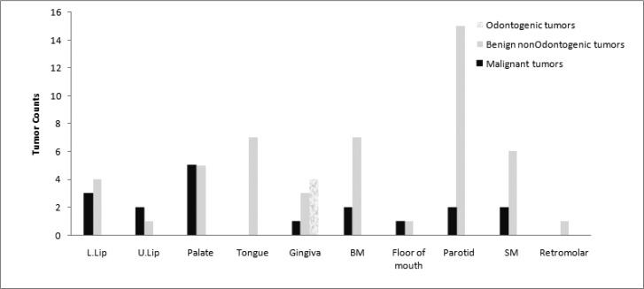
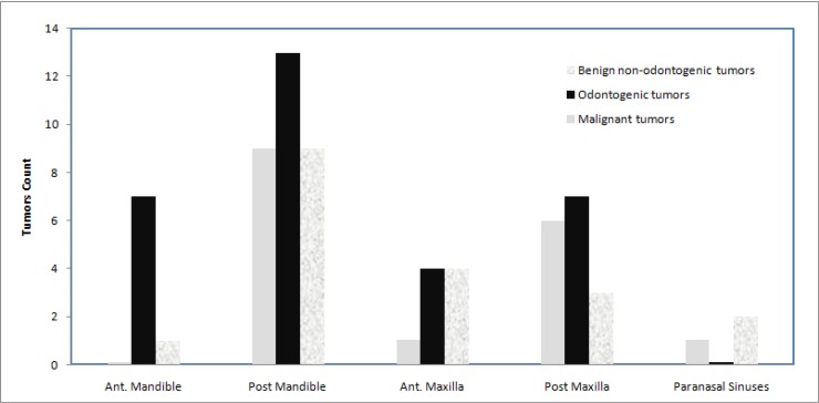
The neoplasms were composed of 107 (50.7%) benign non-odontogenic, 35 (24.6%) odonto-genic and 35 (24.6%) malignant non-odonto-genic tumors. Overall, pleomorphic adenoma was the most common tumor. Parotid gland and the palate were the most frequent affected sites in the extra-oral and intra-oral soft tissue. The most common site of involvement among intrabony tumors was the posterior part of the mandible (Fig 1 and 2).
Fig. 1.
Location of soft tissue tumors
L: Lower, U: Upper, BM: Buccal mucosa, SM: Submandibular, T: tumor
Fig. 2.
Location of intrabony tumors
Ant: Anterior, Post: Posterior, T: Tumor
Odontogenic tumors
There were 35 odontogenic tumors all of which were benign. The majority of the tumors (68.5%) were found in adolescents. M/F ratio was 1.2:1.
Odontoma and ameloblastoma were the most frequent tumors in children and adolescents, respectively. Four cases of peripheral odontogenic fibroma were also seen. (Table 1)
Table 1.
Odontogenic tumors: type, age and gender distribution
| Tumor type | Age group | Total | Male/Female ratio | ||
|---|---|---|---|---|---|
| ≤12 | 13-18 | ≤12 | 13-18 | ||
| Ameloblastoma | 2 | 7 | 9 | 0:2 | 5:2 |
| Adenomatoid Odontogenic Tumor | -- | 7 | 7 | -- | 2:5 |
| Ameloblastic fibroma | 1 | -- | 1 | 0:1 | -- |
| Ameloblastic fibro-odontoma | 1 | -- | 1 | 0:1 | -- |
| Odontoma | 6 | 4 | 10 | 5:1 | 3:1 |
| Odontogenic Fibroma | 1 | 4 | 5 | 1:1 | 2:2 |
| Odontogenic Myxoma | -- | 1 | 1 | -- | 1:0 |
| Cementoblastoma | -- | 1 | 1 | -- | 0:1 |
| Total (%) | 11 (31.4) | 24 (68.5) | 35(100) | 1.2:1 | 1.2:1 |
Benign non-odontogenic tumors
There were 72 (50.7%) benign non-odontogenic tumors, most of which (53%) were mesenchymal followed by epithelial and bone tumors. These neoplasms were more common in adolescents and M/F ratio was 1.3:1.
Hemangioma and pleomorphic adenoma were the most common tumors in children and adolescents, respectively (Table 2).
Table 2.
Benign non-odontogenic tumors: type, age and gender distribution
| Tumor type | Age group | Total | Male/Female ratio | ||
|---|---|---|---|---|---|
| ≤12 | 13-18 | ≤12 | 13-18 | ||
| Pleomorphic adenoma | 1 | 17 | 18 | 0:1 | 8:9 |
| Monomorphic Adenoma | 2 | 2 | 0:2 | ||
| Warthin's Tumor | 2 | 2 | 1:1 | ||
| Nevus | 1 | 1 | 0:1 | ||
| Hemangioma | 5 | 12 | 17 | 3:2 | 9:3 |
| Lymphangioma | 2 | 2 | 4 | 1:1 | 1:1 |
| Fibrous Histiocytoma | 1 | 3 | 4 | 0:1 | 3:0 |
| Schwannoma | 4 | 2 | 6 | 4:0 | 2:0 |
| Neurofibroma | 3 | 4 | 7 | 0:3 | 4:0 |
| Osteoma | 1 | 1 | 1:0 | ||
| Central ossifying fibroma | 3 | 5 | 8 | 3:0 | 1.4 |
| Desmoplastic Fibroma | 2 | 2 | 1:1 | ||
| Total (%) | 33(24) | 48(67) | 100(72) | 1.2:1 | 1.4:1 |
Malignant tumors
Among 142 orofacial neoplasms, 35 (24.6%) were malignant. These tumors were composed of 12 (34.2%) cases of carcinoma, 10 (28.8%) cases of sarcoma and 10 (28.8%) cases of hematopoietic malignancy consisting of 7 cases of lymphoma and 3 cases of eosinophilic granuloma. Definitive diagnosis of 3 cases of round cell tumor had not been determined (Table 3). Lymphoma and rhabdomyosarcoma were the most common malignancy in children and adolescents, respectively. Among carcinomas, salivary gland tumors constituted 66.7% of the cases. M/F ratio in this group of tumors was 1.7:1.
Table 3.
Malignant tumors: type, age and gender distribution
| Tumor type | Age group | Total | Male/Female ratio | ||
|---|---|---|---|---|---|
| ≤12 | 13-18 | ≤12 | 13–18 | ||
| Squamous cell carcinoma | 4 | -- | 4 | 2.2 | -- |
| Mucoepidermoid carcinoma | 2 | 2 | 4 | 1:1 | 1:1 |
| Adenoid cystic carcinoma | 1 | 3 | 4 | 1:0 | 2:1 |
| Rhabdomyosarcoma | -- | 5 | 5 | -- | 4:1 |
| Malignant fibrous histiocytoma | -- | 1 | 1 | -- | 1:1 |
| Osteosarcoma | 1 | -- | 1 | 1:0 | -- |
| Ewing sarcoma | -- | 2 | 2 | -- | 2:0 |
| Chondrosarcoma | -- | 1 | 1 | -- | 0:1 |
| Langerhans cell histiocytoma | 2 | 1 | 3 | 1:1 | 0:1 |
| Lymphoma | 6 | 1 | 7 | 3:3 | 1:0 |
| Undiagnosed | 2 | 1 | 3 | 1:1 | 1:0 |
| Total (%) | 51.4(18) | 17(48.6) | 100(35) | 1.2:1 | 2.4:1 |
Discussion
Learning clinical features of pathologic disorders such as tumors including primary site, age and sex distribution is important for early diagnosis. These features may be very different among various populations. It has been reported that 10% of tumors among Iranian children and adolescents are located in the head and neck region [13]. But to the present time, data on oral and maxillofacial neoplasms in this group are not available. We found that 16.5% of all the neoplasms in these locations occurred in children and adolescents. This percentage has a wide range in various studies from 7-28% [4, 10].
This discrepancy is partly due to the varied age and pathologic lesions included, as reactive and viral lesions considered as neoplasm in some researches [3, 5, 10, 11].
Most of the tumors were benign non-odontogenic, occurring more commonly in adolescents. Others have found the same figure in Jordanian and African populations [2, 5, 6, 10].
From benign non-odontogenic tumors, mesenchymal origin was found more frequently. Many authors have confirmed this finding[2–4, 6, 8]. However, malignant tumors have had a more common frequency in some African populations due to the high prevalence of endemic Burkitt's lymphoma among pediatric patients [5, 10, 14].
Our results showed that hemangioma was the most frequent benign non-odontogenic tumor in children as mentioned in most of the previous and recent studies [1, 2, 10, 13, 15]. This lesion was observed commonly in the lip. In the adolescents' group in the same line with the present study, Elarbi and Arbegosela et al have noted that pleomorphic adenoma constituted a high percentage in African population [2, 10].
Odontogenic tumors were found more commonly in adolescents. Most of the studies have supported this result [2, 3, 6, 16]. Odontogenic tumors arise from the rest of the tooth germ.
Crown formation terminates at 4 or 5 years of age in most of the permanent teeth; therefore many of these tumors develop after the age of 6[17]. In this group similar to our findings odontoma has been noted as the most frequent odontogenic tumor in many studies [3, 6, 12, 16]. In other studies that have considered this entity as a hamartoma, ameloblastoma has been the most common [4, 5]. In our series, the majority of ameloblastomas were unicystic type. This figure was expected because conventional ameloblastoma is uncommon in pediatrics, whereas about 50% of all the unicystic types are diagnosed during the second decade of life [12].
Malignant tumors accounted for 24.6% of all the neoplasms. In previous studies, 3-12.5% of the tumors have been malignant. This significant discrepancy is partly due to this fact that reactive and tumor-like lesions recorded as tumor in these publications[2, 3, 6]. The studies done in Africa reported that 43-67% of all the tumors were malignant with a high incidence of Burkitt's lymphoma[5, 9, 10].
In our series, the majority (34%) of malignancies were carcinomas, followed by sarcomas (28.5%) and lymphomas (20%). About 66% of the carcinomas were salivary gland tumors including adenoid cystic carcinoma and mucoepidermoid carcinoma. In comparison with other studies, salivary gland carcinomas seem to be more common in our group; however, these neoplasms are rare in patients during the first and second decades of life [18]. In agreement with our results, Jones has stated that carcinomas were more common than sarcomas in England[11]. But others in North Jordan, Nigeria and Libya reported that sarcomas had a higher incidence [6, 10, 19]. Al-Khateeb has classified lymphomas in the category of sarcomas [6]. The most common type of sarcomas was rhabdomyosarcoma as reported in previous studies [1, 11, 12].
Our findings showed that neoplasms are more common in boys. Malignant tumors revealed a higher M/F ratio similar to most of the studies [2, 4, 6, 10 14]. The high M/F ratio of malignancies in adults is attributed to smoking, alcohol consumption and UV radiation [1, 12], but these results reveal that other potential causative factors like genetic predisposition and hormones influence the tendency of malignancies toward the males.
Overall, 52% of all the tumors occurred in the soft tissue and 48% in the jaws. The palate and posterior region of the mandible were the most commonly affected sites in the soft tissue and bone. The lip, palate and tongue have been presented as the most common location in other publications, and of bony tumors the posterior part of the mandible [2, 3, 6, 10].
These discrepancies are attributed to racial and geographic variations, and some differences in the design of the studies, for example the referral nature of the research center. These instances obstruct precise comparison of different studies. However, data on characteristics of the special lesions in any population could help the physicians to more accurate evaluation and early diagnosis.
Conclusion
Our findings demonstrated that the orofacial tumors in children and adolescents have special characteristics in Southern Iran which are different in some aspects from those in other parts of the world. The salivary gland tumors were more common and male's propensity was more predominant.
Acknowledgment
The ethics committee of Shiraz University of Medical Sciences approved this study. The authors would like to thank Dr. Nasrin Shokripour, at center for development of clinical research of Namazi hospital for editorial assistance.
Conflict of Interest
None
References
- 1.Regezi JA, Sciubbaj J, Jordan RCK. Oral pathology-clinical pathologic correlations. Odontogenic tumors. [Google Scholar]
- 2.Elarbi M, Gl-Gehani R, Subhashraj K, et al. Orofacial tumors in Libyan children and adolescents. A descriptive study of 213 cases. Int J of Pediatr Otorhinolaryngol. 2009;73(2):237–42. doi: 10.1016/j.ijporl.2008.10.013. [DOI] [PubMed] [Google Scholar]
- 3.Tanaka N, Murata A, Yamaguchi A, et al. Clinical features and management of oral and maxillofacial tumors in children. Oral Surg Oral Med Oral Pathol Oral Radiol Endod. 1999;88(1):11–5. doi: 10.1016/s1079-2104(99)70186-1. [DOI] [PubMed] [Google Scholar]
- 4.Ulmasky M, Lustmann J, Balkin N. Tumors and tumor-like lesions of oral cavity and related structures in Israeli children. Int J Oral Maxillofac Surg. 1999;28(4):291–4. [PubMed] [Google Scholar]
- 5.Adebayo ET, Ajike SO, Adekeye EO. Tumors and tumor-like lesions of the oral and perioral structures of Nigerian children. Int J Oral Maxillofac Surg. 2001;30(3):205–8. doi: 10.1054/ijom.2001.0052. [DOI] [PubMed] [Google Scholar]
- 6.Al-Khateeb T, Al-Hadi Hamasha A, Almasri NM. Oral and maxillofacial tumors in North Jordanian children and adolescents: a retrospective analysis over 10 years. Int J Oral Maxillofac Surg. 2003;32(1):78–83. doi: 10.1054/ijom.2002.0309. [DOI] [PubMed] [Google Scholar]
- 7.Tanrikulu R, Erol B, Haspolat K. Tumors of the maxillofacial region in children: retrospective analysis and long-term follow-up outcomes of 90 patients. Turk J Pediatr. 2004;46(1):1–2. [PubMed] [Google Scholar]
- 8.Arotiba GT. A study of orofacial tumors in Nigerian children. J Oral Maxillofac Surg. 1996;54(1):34–8. doi: 10.1016/s0278-2391(96)90299-2. [DOI] [PubMed] [Google Scholar]
- 9.Parkins GEA, Armah G, Ampofo P. Tumors and tumor-like lesions of the lower face at Korle Bu Teaching Hospital, Ghana-an eight year study. World J Surg Oncology. 2007;7(5):48. doi: 10.1186/1477-7819-5-48. [DOI] [PMC free article] [PubMed] [Google Scholar]
- 10.Aregbesola SB, Ugboko V, Akinwande J, et al. Orofacial tumors in suburban Nigerian children and adolescents. Brit J Oral Maxillofac Surg. 2005;43(3):226–31. doi: 10.1016/j.bjoms.2004.11.006. [DOI] [PubMed] [Google Scholar]
- 11.Jones AV, Franklin CD. An analysis of oral and maxillofacial pathology found in children over a 30-year period. Int J Pediatr Dentistry. 2006;16(1):19–30. doi: 10.1111/j.1365-263X.2006.00683.x. [DOI] [PubMed] [Google Scholar]
- 12.Neville BW, Damm DD, Allen CM, et al. 3rd ed. Philadelphia: Saunders; 2009. Oral and maxillofacial pathology. [Google Scholar]
- 13.Khademi B, Taraghi A, Mohammadianpanah M. Anatomical and histopathological profile of head and neck neoplasms in Persian pediatric and adolescent population. Int J Pediatr Otorhinolaryngol. 2009;73(9):1249–53. doi: 10.1016/j.ijporl.2009.05.017. [DOI] [PubMed] [Google Scholar]
- 14.Asamoa EA, Ayanlere AO, Olaitan AA, et al. Pediatric tumors of the jaws in northern Nigeria. J Cranio Maxillofac Surg. 1990;18(3):130–5. doi: 10.1016/s1010-5182(05)80330-0. [DOI] [PubMed] [Google Scholar]
- 15.Maitra A, Kumar V. Genetic and pediatrics diseases. In: Kumar V, Cotran R, Robbins A, editors. 7th ed. Philadelphia: Saunders; 2003. p. 251. Robbin's basic pathology. [Google Scholar]
- 16.Mehran M, Eslami M, Jalayernaderi N, et al. An investigation on the prevalence of odontogenic tumors among children referred to the oral and maxillofacial pathology department, Faculty of Dentistry, Tehran University of Medical Sciences (1962–2002) J Islamic Dent Associat Iran. 2006;18(1):7–12. [Google Scholar]
- 17.Sato M, Tanaka N, Sato T, et al. Oral and maxillofacial tumors in children: a review. Br J Oral Maxillofac Surg. 1997;35(2):92–5. doi: 10.1016/s0266-4356(97)90682-3. [DOI] [PubMed] [Google Scholar]
- 18.Dahlquist A, Ostberg Y. Malignant salivary gland tumors in children. Acta Otolaryngol. 1982;94(1-2):175–9. doi: 10.3109/00016488209128902. [DOI] [PubMed] [Google Scholar]
- 19.Ajayi OF, Adeyemo WL, Ladeinde AL, et al. Malignant orofacial neoplasms in children and adolescents: a clinicopathologic review of cases in a Nigerian tertiary hospital. Int J Pediatr Otorhinolaryngol. 2007;71(6):959–6. doi: 10.1016/j.ijporl.2007.03.008. [DOI] [PubMed] [Google Scholar]


