Abstract
Many medical schools use learning management systems (LMSs) to give students access to online lecture notes, assignments, quizzes, and other learning resources. LMSs can also be used to provide access to digital radiology images, potentially improving preclinical teaching in anatomy, physiology, and pathology while also allowing students to develop interpretation skills that are important in clinical practice. However, it is unclear how radiology images can best be stored, imported, and displayed in an LMS. We developed University of Saskatchewan Radiology Courseware (USRC), a new web application that allows course designers to import images into pages linked to BlackBoard Learn, a popular LMS. Page content, including images, annotations, captions, and supporting text, are stored as teaching cases on a MIRC (Medical Imaging Resource Center) server. Course designers create cases in MIRC, and then create a corresponding page in BlackBoard by modifying an HTML template so that it holds the URL of a MIRC case. When a user visits the page in BlackBoard, the page requests content from the MIRC case, reformats the text for display in BlackBoard, and loads an image viewer plug-in that allows students to view and interact with the images stored in the case. The USRC technology can be used to reformat MIRC cases for presentation in any website or in any learning management system that supports custom pages written in HTML with embedded JavaScript.
Keywords: Preclinical medical education, Digital teaching files, Web technology, MIRC, Radiology teaching files, Learning management systems, BlackBoard Learn
Background
Campus-wide Learning Management Systems (LMSs), such as BlackBoard Learn (BlackBoard Inc., http://www.blackboard.com/), Moodle (Modular Object-Oriented Dynamic Learning Environment, http://moodle.org/), and ILIAS (Integriertes Lern-, Informations- und Arbeitskooperations-System, http://www.ilias.de/), are used at many universities and medical colleges. LMSs provide a common location for students to access digital course content, which can include course notes, supplemental readings, and assignments. Instructors are given administrative capabilities, allowing them to develop, manage, and monitor the course. Common features found in most LMSs include communication support, assignment submission, student assessment, and class calendars [1].
Current LMSs do not provide explicit support for adding radiology images to courses. However, most systems have content editors that allow instructors to embed standard images, such as JPGs and PNGs (e.g., BlackBoard Learn, Moodle). Standard formats are adequate in many situations, but using static images prevents user interaction, so students cannot gain experience with digital image viewing techniques such as window/level, cine loop, and filtering.
A possible solution for allowing students to interact with images would be to use an embedded DICOM viewer in LMS pages. This solution provides significant flexibility, allowing instructors to use a range of image formats, and can potentially support a range of features such as zooming, panning, and editing. However, adding a DICOM viewer as a browser plug-in requires the use of custom HTML tags and the explicit linking of image URLs, which requires a level of technical expertise that makes it impractical for most course designers. Further, providing students with access to DICOM images requires careful vetting of the images, with full anonymization of pixels and DICOM headers, to satisfy national and regional privacy laws (e.g., HIPAA, PIPEDA, etc.).
To address the challenges involved in importing radiology content into LMSs, we developed University of Saskatchewan Radiology Courseware (USRC), a new web application that allows students to view and interact with DICOM images. USRC stores page content as cases on the MIRC digital teaching file server (Medical Imaging Resource Center, Radiological Society of North America, http://mircwiki.rsna.org), and an LMS page is created by modifying an HTML template so that it contains the URL for a MIRC case. When a student accesses the page in the LMS, the content is dynamically created by retrieving case data from a MIRC server, reformatting and displaying text content, and inserting an image viewer plug-in that provides access to all of the images in the case including source images, annotated images, thumbnails, and captions. Our implementation was developed for BlackBoard Learn, but USRC can be used in any LMS that allows custom pages to be created in HTML with embedded JavaScript.
Technical Overview
University of Saskatchewan Radiology Courseware was developed to allow interaction between two pieces of software running on separate servers: RSNA’s MIRC and BlackBoard Learn. One of the main technical challenges involved in combining content from the two servers is that modern browsers enforce a security policy that requires content to originate on the same server (the “same origin” policy [2]). Therefore, a webpage on BlackBoard Learn (or any web-based LMS) would not normally be able to access content on MIRC. USRC uses a technology called JavaScript Object Notation with padding (JSONP; [3]), which allows cross-server page requests and is supported on modern and legacy browsers. JSONP sends requests to a remote server process (USRC uses a Java servlet), which in turn sends a properly formatted response to the browser.
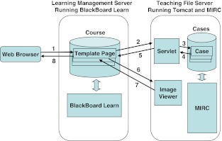
USRC consists of three main software components: (1) an HTML template page, (2) a translation servlet that handles requests for MIRC cases, and (3) an image viewer plug-in that allows students to view and interact with images stored in a MIRC case (see Fig. 1). The template page is used by course instructors to add case content to BlackBoard.
Fig. 1.
Overview of USRC architecture and transactions. 1 Student requests a page from the BlackBoard server. 2 The JavaScript code in the BlackBoard page makes a JSONP request to the translation servlet running on the teaching file server. 3,4 The servlet accesses the requested case in MIRC. 5 The servlet processes the file and returns text and image content in JSON format. 6 The JavaScript code running in the BlackBoard page sends image captions and URLs for thumbnails, source images, and annotated images to the image viewer. 7 The BlackBoard page begins to load the image viewer. 8 The BlackBoard server sends the page to the student’s web browser, and the image viewer continues to load
The servlet, which receives and responds to JSONP requests from pages based on the HTML template, and the image viewer plug-in must be copied into the MIRC directory that holds the course content (i.e., the directory for the proper MIRC Storage Service). The current version of USRC was developed and deployed using MIRC version T36c, which relies on the Apache Tomcat web application server, so it does not follow the recently redesigned servlet plug-in architecture used in CTP/MIRC software, which packages MIRC with the RSNA’s Clinical Trial Processor (http://mircwiki.rsna.org/index.php?title=MIRC).
HTML Template
The HTML template is a file that contains the code needed to support USRC transactions on the LMS, and all pages that import content from the MIRC server must be based on the template. It contains embedded JavaScript, with JQuery and JSONP calls, and it is linked to an external Cascading Style Sheet that formats content according to the visual style used in BlackBoard Learn. When a browser loads an HTML page based on the template, it makes a JSONP request to the servlet and evaluates the response. The page then makes a call to the image viewer plug-in, passing it the URLs of images contained in the case. The browser creates the final version of the page, inserting content using JQuery and loading the viewer plug-in.
At minimum, the course designer must modify a single variable in the template so that it contains the URL of the desired MIRC case. However, the template also supports other optional settings, allowing designers to further customize the page. For example, variables can be changed to modify the behavior of the DICOM viewer, or to control whether the case title, image captions, or section titles (e.g., history, findings, references, etc.) will be displayed. Course designers can also control the order in which imported case content will be displayed.
Translation Servlet
The translation servlet was written in Java, and it runs concurrently with MIRC in the Tomcat application server. It receives JSONP requests for cases from template pages running in BlackBoard Learn. Each MIRC case is stored as an XML file that follows the MIRC Document Schema [4]. The standardization of the MIRC document format makes it possible for the servlet to interpret any MIRC-compliant case regardless of the content. The servlet reads and parses the requested XML case file, and gathers data for all text sections (e.g., history, references, etc.) and images. The data are then converted to JSON format (JavaScript Object Notation, a data format required for JSONP calls) and are sent to the web browser that initiated the JSONP call.
Image Viewer Plug-in
A new image viewer was developed to display images stored in MIRC cases. The viewer was implemented as a Java applet and is stored on the server running MIRC. Template pages invoke the viewer by sending it image captions and URLs for images in a case, including source images, which can be in DICOM, JPG, JPEG2000, GIF, PNG, BMP, or TIF formats; annotated images; and thumbnail images. The applet runs as a browser plug-in, embedded in the webpage, and users are given the option of launching it in a separate browser tab to allocate more screen space for the images.
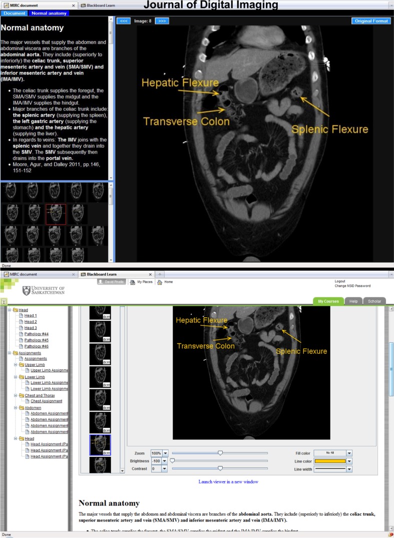
The plug-in was designed to provide basic image viewing and editing capabilities. Users can navigate images by clicking the thumbnails that are shown on the left (see Fig. 2), or by using cine loop or next/previous image controls. When annotations are available, the main view can be switched between the source image and the annotated image using a toggle button. Other viewing features include zoom, pan, brightness, and contrast controls. Editing features allow users to modify the on-screen version of the image, but do not allow them to save changes made to the image. Supported editing features include drawing simple shapes; adding text; and customizing line width, line color, and fill color.
Fig. 2.
Case in MIRC and BlackBoard Learn. Top MIRC case covering normal anatomy of the abdomen and containing explanatory text and an annotated CT series. Bottom The same case displayed in BlackBoard Learn using USRC technology. The CT series is shown using the image viewer, and the text is reformatted and displayed at the bottom of the screen
Deployment Experiences
University of Saskatchewan Radiology Courseware is being used to support Form and Function, a first-year course in the College of Medicine at the University of Saskatchewan. The course covers several scientific and clinical topics, including gross anatomy, physiology, histology, and embryology. Students enrolled in Form and Function use the campus-wide BlackBoard Learn system to access lecture notes and supplementary readings. However, prior to the deployment of USRC, radiology images were not available in BlackBoard Learn.
Our primary goal was to improve existing BlackBoard Learn content by adding digital imaging support to the gross anatomy portion of the course. We began by selecting DICOM images that provide coverage of the normal anatomy and the pathologies listed in the course outline. Since the images would be accessed over the Internet, we used a resize tool on large images to downscale image dimensions, which resulted in a corresponding reduction in file size (each file <5 Mb), and allowed students to download the images in a reasonable amount of time.
We used an in-house teaching file authoring application to create a case for each page we planned to add to BlackBoard. For example, we created cases on normal shoulder anatomy, rotator cuff injuries, shoulder dislocations, shoulder separations, etc. We wrote supporting text, providing background information on the course topic covered in the case. We also included a discussion of the accompanying images, giving students information to help with image interpretation, and a list of related textbook pages. We uploaded the cases to our departmental MIRC server.
For each case we created on the MIRC server, we added a corresponding HTML page in BlackBoard Learn to import, reformat, and display the case content. Each BlackBoard page was based on the USRC HTML template, which was modified to hold the URL of the XML file for the desired MIRC case. The BlackBoard pages were arranged in a sequence that corresponded to the Form and Function course outline.
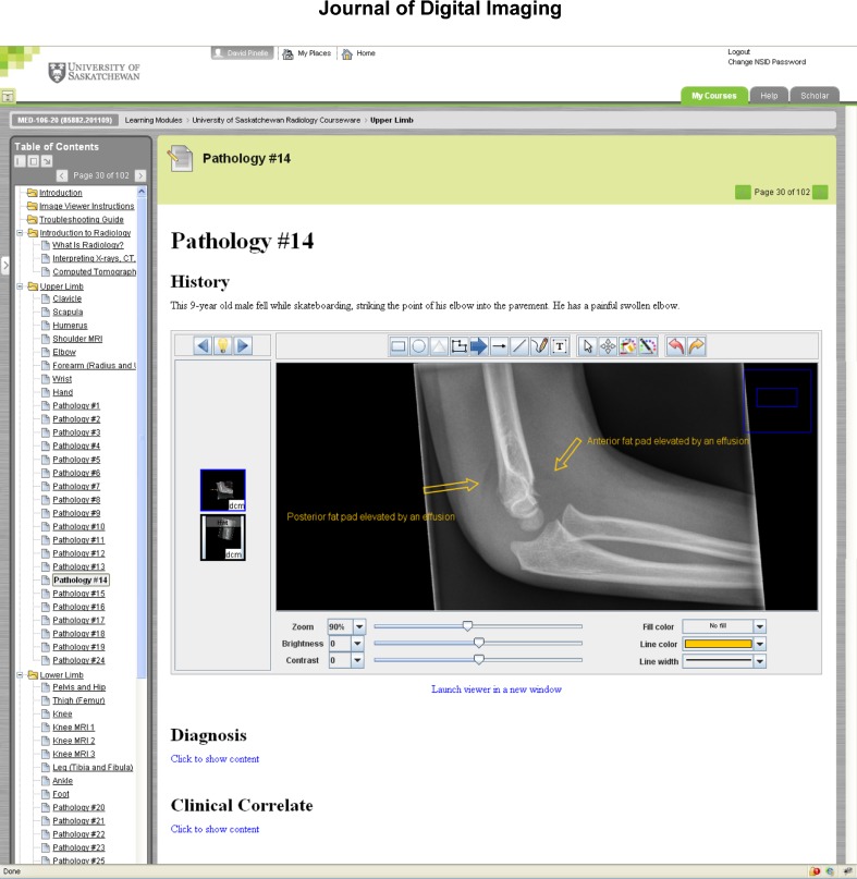
Pages were customized further to allow students to explore and interact with the content. For example, all pathologies were listed as unknowns, and after students viewed the accompanying images, they could click on internal hyperlinks to reveal the diagnosis and a clinical correlate (see Fig. 3). Several assignments were also developed, and they required students to find and label anatomical structures using the image viewer’s editing capabilities. To complete the assignments, the students take screen captures of the images after they make the requested changes, paste the images into a Microsoft Word document, and return the printed report to their instructor.
Fig. 3.
BlackBoard Learn page showing pathology as an unknown. The diagnosis and clinical correlate are hidden but can be viewed by clicking internal hyperlinks. A list of USRC content for the course is shown on the left sidebar
We deployed the system, linking the newly created template pages to the Form and Function section on the University of Saskatchewan’s BlackBoard Learn system. We provided students with a brief 20-minute orientation to the technology in the classroom, and answered their initial questions during that time. All students in the course had previous experience with using BlackBoard Learn for other courses, so they already had basic familiarity with web-based technologies.
USRC is currently being used by all first-year medical students. The technical support needs have been minimal, and have primarily involved providing students with assistance with configuring their browsers to support Java applets (fewer than 2 % of students have requested assistance). Initial feedback from students and instructors has been positive. Students commented that the technology is easy to use, and the instructor said that all students were able to successfully complete course assignments using the image viewer plug-in. We are currently conducting a more detailed longitudinal evaluation on the usability and the utility of the system.
Discussion
The organization and presentation style used in most digital teaching file (DTF) systems, such as MIRC, Radiology Teacher [5], and MyPACS (McKesson Corporation, http://www.mypacs.net), is not always well-suited to the needs of students and instructors. Most DTF systems provide a non-sequential organization structure, grouping cases into libraries or folders, and users often rely on a built-in search feature to find cases of interest. While preclinical medical students do benefit from opportunities to learn in a self-directed fashion, institutions are also concerned with making sure they have adequate depth and breadth coverage on required classroom topics. Course content is usually designed accordingly, often following a fixed sequence, where an understanding of material in early lectures is needed prior to moving to more complex topics covered later.
USRC transforms cases stored in MIRC so that they can be displayed in a learning management system. This enables the LMS to impose a sequential organization on the cases so that they can be used to support established preclinical medical school courses. Importing cases into the LMS also allows instructors to use built-in features to enhance their value as learning resources. They can add communication and assessment tools to the radiology content, and can embed other related resources, such as supplementary readings and multimedia files.
Since USRC content is stored as MIRC cases, course development primarily involves the creation of digital teaching files rather than the creation of content in the LMS. MIRC supports case creation using a web interface, but the case creation process can be streamlined significantly by using other case authoring applications, such as RadPix (Weadock Software, http://www.radpix.com/), TCE Selector (Klinik und Poliklinik für Diagnostische und Interventionelle Radiologie, Johannes Gutenberg-Universität Mainz, www.unimedizin-mainz.de/radiologie/), or RadXtreme (Tesla9, http://radxtreme.org/). These applications are designed to simplify case creation by reducing the effort needed to import images and to add text and attachments to cases.
We plan to refine USRC in the future to improve its integration with evolving technical standards and to address feedback from students and instructors. When we upgrade our digital teaching file server to the CTP/MIRC version, we will likely need to modify the translation servlet so that it will run in MIRC’s new embedded servlet container. We also plan to develop a lightweight version of the image viewer using HTML 5, which is still under development, because of its extensive support for graphics and multimedia. The HTML 5 approach would reduce the need for technical support related to browser plug-ins (e.g., Java Runtime Environment, Adobe Flash Player) since all content would be interpreted directly in the browser, without the need for external technologies. We also plan to continue to elicit suggestions and recommendations from students and instructors, and to improve USRC based on their feedback.
Online Learning Resources for Preclinical Training
There are many websites that provide access to radiology images and teaching cases. However, the presentation and organization of the content varies depending on the target audiences. Many sites use a loose organizational structure, allowing users to issue search queries against teaching file archives. For example, the MyPACS web portal [6] provides access to an extensive teaching file archive, and the RSNA’s MIRC portal [7] gives access to teaching file archives that are maintained by external hospitals and academic institutions.
Other sites provide structured educational content that is designed for radiology trainees, but that is not necessarily appropriate for medical students who are still in the early stages of training [8–10]. For example, COX (Case of the Day, developed by the Institute for Diagnostic and Interventional Radiology at the University Hospital of Johann Wolfgang Goethe University) is an e-learning system that holds approximately 1,450 radiological cases [11]. The cases are organized to support continuing medical education and radiology training, but they can also be exported in SCORM [12] and MIRC format so can be reused to support educational goals in other organizations. Radiology-Integrated Training Initiative, developed by the United Kingdom National Health Service in collaboration with the Royal College of Radiologists is a similar e-learning system that contains validated e-learning lessons that are designed for radiology trainees [13, 14]. The content is presented through self-contained sessions, and each contains a series of pages, made up of text, static images, and animations. Radiolopolis is an online radiology community that also allows users to create teaching cases, and they can browse and discuss a repository of over 12,000 cases [15].
There are few published examples of new technologies that were developed to add radiology content to established preclinical medical school courses. Schütze et al. [16] added radiology content to the ILIAS learning management system at Johannes Gutenberg-Universität Mainz. They created LMS pages that contain hyperlinks to their teaching file server. This allowed students to open and view imaging content directly in MIRC [17]. Reilly et al. [18] describe online course content that they developed for first-year medical and dental students. They converted digital radiographs covering head and neck anatomy into Adobe Flash format and used it to complement classroom and dissection activities in an anatomy course.
Conclusions
University of Saskatchewan Radiology Courseware provides a new way to add radiology images to learning management systems. Instructors create new pages in the LMS using an HTML template that points to the URL of a corresponding MIRC case. When a student accesses a page based on the template, the case content is imported—the text is reformatted so that it adheres to the look and feel of the LMS, and an image viewer is loaded that provides access to all images, thumbnails, annotations, and captions in the case. The main benefits of USRC are that it:
Allows DICOM, JPG, PNG, GIF, BMP, TIF, and JPEG2000 images to be displayed in LMSs
Enables students to interact with the images using zoom, pan, filter, and editing features
Allows imported content from MIRC to be organized within the LMS to fit the teaching objectives of the course, and
Reduces the effort and technical expertise needed to display imaging content in the LMS
USRC is being used by first year students in the College of Medicine at the University of Saskatchewan. We are continuing to evaluate the system, and plan to develop content for additional courses and for clinical training.
Acknowledgments
We thank SaskTel and the Royal University Hospital Foundation for generously supporting this project. We also thank Greg Malin and Paul Babyn for their feedback and suggestions.
References
- 1.Barchino R, Gutiérrez JM, Otón, S: An Example of Learning Management System. in Proceedings of IADIS Virtual Multi Conference on Computer Science and Information Systems (MCCSIS 2005), IADIS Press: 140–141, 2005.
- 2.Jackson C, Bortz A, Boneh D, Mitchell JC: Protecting browser state from web privacy attacks. in Proceedings of the 15th international conference on World Wide Web (WWW '06). ACM Press, New York: 737–744, 2006.
- 3.JSONP. Available at http://en.wikipedia.org/wiki/JSONP. Accessed 15 November 2011.
- 4.The MIRCdocument Schema. Available at http://mircwiki.rsna.org/index.php?title=The_MIRCdocument_Schema. Accessed 15 November 2011.
- 5.Talanow R. Radiology Teacher: a free, Internet-based radiology teaching file server. Journal of the American College Radiology. 2009;6(12):871–875. doi: 10.1016/j.jacr.2009.08.001. [DOI] [PubMed] [Google Scholar]
- 6.MyPACS Teaching File Software. Available at http://www.mypacs.net. Accessed 20 July 2011.
- 7.The RSNA Teaching File. Available at http://mirc.rsna.org/. Accessed 20 July 2011.
- 8.Radmoodle.org. Available at http://www.radmoodle.org/. Accessed 21 November 2011.
- 9.Sparacia G, Cannizzaro F, D'Alessandro DM, D'Alessandro MP, Caruso G, Lagalla R. Initial experiences in radiology e-learning. Radiographics. 2007;27(2):573–581. doi: 10.1148/rg.272065077. [DOI] [PubMed] [Google Scholar]
- 10.Hoa D, Micheau A, Gahide G: Creating an interactive Web-based e-learning course: a practical introduction for radiologists. RadioGraphics 26(6): e25; quiz e25, 2006. [DOI] [PubMed]
- 11.COX. Case of the Day, The Open Source X-Ray Gallery. Available at http://cox-radiology.org/. Accessed 21 November 2011.
- 12.Njuguna N, Flanders AE, Kahn CE., Jr Informatics in radiology: envisioning the future of e-learning in radiology: an introduction to SCORM. RadioGraphics. 2011;31:1173–1179. doi: 10.1148/rg.314105191. [DOI] [PubMed] [Google Scholar]
- 13.Ramsden WH. The Radiology Integrated Training Initiative (R-ITI) Med Educ. 2009;43(5):494–495. doi: 10.1111/j.1365-2923.2009.03350.x. [DOI] [PubMed] [Google Scholar]
- 14.R-ITI: Radiology-Integrated Training Initiative. Available at http://www.e-lfh.org.uk/projects/radiology/. Accessed 21 November 2011.
- 15.Radiolopolis: The International Radiology Community and Professional Radiology Network. Available at http://www.radiolopolis.com. Accessed 20 February, 2012.
- 16.Schütze B, Mildenberger P, Kämmerer M. E-learning in radiology: the practical use of the content management system ILIAS [in German] Rofo Fortschr Geb Rontgenstr Neuen Bildgeb Verfahr. 2006;178(5):525–530. doi: 10.1055/s-2006-926628. [DOI] [PubMed] [Google Scholar]
- 17.Mildenberger P, Brüggemann K, Rösner F, Koch K, Ahlers C. PACS infrastructure supporting e-learning. European Journal of Radiology. 2011;78(2):234–238. doi: 10.1016/j.ejrad.2010.05.006. [DOI] [PubMed] [Google Scholar]
- 18.Reilly F, Kamsala M, Davis A, Altemus J, Reddy S: Introductory Head & Neck Radiology for Preclinical Learners. MedEdPORTAL: 2008. Available at www.mededportal.org/publication/1566.


