Abstract
This study aimed to detect, by radiographic examination, the evolution of osteochondral lesions in the tarsocrural and femoropatellar joints of Lusitano foals. Within 1 month of age, 76.08% of foals had radiographic signs of osteochondrosis, but only 16.20% had lesions at 18 months. The radiographic signs resolved by 5 mo of age in most foals, but some cases that involved either joint, were not resolved until 12 mo of age. It is thought that the “age of no return” is 5 mo for the tarsocrural and 8 mo for the femoropatellar joint but this study demonstrated regression of osteochondral lesions in both joints of Lusitano foals up to 12 months of age.
Résumé
Développement de l’ostéochondrose chez les poulains Lusitaniens : une étude radiographique. Cette étude visait à détecter, par examen radiographique, l’évolution des lésions ostéochondrales dans les articulations tarso-crurale et fémoro-patellaire des poulains Lusitaniens. À l’âge de 1 mois, 76,08 % des poulains présentaient des signes radiographiques d’ostéochondrose, mais seulement 16,20 % avaient des lésions à l’âge de 18 mois. Les signes radiographiques se résorbaient à l’âge de 5 mois chez la plupart des poulains, mais, dans certains cas qui touchaient l’une ou l’autre des articulations, ils n’étaient pas résolus jusqu’à l’âge de 12 mois. On croit que l’«âge de non-retour» est de 5 mois pour l’articulation tarso-crurale et de 8 mois pour l’articulation fémoro-patellaire, mais cette étude a démontré la régression des lésions ostéochondrales chez les poulains Lusitaniens jusqu’à l’âge de 12 mois.
(Traduit par Isabelle Vallières)
Introduction
The appearance of orthopedic developmental diseases, such as osteochondrosis (OC), physitis, angular deformities, and flexural deformities, is an important factor in the first year of life of horses, especially those that will engage in athletic activities. Among these disorders, OC has the highest economic impact (1), due to treatment costs, time lost to rehabilitation, reduction in breeding potential of affected stallions, and market value of well-performing athletic horses (2).
The prevalence of OC ranges between 8% and 79% (3,4), depending on the horse population and joints studied. Moreover, the incidence of OC has increased considerably in recent decades (5).
In horses, OC is predominantly found in the femoropatellar and tarsocrural joints. The lesions are usually located in the cranial aspect of the intermediate ridge of the distal tibia, the distal aspect of the lateral trochlear ridge of the talus, and the central region of the lateral trochlear ridge of the femur (6,7). Other common locations include the metacarpophalangeal, metatarsophalangeal, and scapulohumeral joints (8).
Most lesions likely form in the first months of life (9), but clinical signs may not be apparent at this time; they are frequently diagnosed at a later age, when training begins, or fragments become loose. Lesions in racing Thoroughbreds and Standardbreds are usually recognized by 2 y of age, but in Warmbloods that are older when they begin training, clinical signs may not be seen until the horse is 5 to 6 y of age (8).
Some radiographic lesions, such as intermediate ridge lesions, at both tarsocrural joint sites are visible in lateromedial radiographic views; other lesions are more evident in dorsomedial-plantarolateral oblique views (7). Lesions in the center of the lateral trochlear ridge of the femur are visible on lateromedial radiographs and are often most obvious in caudolateral-craniomedial views (6,7).
Subchondral contour changes must be carefully analyzed in foals, especially in the areas where ossification occurs later, such as in the trochlear ridges of the femur or intermediate ridge of tibia (8). Complete ossification of growth plates, or the arrest of endochondral ossification process, occurs at different times in different bones. In the proximal tibial physis and distal femoral physis closure is reported at 24 to 30 mo and in the distal tibial physis closure occurs between 17 and 24 mo of age, whereas most of the physes of more distally located bones do not close until at least 18 mo of age (10).
Dik et al (6) found that osteochondral lesions in the tarsocrural and femoropatellar joints develop early in Dutch Warmblood foals, but most of these lesions are temporary. However, lesions that are still present in the tarsocrural joint at 5 mo of age and in the lateral ridge of the femoral trochlea at 8 mo of age will likely be permanent. Radiographic screening of foals for OC in the tarsocrural and femoropatellar joints before the age of 8 mo, therefore, seems contraindicated, as new lesions may develop and existing ones may disappear. Rather, 12 mo is a more realistic and practical age for the radiographic diagnosis (6).
According to Ekman et al (5), recent research has shown that repair in foals begins almost immediately after the osteochondral lesion is formed. It is clear that OC has a very dynamic character, and lesions may develop or regress spontaneously. The intensity and effectiveness of the repair are determined by the turnover rates of cartilage extracellular matrix components, which are strongly related to age and decrease rapidly after birth (5). Assuming that some breeds mature more rapidly than other breeds, the appropriate age at which these horse breeds should be subjected to radiographic examination may vary.
On farms with a high incidence of OC, radiographic examinations are important for the early diagnosis of lesions, but should be interpreted bearing in mind the time during which these lesions are still capable of regression; in this period they are still amenable to clinical intervention such as changes in feeding practices and exercise (11). Precocious recognition of osteochondral lesions can, therefore, alleviate clinical signs as well as modify the course of disease, in cases in which regression would not occur spontaneously.
After the original study by Dik et al in 1999 (6), very little information has been gathered to confirm or reject their findings, especially in other breeds. The aim of the study herein was to determine the presence and evolution of osteochondral lesions in the tarsocrural (cranial aspect of the intermediate ridge of the distal tibia and the distal aspect of the lateral trochlear ridge of the talus) and femoropatellar (central lateral trochlear ridge of the femur) joints of Lusitano foals through radiographic examination.
Materials and methods
Animals
The study was conducted on 38 randomly selected Lusitano foals that were reared in a semi-stabling environment on 4 farms. After birth, all foals remained with their dams until weaning at 6 mo, after which they remained on pasture, receiving concentrates according to age, and with no training until 18 mo of age.
None of the animals showed lameness or joint effusion. Some foals were sold before the end of the study; therefore, only 23 foals were available for assessment of the evolution of the radiographic appearance of the tarsocrural and femoropatellar joints. Tarsocrural and femoropatellar joint radiographic examinations (n = 120) were performed in 30 foals at 1 mo of age; 644 examinations were performed in 23 foals that were 3 to 18 mo of age.
Radiographic examinations
Radiographic examinations were done at 1, 3, 5, 8, 12, 14, 16, and 18 mo of age. The surveys included dorsomedial-plantarolateral oblique, lateromedial, and dorsoplantar views of both tarsocrural joints to evaluate the cranial aspect of the intermediate ridge of the distal tibia and the distal aspect of the lateral trochlear ridge of the talus. Lateromedial and caudolateral-craniomedial views of both femoropatellar joints were included to evaluate the central lateral trochlear ridge of the femur.
The cranial aspect of the intermediate ridge of the distal tibia, the distal aspect of the lateral trochlear ridge of the talus, and the central region of the lateral trochlear ridge of the femur of each foal were scaled from 0 to 4 according to Dik et al (6) (Table 1).
Table 1.
Radiographic classification of osteochondral abnormalities of the intermediate ridge of the distal tibia, the distal aspect of the lateral trochlear ridge of the talus, and the central region of the lateral trochlear ridge of the femur
| Grade | Contour | Bone texture | Subchondral bone | Fragment(s) |
|---|---|---|---|---|
| 0 | normal | Rounded | with diffuse density | absent |
| 1 | minimal | Smoothly flattened | with obscure lucency | absent |
| 2 | mild | Irregularly flattened | with obvious lucency, limited and poorly defined border | absent |
| 3 | moderate | Small, rounded, or irregular concavity | with obvious, well-defined local lucency | Small fragment(s) |
| 4 | severe | Large, rounded or irregular concavity | with obvious, well-defined extensive lucency | Large fragment(s) |
The radiographic images were evaluated in a blind manner by 2 radiologists and 1 equine surgeon, each in a separate session. Scoring at 1, 3, 5, 8, 12, and 18 mo was compared in the joints that were monitored over 18 mo. Statistics were performed using Minitab 14.1 statistical software (Minitab, State College, Pennsylvania, USA). The Pearson correlation was used to establish the influence of age on frequency of occurrence of osteochondrosis and statistical significance was set at a P < 0.05.
Results
A total of 76.1% of 1-month-old foals had osteochondral abnormalities in their radiographic examinations in at least 1 of their tarsocrural or femoropatellar radiographic images. Radiographic images showed that 43.3%, 30.0%, and 26.7% of 1-month-old Lusitano foals exhibited changes in the intermediate ridge of the tibia, lateral trochlear ridge of the talus, and central region of the lateral trochlear ridge of the femur, respectively (Table 2).
Table 2.
Percentages of Lusitano foals with abnormal radiographic appearances of the cranial aspect of the intermediate ridge of the distal tibia, distal aspect of the lateral trochlear ridge of the talus, and central lateral trochlear ridge of the femur at 1 to 3, 5 to 8, and 12 to 18 months of age
| Cranial aspect of the intermediate ridge of the distal tibia | Distal aspect of the lateral trochlear ridge of the talus | Central lateral trochlear ridge of the femur | |||||||
|---|---|---|---|---|---|---|---|---|---|
| Age (months) |
1 to 3 (n = 30) |
5 to 8 (n = 38) |
12 to 18 (n = 37) |
1 to 3 (n = 30) |
5 to 8 (n = 38) |
12 to 18 (n = 37) |
1 to 3 (n = 30) |
5 to 8 (n = 38) |
12 to 18 (n = 37) |
| Percentage of foalsa (n) |
43.3% (13) |
21% (8) |
10.8% (4) |
30% (9) |
23.7% (9) |
5.4% (2) |
26.7% (8) |
7.9% (3) |
0% |
Percent of foals with radiographic lesions.
At 18 mo of age, only 16.2% of the foals had persistent lesions. All of the persistent lesions were observed in the tarsocrural joint (10.8% were observed in the intermediate ridge of the tibia, and 5.4% were observed in the lateral trochlear ridge of the talus). No persistent lesions were observed in the femoropatellar joint (Table 2).
Radiographic lesions strongly tended towards regression (i.e., a reduction in the number of foals affected by OC) in relation to age (r = 0.906, P < 0.05). The progression or development of lesions in joints without previous radiographic signs was less common.
There were no differences in radiographic images of tarsocrural or femoropatellar joints between 1 and 3 mo, 5 and 8 mo, and between 12 and 18 mo of age.
When evaluating tarsocrural joints radiographed in the first month of life (n = 46), we found that 30.4% (n = 14) of the joints had lesions on the intermediate ridge of the tibia (grades 1–3: 28%; grade 4: 2%). Considering the subsequent development of the lesions during the first 5 mo (Table 3), the majority of the initially normal appearances remained as such. Lesions initially graded as 1 on the intermediate ridge of the tibia showed a marked tendency for resolution (n = 11), and the only initial grade 4 lesion resolved completely. On the other hand, lesions that were consistent with OC developed in joints that had no changes in the first month: 15.6% (n = 5) of the initially healthy tarsocrural joints had changes in the intermediate ridge of the tibia (Table 3).
Table 3.
Development of radiographic appearances of the cranial aspect of the intermediate ridge of the distal tibia from 1 to 5 months
| Classification at 1 month | n | Subsequent development | ||
|---|---|---|---|---|
|
| ||||
| Stationary | Progressed | Resolved | ||
| Grade | ||||
| 0 | 32 | 27 | 5 | 0 |
| 1 | 12 | 1 | 0 | 11 |
| 2 | 0 | 0 | 0 | 0 |
| 3 | 1 | 1 | 0 | 0 |
| 4 | 1 | 0 | 0 | 1 |
| Total | 46 | 29 | 5 | 12 |
n = number of joints.
In the following months, almost all normally appearing tarsocrural joints (n = 34) and 1 with a grade 3 lesion were stationary. They had the same lesions in the intermediate ridge of the tibia at 12 and 18 mo that were found at 5 and 8 mo; 8.1% (n = 3) of the joints with normal appearance in the intermediate ridge of the tibia at 5 and 8 mo showed lesions at 12 and equally at 18 mo, and 6 lesions graded as 1 showed resolution (Table 4).
Table 4.
Development of radiographic appearances of the cranial aspect of the intermediate ridge of the distal tibia from 5 to 18 months
| Classification at 5 months | n | Subsequent development | ||
|---|---|---|---|---|
|
| ||||
| Stationary | Progressed | Resolved | ||
| Grade | ||||
| 0 | 37 | 34 | 3 | 0 |
| 1 | 6 | 0 | 0 | 6 |
| 2 | 0 | 0 | 0 | 0 |
| 3 | 1 | 1 | 0 | 0 |
| 4 | 0 | 0 | 0 | 0 |
| Total | 44 | 35 | 3 | 6 |
n = number of joints.
The incidence of normal appearances increased from 1 to 5 mo, and the incidence of grade 1–4 abnormalities decreased. After age 5 mo progression and resolution were also observed in the intermediate ridge of the tibia, but less commonly.
At age 1 mo 15.2% (n = 7) of the tarsocrural joints had lesions on the lateral trochlear ridge of the talus (all grades 1–3) and most of the radiological images were normal (n = 39). However, 4 initially normal radiographs (10.2%) showed changes in the lateral trochlear ridge of the talus at 5 and 8 mo. Four grade 1 and 1 grade 3 lesions on the lateral trochlear ridge of the talus showed resolution (Table 5).
Table 5.
Development of radiographic appearances of the distal aspect of the lateral trochlear ridge of the talus from 1 to 5 months
| Classification at 1 month | n | Subsequent development | ||
|---|---|---|---|---|
|
| ||||
| Stationary | Progressed | Resolved | ||
| Grade | ||||
| 0 | 39 | 35 | 4 | 0 |
| 1 | 5 | 1 | 0 | 4 |
| 2 | 0 | 0 | 0 | 0 |
| 3 | 2 | 1 | 0 | 1 |
| 4 | 0 | 0 | 0 | 0 |
| Total | 46 | 37 | 4 | 5 |
n = number of joints.
All lesions graded as 1–3 at 5 and 8 mo in the lateral trochlear ridge of the talus showed resolution (n = 6). Only 2 subjects (5.3%) with normal radiographs at 5 and 8 mo had lesions in the lateral trochlear ridge of the talus at 12 and equally at 18 mo (Table 6).
Table 6.
Development of radiographic appearances of the distal aspect of the lateral trochlear ridge of the talus from 5 to 18 months
| Classification at 5 months | n | Subsequent development | ||
|---|---|---|---|---|
|
| ||||
| Stationary | Progressed | Resolved | ||
| Grade | ||||
| 0 | 38 | 36 | 2 | 0 |
| 1 | 5 | 0 | 0 | 5 |
| 2 | 0 | 0 | 0 | 0 |
| 3 | 1 | 0 | 0 | 1 |
| 4 | 0 | 0 | 0 | 0 |
| Total | 44 | 36 | 2 | 6 |
n = number of joints.
Although lesions in the lateral trochlear ridge of the talus showed resolution as well as progression from 1 to 5 mo, the incidence of normal appearances, grade 1–3 and grade 4 lesions did not show significant changes. After age 5 mo progression and resolution were also observed.
Despite the appearance of new lesions by 5 to 12 mo of age, the number of joints affected by tarsocrural OC on the intermediate ridge of the tibia (r = 0.867; P < 0.05) or lateral trochlear ridge of the talus (r = 0.932; P < 0.01) decreased with increasing age of the foals.
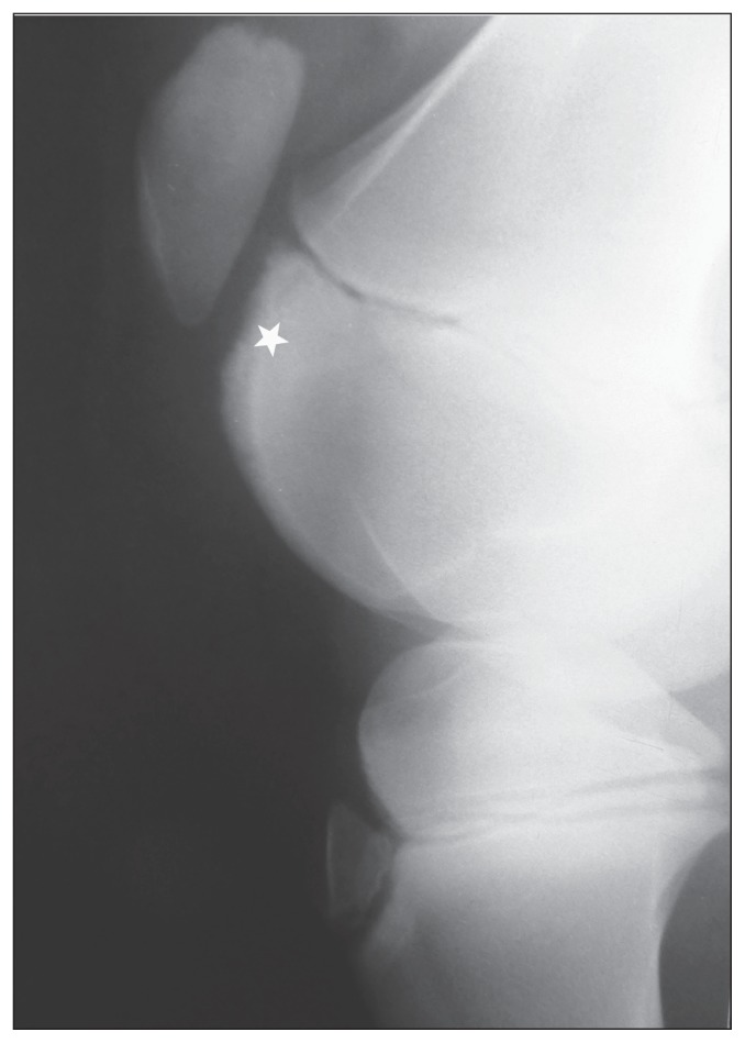
Concerning the femoropatellar joints, 23.9% (n = 11) had lesions in the center of the lateral trochlear ridge of the femur at 1 mo of age. At this age, 72.7% of the femoral trochlear ridges showed irregular contour and granular subchondral bone opacities in their proximal part (Figure 1).
Figure 1.
Lateromedial view of the femoropatellar joint of a normal 1-month-old Lusitano foal. Note the irregular contour and granular subchondral opacity of the proximal part of the femoral trochlea (star).
In the following months, most of the initially radiologically normal joints remained so (n = 34), and 1 normal joint developed a grade 2 lesion at age 5 mo. Also, 3 initial grade 1–3 lesions were stationary (Table 7).
Table 7.
Development of radiographic appearances of the central lateral trochlear ridge of the femur from 1 to 5 months
| Classification at 1 month | n | Subsequent development | ||
|---|---|---|---|---|
|
| ||||
| Stationary | Progressed | Resolved | ||
| Grade | ||||
| 0 | 35 | 34 | 1 | 0 |
| 1 | 7 | 1 | 0 | 6 |
| 2 | 1 | 1 | 0 | 0 |
| 3 | 3 | 1 | 0 | 2 |
| 4 | 0 | 0 | 0 | 0 |
| Total | 46 | 37 | 1 | 8 |
n = number of joints.
Finally, all the lesions on the lateral trochlear ridge of the femur that were observed at 5 and 8 mo of age had completely regressed by 12 mo and were normal at 18 mo. The development of new lesions between 5 and 18 mo of age was not observed (Table 8).
Table 8.
Development of radiographic appearances of the central lateral trochlear ridge of the femur from 5 to 18 months
| Classification at 5 months | n | Subsequent development | ||
|---|---|---|---|---|
|
| ||||
| Stationary | Progressed | Resolved | ||
| Grade | ||||
| 0 | 40 | 40 | 0 | 0 |
| 1 | 1 | 0 | 0 | 1 |
| 2 | 2 | 0 | 0 | 2 |
| 3 | 1 | 0 | 0 | 1 |
| 4 | 0 | 0 | 0 | 0 |
| Total | 44 | 40 | 0 | 4 |
n = number of joints.
In summary, the incidence of normal appearances on the lateral trochlear ridge of the femur increased from 1 to 5 mo and the incidence of grade 1–3 lesions decreased. Lesions showing resolution were observed after 5 mo of age. All radiologically abnormal joints had returned to normal at age 12 mo. The number of joints affected by femoropatellar OC (r = 0.859; P < 0.05) decreased with increasing age of the foals.
Discussion
Radiological monitoring of osteochondral lesions on tarsocrural and femoropatellar joints showed that OC is a dynamic disease. Lesions became apparent and subsequently progressed, regressed or completely disappeared until a certain age referred to as “point of no return,” which is not identical for all joints (12) and all horse breeds (11). In the first and third months of life, several of the Lusitano foals had radiographic changes that were consistent with OC, but there was no lameness.
According to Dik et al (6), in the first month of life, the intermediate ridge of the tibia often appears abnormal, but abnormal appearances of the lateral trochlear ridge of the talus and the lateral trochlear ridge of the femur are uncommon. However, in the present study 43.3%, 30%, and 26.7% of the Lusitano foals had abnormalities on the intermediate ridge of the tibia, the lateral trochlear ridge of the talus, and the center of the lateral trochlear ridge of the femur, respectively.
In this study, most femoropatellar lesions became radiographically apparent at the first examination at 1 mo of age in Lusitano foals, unlike others in which femoropatellar lesions became radiographically apparent only in the 3rd to 5th months (6,13). Because of the normal irregular ossification pattern, the radiographic contours and texture of both ridges of the femoral trochlea appear very irregular on radiographic images. Due to these irregularities, radiographic assessment of the outline of the femoral ridges and interpretation of subchondral lucencies can be unreliable (6). These findings suggest that the first trimester of life is not a suitable period for radiographic survey of OC in femoropatellar joints of Lusitano foals.
Perhaps the radiographic changes at age 1 mo could be classified as the manifestation of physiological variation in the process of endochondral ossification, and several external (e.g., feeding practices and exercise conditions) and intrinsic (e.g., potential growth rate) factors may determine whether these radiographic abnormalities observed at 1 month of age regressed, or progressed toward pathological entities that may later become clinically manifest (13).
Several studies have shown that permanent osteochondral lesions develop before 7 mo of age (12,14–18). Lesions of the intermediate ridge of the tibia often vary from minimal to moderate (grades 1–3); severe lesions (grade 4) are rare. Although radiographic lesions of the distal aspect of the lateral trochlear ridge of the talus and central lateral trochlear ridge of the femur are less common than at the intermediate ridge of the tibia a similar gradation is observed. Most of these lesions gradually disappeared, but healthy joints were not exempt from developing OC (6). Our findings corroborate this concept. In the current study the regression of lesions in the central trochlear ridge of the femur and cranial aspect of the intermediate ridge of the tibia mainly occurred prior to 5 mo of age. However, a decline continued to be observed, albeit at a lower frequency, in both joints until 12 mo of age. Regarding the distal aspect of the lateral trochlear ridge of the talus, identical extent of regression was observed between 1 to 5 mo and 5 to 12 mo.
According to Lepeule et al (11) a large pasture area at an early age (> 1 ha at less than 2 wk of age or > 6 ha at less than 2 mo of age) is a risk factor for OC; this means that the severity of osteochondral lesions increases with the exercise area. However, van Weeren and Barneveld (1) suggest that osteochondral lesions observed in tarsocrural and/or femoropatellar are less severe for foals which exercise freely in pastures than for foals kept in a box or kept in a box and exercised once a day.
After weaning, all Lusitano foals remained on pasture (between 1 and 3 ha). At this level of exercise, the number of joints affected by tarsocrural OC on the intermediate ridge of the tibia, or lateral trochlear ridge of the talus significantly decreased with the increasing age of the foals. A similar pattern was observed with femoropatellar joints.
In the radiographic studies performed by Gondahl and Dolvik (9), 753 trotter foals (between 6 and 20 mo of age) were evaluated, and OC was diagnosed in the intermediate ridge of the tibia and/or lateral trochlea of the talus in 108 (14.3%) of the animals. This value is very close to that found in our study, in which OC was diagnosed in 16.20% of the foals by 12 mo of age. Of the 753 animals in the study by Gondahl and Dolvik (9), 79 were radiographed again 6 to 18 mo after the first examination. Only 1 animal that had previously been OC-free (as determined by radiographic analysis) developed signs of OC after 1 y of age. This corresponds to an incidence of OC of 0.7% in the tarsocrural joint with no previous signs of OC. Our results closely agrees with this value because none of the Lusitano foals that had previously been OC-free in the tarsocrural joint developed signs of OC after 1 y of age.
van Grevenhof et al (4) found higher percentages of radiographic lesions consistent with OC in Dutch Warmblood horses, with a mean age of 12 ± 2.6 mo, when compared with the findings of Gondahl and Dolvik (9) in trotter foals. These values were 39.3% and 31.4% of femoropatellar and tarsocrural joints, respectively, and were also higher than those found in our study, which were, at 12 mo of age, 16.2% in tarsocrural joints and 0% in femoropatellar joints.
Among the 378 foals in the study of Lepeule et al (11), 36% were affected by OC as detected by radiographic examination. The study sample consisted of foals from 3 breeds: French Warmblood, French trotter Standardbred, and Thoroughbred, followed from birth to 6 mo of age. Prevalence of OC varied significantly among breeds and most of the affected foals were Warmbloods. In the Lusitano foals we found similar percentages, that is 31.6 % (12 of 38 foals) affected at 5 mo of age. Different results among these studies may be related to the use of preselected datasets, differences in breeds, or in the number of predilection sites screened.
According to Dik et al (6), any osteochondral lesions that are still present in the tarsocrural joint at 5 mo of age and on the lateral trochlear ridge of the femur at 8 mo of age are permanent. Subsequent studies have supported this concept that the “age of no return” is 5 mo for the tarsocrural and 8 mo for the femoropatellar joint (4,8,11,13). In contrast, our study demonstrated both the regression and development of osteochondral lesions in the tarsocrural joint of Lusitano foals between 5 and 12 mo of age and the regression of osteochondral lesions in the femoropatellar joint up to 12 mo of age. The definitive presence of OC in the tarsocrural and femoropatellar joints was confirmed at 12 mo of age.
We also observed that fragmentation of the intermediate tibial ridge, lateral trochlear ridge of the talus and central lateral trochlear ridge of the femur did not always result in a permanent lesion, which is in agreement with the findings reported by Dik et al (6).
Our findings highlight the importance of understanding the evolution of OC and emphasize the importance of age in the irreversible establishment of OC in different breeds. Identifying these key ages is important for preventing delays in administering therapies and minimizing decision-making errors when buying and selling foals. Recent work from de Grauw et al (12), using several biomarkers in synovial fluid of foals with or without OC, provides insight into the dynamic disease mechanisms in the first year of life (12). Insulin Growth Factor 1 (IGF-1) is a useful synovial fluid biomarker that allows early recognition of affected cartilage; IGF-1 concentrations are significantly lower in OC joints until 5 mo of age (12,19,20), and the expected result is reduced local proteoglycan and collagen II synthesis (12) as well as impaired longitudinal bone growth, chondrocyte proliferation and differentiation.
In conclusion, results of this study suggest 12 mo as the “age of no return” for the diagnosis of osteochondral lesions in Lusitano foals. However, this age is late for introduction of prophylactic measures. Chondrocytes from early osteochondral lesions (5 mo old foals) although impaired, retain their vitality, being capable after proper stimulation of regaining the same level of metabolic activity as their healthy counterparts. This is not true for affected chondrocytes from 11-month-old foals, whose low metabolic level cannot be reversed (21).
In valuable offspring and/or those with remarkable genetic or athletic potential, an early definite diagnosis of OC is not possible based only on radiographic examination; by the time radiology proves itself useful in establishing with certainty a positive diagnosis of OC, another important time window has closed: that is the one for prophylactic or management intervention. CVJ
Figure 2.
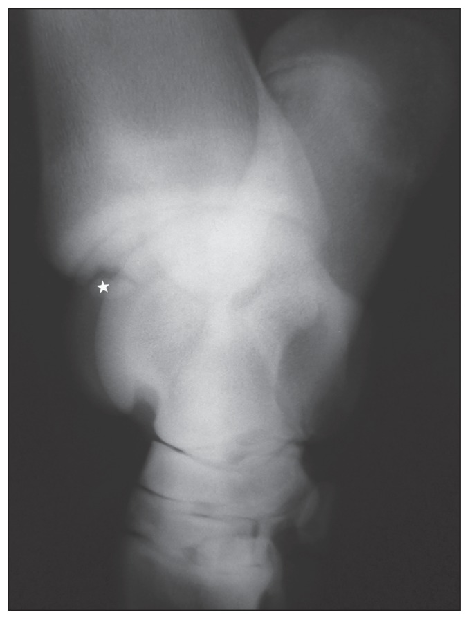
Dorsomedial-plantarolateral oblique radiographic view of the right tarsocrural joint of an 18-month-old Lusitano foal. There is a single large osteochondral fragment of the distal intermediate ridge of the tibia (star).
Footnotes
Use of this article is limited to a single copy for personal study. Anyone interested in obtaining reprints should contact the CVMA office (hbroughton@cvma-acmv.org) for additional copies or permission to use this material elsewhere.
This study was funded by Fundação de Amparo a Pesquisa do Estado de São Paulo (Fapesp).
References
- 1.van Weeren PR, Barneveld A. The effect of exercise on the distribution and manifestation of osteochondrotic lesions in the Warmblood foal. Equine Vet J. 1999;31:16–25. doi: 10.1111/j.2042-3306.1999.tb05309.x. [DOI] [PubMed] [Google Scholar]
- 2.van Weeren PR. Etiology, diagnosis and treatment of OC(D) Clin Tech Equine Pract. 2006;5:248–258. [Google Scholar]
- 3.Verwilghen D, Busoni V, Gangl M, et al. Relationship between biochemical markers and radiographic scores in the evaluation of the osteoarticular status of Warmblood stallions. Res Vet Sci. 2009;87:319–328. doi: 10.1016/j.rvsc.2009.02.002. [DOI] [PubMed] [Google Scholar]
- 4.van Grevenhof EM, Ducro BJ, Van Weeren PR, Tartwijk JMFM, Van Den Belt AJ, Bijma P. Prevalence of various radiographic manifestations of osteochondrosis and their correlations between and within joints in Dutch Warmblood horses. Equine Vet J. 2009;4:11–16. doi: 10.2746/042516408x334794. [DOI] [PubMed] [Google Scholar]
- 5.Ekman C, Carlson CS, Weeren PR. Equine Vet J; Third International Workshop on Equine Osteochondrosis; Stockholm. 29–30th May 2008; 2009. pp. 504–507. [DOI] [PubMed] [Google Scholar]
- 6.Dik KJ, Enzerink E, Van Weeren PR. Radiographic development of osteochondral abnormalities in the hock and stifle of Dutch Warmblood foals, from age 1 to 11 months. Equine Vet J. 2009;Supplement 31:9–15. doi: 10.1111/j.2042-3306.1999.tb05308.x. [DOI] [PubMed] [Google Scholar]
- 7.Relave F, Meulyzer M, Alexander K, Beauchamp G, Marcoux M. Comparison of radiography and ultrasonography to detect osteochondrosis lesions in the tarsocrural joint: A prospective study. Equine Vet J. 2009;41:34–40. doi: 10.2746/042516408x343019. [DOI] [PubMed] [Google Scholar]
- 8.Richardson DW. Diagnosis and management of osteochondrosis and osseus cyst-like lesions. In: Ross MW, Dyson SJ, editors. Diagnosis and Management of Lameness in the Horse. St. Louis, Missouri: WB Saunders; 2003. pp. 549–554. [Google Scholar]
- 9.Grondahl AM, Dolvik NI. Heritability estimations of osteochondrosis in the tibiotarsal joint and of bony fragments in the palmar/plantar portion of the metacarpo- and metatarsophalangeal joints of horses. J Am Vet Med Assoc. 1993;203:101–104. [PubMed] [Google Scholar]
- 10.Butler AJ, Colles CM, Dyson SJ, Kold SE, Poulos PW. Clinical Radiology of the Horse. 3rd ed. Oxford, UK: Wiley-Blackwell; 2008. [Google Scholar]
- 11.Lepeule J, Bareille N, Robert C, et al. Association of growth, feeding practices and exercise conditions with the prevalence of Developmental Orthopaedic Disease in limbs of French foals at weaning. Prev Vet Med. 2009;89:167–177. doi: 10.1016/j.prevetmed.2009.02.018. [DOI] [PubMed] [Google Scholar]
- 12.de Grauw JC, Donabedian M, van de Lest CHA, et al. Assessment of synovial fluid biomarkers in healthy foals and in foals with tarsocrural ostochondrosis. Vet J. 2011 doi: 10.1016/j.tvjl.2010.12.001. In press. [DOI] [PubMed] [Google Scholar]
- 13.Barneveld A, van Weeren PR. Conclusions regarding the influence of exercise on the development of the equine musculoskeletal system with special reference to osteochondrosis. Equine Vet J. 1999;31:112–119. doi: 10.1111/j.2042-3306.1999.tb05323.x. [DOI] [PubMed] [Google Scholar]
- 14.Carlson CS, Cullins LD, Meuten DJ. Osteochondrosis of the articular-epiphyseal cartilage complex in young horses: Evidence for a defect in cartilage canal blood supply. Vet Pathol. 1995;32:641–647. doi: 10.1177/030098589503200605. [DOI] [PubMed] [Google Scholar]
- 15.Shingleton WD, Mackie EJ, Cawston TE, Jeffcott LB. Cartilage canals I equine articular/epiphyseal growth cartilage and a possible association with dyschondroplasia. Equine Vet J. 1997;29:360–364. doi: 10.1111/j.2042-3306.1997.tb03139.x. [DOI] [PubMed] [Google Scholar]
- 16.Fortier LA, Kornatowski MA, Mohammed HO, Jordan MT, O’Cain LC, Stevens WB. Age related changes in serum insulin-like growth factor-I, insulin-like growth factor-I biding protein-3 and articular cartilage structure in Thoroughbred horses. Equine Vet J. 2005;37:37–42. doi: 10.2746/0425164054406838. [DOI] [PubMed] [Google Scholar]
- 17.Olstad K, Ytrehus B, Ekman S, Carlson CS, Dolvik NI. Early lesions on osteochondrosis in the distal tibia of foals. J Orthop Res. 2007;25:1094–1105. doi: 10.1002/jor.20375. [DOI] [PubMed] [Google Scholar]
- 18.Olstad K, Ytrehus B, Ekman S, Carlson CS, Dolvik NI. Early lesions on osteochondrosis in the distal femur of foals. Vet Pathol. 2011;48:1165–1175. doi: 10.1177/0300985811398250. [DOI] [PubMed] [Google Scholar]
- 19.Sloet van Oldruitenborgh-Oosterbaan MM, Mol JA, Barneveld A. Hormones, growth factors and other plasma variables in relation to osteochondrosis. Equine Vet J. 1999;31:45–54. doi: 10.1111/j.2042-3306.1999.tb05313.x. [DOI] [PubMed] [Google Scholar]
- 20.Baccarin RYA, Pereira MA, Roncati NV, Furtado PV, Oliveira CA, Hagen SCF. Identification of insulin-like growth factor-1 serum levels in foals with osteochondrosis. Pesq Vet Bras. 2011;31:677–682. [Google Scholar]
- 21.van den Hoogen BM, van de Lest CHA, van Weeren PR, van Golde LMG, Barneveld A. Changes in proteoglycan metabolism in osteochondrotic articular cartilage of growing foals. Equine Vet J. 1999;31:38–44. doi: 10.1111/j.2042-3306.1999.tb05312.x. [DOI] [PubMed] [Google Scholar]


