Abstract
Intestinal epithelial cell damage is frequently seen in the mucosal lesions of infectious or inflammatory bowel diseases such as ulcerative colitis or Crohn's disease. Complete remission of these diseases requires both the disappearance of inflammation and the repair of damaged epithelium. Saccharomyces boulardii (Sb, Biocodex) is a non-pathogenic yeast widely used as a preventive and therapeutic probiotic for the prevention and treatment of diarrhea and other gastrointestinal disorders. We recently showed that it enhances the repair of intestinal epithelium through activation of α2β1 integrin collagen receptors. In the present study, we demonstrated that α2β1 integrin is not the sole cell-extracellular matrix receptor involved during Sb-mediated intestinal restitution. Indeed, by using cell adhesion assays, we showed that Sb supernatant contains heat sensitive molecule(s), with a molecular weight higher than 9 kDa, which decreased αvβ5 integrin-mediated adhesion to vitronectin by competing with the integrin. Moreover, Sb-mediated changes in cell adhesion to vitronectin resulted in a reduction of the αvβ5signaling pathway. We used a monolayer wounding assay that mimics in vivo cell restitution to demonstrate that down-modulation of the αvβ5 integrin-vitronectin interaction is related to Sb-induced cell migration. We therefore postulated that Sb supernatant contains motogenic factors that enhance cell restitution through multiple pathways, including the dynamic fine regulation of αvβ5 integrin binding activity. This could be of major importance in diseases characterized by severe mucosal injury, such as inflammatory and infectious bowel diseases.
Introduction
The colonic epithelium forms a continuous physical and functional barrier that protects the internal environment of the body from the fluctuating external milieu [1]. A variety of inflammatory gastrointestinal disorders, including infectious colitis and inflammatory bowel disease, result in the breakdown of the intestinal epithelial barrier and subsequent erosion and ulceration [1], [2], [3], [4]. The colonic epithelium possesses an innate ability to rapidly reseal superficial wounds, critical for the maintenance of barrier function and homeostasis. This process is dependent on the precise balance of migration, proliferation and differentiation of epithelial cells adjacent to the wounded area [1]. As with other epithelia of the gastrointestinal tract, the repair of damaged colonic mucosa initially requires cell restitution. This process is identified by stages of cell spreading and migration into the wound to restore epithelial continuity [1]. Restitution is followed by the proliferation and subsequent maturation and differentiation of the cells, allowing restoration of normal architecture and absorptive/secretory functions.
Colonic restitution has been found to be influenced by a broad spectrum of factors derived from the gastrointestinal environment, including host epithelial and lamina propria cells, resident microbiota, and both dietary and non-dietary components present in the gastrointestinal lumen [4], [5]. Both in vitro and in vivo studies have unveiled that adhesion-mediated signaling between cells and the extracellular matrix (ECM) is critical in the regulation of cell restitution [1], [6], [7], [8]. Moreover, in vitro studies have demonstrated that restitution is enhanced in the presence of ECM proteins [9], [10].
Interactions between cells and ECM are mainly mediated by cell surface adhesion molecules termed integrins. Integrins are glycosylated heterodimers composed of non covalently associated type I transmembrane α and β subunits [11]. In mammals, 18 α and 8 β subunits combine to form 24 distinct integrin receptors that bind various ECM ligands with different affinities [11]. Integrins allow a bi-directional flow of mechanochemical information across the plasma membrane and facilitate interactions between the ECM and the actin cytoskeleton. These integrin-mediated interactions are dynamically linked between either sides of the plasma membrane. The cytoskeleton controls the functional state of the integrins thus modulating their interaction with the ECM. Meanwhile integrin binding to the ECM changes the cell shape and the composition of the cytoskeleton beneath [11]. Integrin expression within the intestinal epithelium has been shown to vary, depending on their position along the crypt-villus-axis [12]. This suggests that these molecules are involved in epithelial cell migration. Moreover, during restitution, some integrins undergo a significant level of reorganization [13].
The probiotic yeast Saccharomyces boulardii (Sb) is widely used in a lyophilized form to treat and prevent antibiotic-associated and infectious diarrhea [14]. Recent in vitro and in vivo studies indicate that this probiotic interacts with pathogenic micro-organisms and resident microflora, as well as intestinal mucosa [15], [16], [17]. In addition, clinical trials have suggested that Sb can be effective in the treatment of inflammatory bowel diseases (IBD) [18], [19], [20] via modulation of host cell signaling pathways implicated in the pro-inflammatory response [3], [21], [22]. Furthermore, we have recently shown both in vitro and in vivo that Sb secretes factors that modulate intestinal epithelial cell restitution. This is in part through the activation of the α2β1 integrin collagen receptor signaling pathway [15].
However α2β1 integrin is not the sole cell-ECM receptor involved in colonic restitution, since α3β1 integrin/laminin and αvβ5 integrin/vitronectin (Vn) interactions are also known to regulate colonic restitution [23], [24]. This led us to determine whether Sb supernatant (SbS) has an impact on cell migration via a α2β1 integrin-independent mechanism. In the present study, we have shown that SbS contained one or more heat-sensitive molecules which blocked the αvβ5-mediated cell-Vn interaction. This change in cell-ECM adhesion could be responsible for the observed increase in cell restitution.
Methods
Cell culture
The human colonic adenocarcinoma cell line HCT-8/E11 (gift of Pr M. Bracke-Ghent, Belgium) was routinely cultured as previously described [25]. Cells were cultured on plastic dishes until they reached confluency. These cellular monolayers consisted of polarized cells joined by tight junctions, which exhibited well developed apical microvilli, allowing the study of processes involved in intestinal epithelial cell physiology. The human colonic adenocarcinoma cell line HT29-D4, established in our laboratory, was cultured as previously described [15].
Yeast culture supernatant
Lyophilized Sb was provided by Biocodex laboratories (Gentilly, France). SbS was prepared as described previously [15], [26]. In brief, Sb (100 mg/ml) was rehydrated in epithelial cell culture media RPMI 1640 without fetal calf serum and incubated overnight at 37°C in aerobiosis condition. Conditioned media were centrifuged at 20,000× g for 15 min to separate the yeast cells from the supernatant and the supernatant collected. The supernatant was passed through 0.22 µm filters (Fisher Scientific) to remove cell debris. Serial dilutions ranging from 1/8 to 1/128 were performed in RPMI. None of the diluted supernatants affected cell viability, as verified by the trypan blue exclusion test. In some experiments, SbS was fractionated through a Pierce concentrator 9 kDa MWCO filter (Thermo Scientific). To confirm that the filtration discriminates between molecules superior and inferior to 9 kDa, fractions were analyzed by SDS PAGE. We never detected >9 kDa molecules in the <9 kDa fraction (not shown).
Cell adhesion assay
Adhesion substrata were prepared by coating flat-bottom 96-well microtiter plates overnight at 4°C with 50 µl of vitronectin (Vn) or fibronectin (Fn) diluted in phosphate-buffered saline (PBS) at the indicated concentrations. Coated wells were blocked with 1% BSA in PBS for 30 min, and then washed twice with PBS. HCT-8/E11 single-cell suspensions (50,000 cells/0.1 ml) prepared in DMEM containing 0.2% BSA (adhesion buffer) were seeded in substratum-coated wells and allowed to adhere for 2 h at 37°C. Unattached cells were removed by 4 gentle washes with adhesion buffer and residual attached cells were fixed by 1% glutaraldehyde. After staining with 0.1% crystal violet, cells were lyzed with 1% SDS and the optical density was measured at 600 nm by a microplate reader. When required, 10 µg/ml of function-blocking anti-integrin mAbs (rat anti- αv subunit, clone 69.6.5; Beckman Coulter or mouse anti -α5 subunit, clone SAM-1; Millipore) were added during the time of cell adhesion.
Cell spreading assay
24-well plates were coated at 4°C overnight with 250 µl of either Vn or Fn at 3.15 µg/ml. Coated wells were blocked with 0.5% BSA in PBS for 30 min and then washed twice with PBS. HCT-8/E11 single-cell suspensions (25,000 cells/0.5 ml) were seeded in substratum-coated wells and allowed to adhere for 2 h at 37°C. Cells were not washed in order to preserve both attached and unattached cells. The ratio of cells that have filopodia and/or lamellipodia over total cells was calculated by counting cells microscopically and was referred to the percentage of cell spreading. Cell spreading was determined by counting cells in 5 microscopic fields per well.
Flow cytometry analysis
HCT-8/E11 cell surface expression of integrins was determined by flow cytometry as previously described [27]. Briefly, sub-confluent cells were harvested and resuspended in DMEM containing 20% fetal calf serum and 1% BSA. The single cell suspension (106 cells/ml) was incubated for 1 h at 4°C in the presence of 10 µg/ml anti-integrin mAbs (rat anti -α6 subunit: clone GOH3, Beckman Coulter; rat anti-αv subunit: clone 69-6-5, Beckman Coulter; mouse anti-β1 integrin: clone K20, Beckman Coulter; mouse anti αvβ5 clone P1F6, Millipore). Cells were rinsed once with ice-cold DMEM containing 0.1% BSA and then incubated for 45 min at 4°C with the appropriate Alexa Fluor 488-conjugated antibody. After washing, cells were fixed with 2% formaldehyde and cell-bound fluorescence was quantified using a Becton-Dickinson FACScan flow cytometer. Non-specific labeling was determined by incubating cells with the secondary Alexa 488-conjugated Ab alone.
Focal adhesion labeling
HCT-8/E11 cell suspensions (50,000 cells/0.5 ml) were either treated with or without SbS and seeded in Vn or Fn-coated wells (3.15 µg/ml). They were allowed to adhere for 2 h at 37°C. Cells were fixed for 20 min at 37°C with 3% formaldehyde in PBS, permeabilized by incubation in PBS/0.1% saponin for 30 min, then incubated for 1 h in PBS containing 4% BSA (w/v). αv integrin subunit, vinculin and tyrosine phosphorylated proteins were detected with 10 µg/ml AMF-7 (Beckman-Coulter), hVIN-1 (Sigma), or PY20 (Millipore) Abs, respectively. After 4 washes in PBS, cells were incubated with Alexa Fluor 488-conjugated sheep Ig (20 µg/ml), raised against mouse and rabbit Igs, for 1 h, then washed and mounted in ProLong Gold® (Invitrogen). Images were captured and analyzed using a SP5 Leica confocal microscope equipped with LAS AF Lite software. Focal adhesion (FA) labeling with anti-vinculin, -αvβ5 and -phosphotyrosine Abs was quantified using ImageJ software by measuring the area of each focal adhesion for all cells. Approximately 40 cells for each condition were analyzed.
Immunohistochemistry
Groups of 6-week old C57BL6J female mice (n = 4) were force-fed daily with 200 µL RPMI 1640 or with 200 µL SbS, for 1 week. After the sacrifice of mice, segments of intestine were frozen in liquid nitrogen and cryosectioning (section thickness: 8 µm) was performed. Samples were fixed in acetone (−20°C for 10 min), rehydrated then blocked for 30 min in PBS containing 4% (w/v) BSA. αvβ5 integrin and E-cadherin were detected by an overnight incubation at 4°C with a rabbit anti-β5 integrin subunit (clone H-96; Santa Cruz Biotechnology) and a rat anti-E-cadherin mAb (clone ECCD-2, Millipore), respectively. After 3 washes in PBS, cells were incubated with Alexa Fluor 594- or 488-conjugated sheep Ig (20 µg/ml), raised against rabbit and rat Igs respectively, for 1 h, then washed and mounted in ProLong Gold® (Invitrogen). Non-specific labeling was determined by incubating cells with a mixture of rabbit Igs and rat Igs followed by an incubation with the secondary Alexa 594-and 488-conjugated Abs. Images were captured and analyzed using a SP5 Leica confocal microscope equipped with LAS AF Lite software.
All animal experiments were performed in accordance with the regulations of our institution's ethics commission. They were conducted following the APS Guiding Principles in the Care and Use of Animals. The study was approved by the Ethics Committee in Animal Experimentation of Centre Méditerranéen de Médecine Moléculaire (C3M), Nice, France (protocol 3/2010).
Cell migration assay
Monolayers of differentiated HCT-8/E11 cells were wounded using a sterile tooth-pick and incubated with or without various dilutions of SbS. Plates were placed in a temperature and CO2-controlled chamber mounted on a Nikon TE2000 inverted microscope. Images were captured every 5 min for a total observation period of 5 h using a Cool SnapHQ camera (Princeton Instruments) through a 10× objective lens. For each wound, 10 measurements of wound width were recorded. To assess the role of the integrins in SbS enhanced enterocyte migration, 10 µg/ml of the following function-blocking anti-integrin mAbs were used: αv integrin (69.6.5, Beckman Coulter), αvβ3integrin (mouse IgG1, LM 609 clone, Millipore), αvβ5integrin (mouse IgG1, P1F6 clone, Millipore), or αvβ6 (mouse IgG2a, 10D5 clone, Millipore). Rat-anti dipeptidyl peptidase IV (clone 5H2, gift of S. Maroux, CNRS, Marseille, [28]), and mouse anti-α5 subunit (mouse IgG2, clone SAM-1; Millipore) were used as isotypic Abs controls. Abs were added both 1 h before wounding and during the period of cell migration.
Detection of tyrosine-phosphorylated FAK
HCT-8/E11 cell suspensions (800,000 cells/1 ml), prepared in adhesion buffer, were seeded onto Vn- or Fn-coated wells and allowed to adhere for 1 h. Both adherent cells and cells in suspension were lyzed as previously published [29]. Equal amounts of cell lysates (25 µg) were resolved by SDS-PAGE and blotted onto a nitrocellulose sheet. Membranes were blocked with PBS containing 4% BSA and probed overnight at 4°C with a mouse anti-Y397-FAK (Invitrogen). Blots were then revealed by chemiluminescence after incubation with the appropriate horseradish peroxidase-conjugated secondary Ab (Amersham). Loading amounts were verified by probing the blot with a rabbit-anti FAK (Ozyme) Ab or with a rabbit-anti-β tubulin (Sigma) Ab.
Knock-down of αv integrin by small interfering RNA
αv integrin suppression in HCT-8/E11 cells was performed as previously described [30].
Statistical analysis
Unless noted, data are presented as the means ± S.D. for three independent experiments performed in triplicate. Comparison between the two conditions was made by using the Mann–Whitney test for sampling<30. P<0.05 was considered statistically significant in all analyses and is indicated by ‘***’ when <0.001, ‘**’ when <0.01 and ‘*’ when <0.05.
Results
αvβ5 integrin supports SbS-mediated cell migration
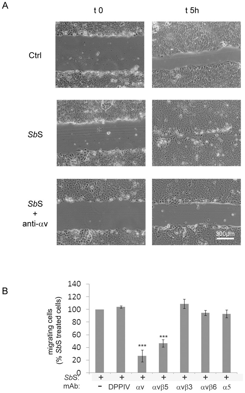
We previously reported both in vitro and in vivo that SbS improves intestinal cell restitution through activation of the α2β1 integrin collagen receptor [15]. However it is clear that α2β1 integrin is not the sole receptor involved in this process since a function-blocking anti-αv subunit integrin mAb partially inhibited SbS-mediated cell migration (figure 1 and supplemental video S1 and S2). Quantification of the surface area recovered by the cells indicated that the anti- αv subunit integrin mAb inhibited the migration capacity of SbS-treated HCT-8/E11 cells by approximately 70% (figure 1 ). To delineate which αvβ integrin was involved in the migratory process we incubated HCT-8/E11 cells with function-blocking anti-αvβ3, anti-αvβ5or anti-αvβ6integrin mAbs during cell migration. As observed in figure 1B , only the anti-αvβ5integrin mAb partially blocked both control and SbS-induced HCT-8/E11 cell migration.
Figure 1. αvβ5 integrin is required for SbS-stimulated cell restitution.
(A) Polarized HCT-8/E11 cell monolayers were pretreated without or with 10 µg/ml of an anti- αv integrin mAb (anti-αv) for 1 h. Cell monolayers were then wounded as described in Methods and incubated for 5 h in the absence (ctrl) or presence of SbS in a medium either containing anti-αv integrin mAb or not. Phase contrast images were acquired at the indicated times. Data shown are from a representative experiment out of 3 performed. Scale bar: 300 µm. (B) HCT-8/E11 cell monolayers were wounded and incubated with SbS. The monolayers were further incubated without (-) or with the following function-blocking anti-mAbs 1 h before wounding and during cell migration: αvintegrin (αv), αvβ3 integrin (αvβ3), αvβ5 integrin (αvβ5), or αvβ6 integrin (αvβ6). Wound closure was determined as described in Methods. Results are expressed as the percentage of cell migration compared to SbS-treated cells without mAbs. Data represent the mean+SD of 5 separate experiments. Rat-anti DPP IV (DPPIV) and mouse anti-α5 subunit (α5) were used as isotypic Abs controls. *** P<0.001.
αvβ5 integrin is redistributed in SbS force-fed mice
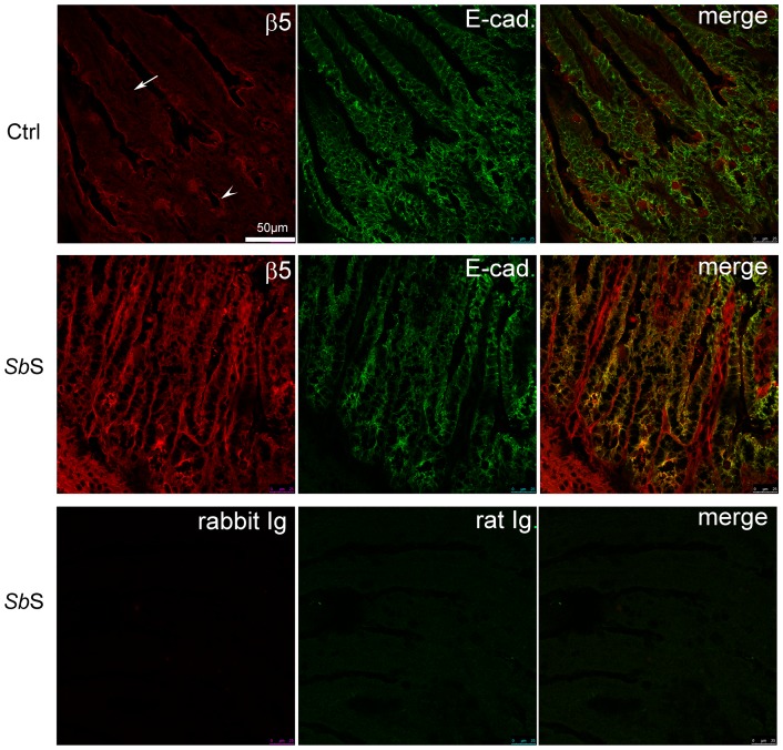
Since αvβ5 integrin is involved in SbS-induced cell migration in vitro, we next evaluated whether SbS affected the distribution of αvβ5 integrin in vivo. C57BL6J mice were force-fed daily with either RPMI 1640 or SbS, for 1 week. We verified that SbS treatment did not alter the mucosal architecture. The distribution of β5 integrin subunit was determined by immunohistochemistry on intestinal tissues (figure 2 ). In control mice, β5 subunit was abundantly detected at the surface of cells located in the crypts and along the apical membrane domain of cell present in the villus. By contrast, in SbS force-fed mice, β5 positive cells were mainly redistributed to the basal surfaces of enterocytes in the villus (figure 2B). The pattern of E-cadherin expression was also analyzed along the crypt-villus axis in order to locate epithelial cells. As observed on figure 2 , E-cadherin positive cells also expressed β5 integrin subunit. Moreover, upon SbS treatment, β5 integrin was re-distributed at the basal side of the epithelial intestinal cells. This suggests that αvβ5integrin participates in SbS-mediated epithelial cell migration. However, it should be noted that SbS increased the β5 subunit integrin expression level of E-cadherin negative cells located into the lamina propria, suggesting that SbS could also act on non-epithelial cells, as recently described [31].
Figure 2. αvβ5 integrin is relocalized in the intestine of SbS force-fed mice.
Mice were daily force-fed for one week with unused culture medium for Sb (Ctrl) or SbS (SbS). Frozen sections of small intestinal tissues were co-stained with a rabbit anti-β5-β integrin subunit (β5) and a rat anti E-cadherin (E-cad). Sections were then incubated with a mixture of Alexa 594-conjugated and Alexa 488-conjugated secondary antibodies against rabbit and mouse IgG, respectively. Rabbit Ig and rat Ig correspond to isotype controls Abs. Colocalized pixels appear in yellow in merged images. Each image is a representative image taken from tissue sections of four mice. The arrow and the arrowhead point out a villus and a crypt, respectively.
SbS modulates αv integrin-mediated adhesion
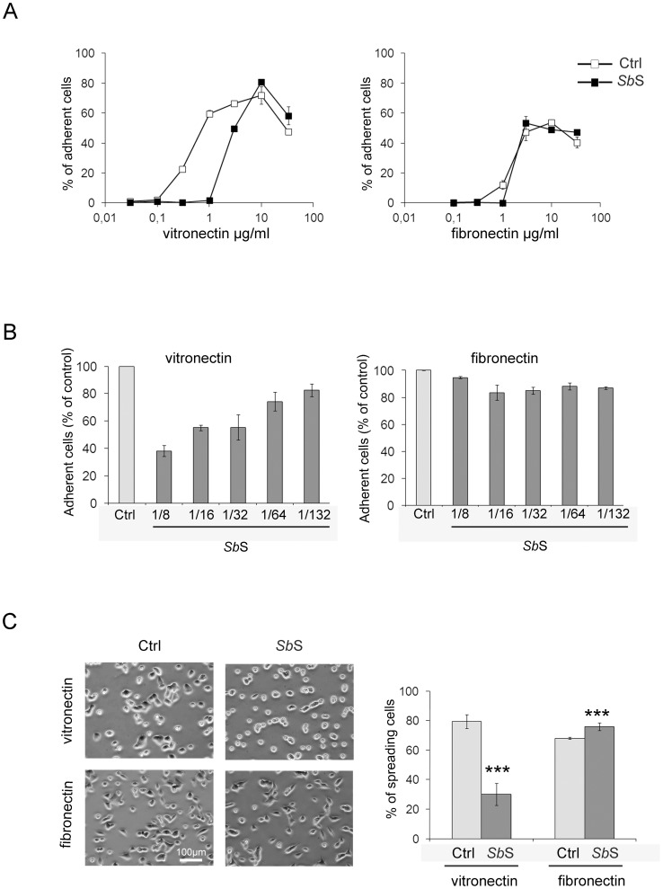
As regulation of cell adhesion and cell spreading are essential for cell migration, we next examined whether SbS altered HCT-8/E11 cell attachment and spreading to both Fn and Vn. Vn is a ligand of αvβ5 integrin whereas Fn is recognized by α5β1 and αvβ3 integrins but not by αvβ5 integrin. However the primary sequence motif for the ligand binding of these integrins is a tripeptide RGD sequence [32]. Cells were either treated with SbS or not, then allowed to attach to increasing amounts of purified Fn or Vn. HCT-8/E11 cells attached to the two matrices tested (figure 3A ). SbS did not alter cell adhesion to Fn. However, the percentage of cells adherent to Vn was dramatically decreased by SbS. As depicted by figure 3B , SbS down-modulated cell attachment to Vn in a dose-dependent manner, with a maximal effect obtained with a 1/8 dilution of SbS. Since cell spreading is initiated immediately after cell contact with matrix proteins, we also determined whether SbS affected cell spreading on both Fn and Vn. As observed in cell adhesion assays, SbS dramatically down-modulated the capacity of cells to spread on Vn. On the other hand, it weakly, albeit significantly, increased HCT-8/E11 cell spreading on Fn (Figure 3C ).
Figure 3. SbS down-modulates cell adhesion to vitronectin.
(A) Isolated HCT-8/E11 cells were treated with or without SbS (dilution 1/8) and plated on either fibronectin or vitronectin at the indicated concentrations. Cell-ECM adhesion was evaluated as described in Methods. Results are expressed as the percentage of cell adhesion. Data represent the mean+SD of 3 separate experiments. (B) HCT-8/E11 cells were treated with or without dilutions of SbS and plated on vitronectin (Vn). Cell-vitronectin adhesion was evaluated as described in Methods. (C) HCT-8/E11 cells were treated with or without SbS, seeded in vitronectin-coated or fibronectin-coated wells and allowed to adhere for 2 h at 37°C. Spreading cells were counted microscopically. Results are expressed as the percentage of spreading cells. Data represent the mean+SD of 3 separate experiments. *** P<0.001.
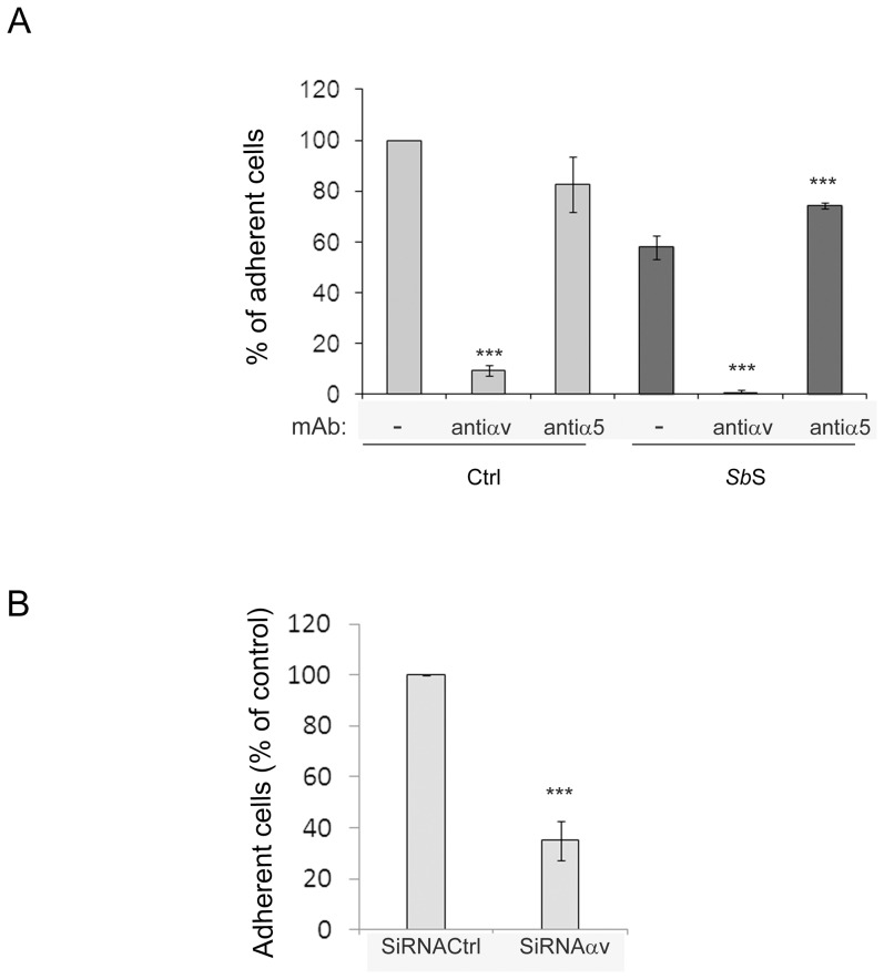
To delineate which α and β integrin subunits were affected by SbS during both cell adhesion and spreading to Vn, we performed adhesion assays in the presence of anti-integrin function-blocking mAbs. The interaction between HCT-8/E11 cells and Vn was mediated solely by the αv integrin subunit (Figure 4A ) whatever the condition tested. An anti- α5integrin subunit that blocked the HCT-8/E11 cell-Fn interaction (not shown) did not affect cell adhesion to Vn. It should be noted that when HCT-8/E11 cells were treated with SbS, blockade of the α5 integrin subunit led to a weak increase in cell adhesion to Vn (Figure 4A ). This may suggest a functional interplay between αv and α5 integrin subunits. As observed in Figure 4B , αv subunit silencing also dramatically blocked cell adhesion to Vn, confirming that the αv integrin subunit is the α subunit receptor for Vn in HCT-8/E11 cells.
Figure 4. αvβ5 integrin is required for HCT-8/E11 cell interaction with vitronectin.
(A) Isolated HCT-8/E11 cells were incubated without (Ctrl) or with SbS (SbS) in the absence (-) or presence of either anti-αv or -α5 integrin mAbs. Cells were then seeded on vitronectin (3,15 µg/ml). Cell adhesion was evaluated as described in Methods. Results are expressed as the percentage of cell adhesion compared to untreated cells (Ctrl). Data represent the mean+SD of 3 separate experiments. (B) HCT-8/E11 cells were transfected with anti -αv-integrin siRNAs (SiRNAαv) or scramble oligos (SiRNACtrl) for 48 h. Cells were then plated on vitronectin for 2 h. Cell-ECM adhesion was evaluated as described in Methods. Results are expressed as the percentage of cell adhesion. Data represent the mean+SD of 3 separate experiments. ***P<0.001.
SbS competes with αv integrin for binding to vitronectin
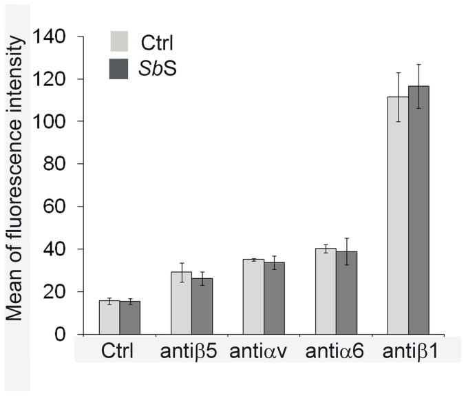
Modulation of integrin expression at the cell surface may alter cell migration. To determine whether SbS modified integrin expression, we quantified the integrin subunits located at the plasma membrane by flow cytometry analysis. As shown in figure 5 , SbS did not significantly alter expression levels of the αv, β5, α6 and β1 integrin subunits.
Figure 5. Impact of SbS on integrin expression.
Sub-confluent HCT-8/E11 cells were incubated for 5 h with (dark bars) or without (light bars) SbS, then harvested and resuspended in the presence of anti-integrin subunit mAbs. After incubation with the appropriate secondary Alexa 488-conjugated Ab, cell-bound fluorescence was quantified using a Becton-Dickinson FACScan flow cytometer. Non-specific labeling was determined by incubating cells with the secondary Alexa 488-conjugated Ab alone (Ctrl). Data represent the mean+SD of 6 separate experiments.
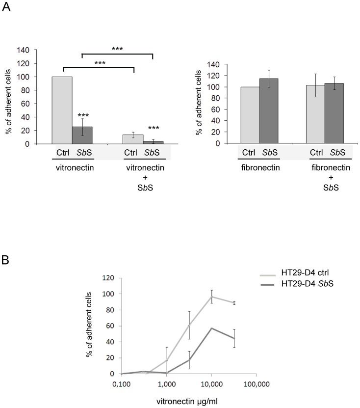
Since SbS inhibited cell adhesion to Vn without modulation of αv integrin subunit expression, we postulated that it blocked αv integrin-Vn interaction. To confirm this hypothesis, ECM-coated plates were incubated in the presence of SbS prior to addition of cells. We first determined that cells did not adhere to SbS coated plates (not shown). Addition of SbS to Vn-coated plates impaired cell adhesion (Figure 6A ). Moreover, the same results were obtained when SbS-coated plates were incubated with Vn prior to cell adhesion (not shown). However, when the same experiments were performed using Fn instead of Vn, no change in cell attachment was found (Figure 6A ). This suggests that SbS specifically interacts with Vn and blocks αv-dependent HCT-8/E11 adhesion. Furthermore, similar results were observed using another intestinal cell line HT29-D4 (Figure 6B ), indicating that the observed phenomenon could be common to intestinal cell lines.
Figure 6. SbS blocked αvβ5 integrin interaction with vitronectin.
(A) Vitronectin- or Fibronectin-coated plates were incubated in the absence (Vitronectin and Fibronectin) or presence of SbS (vitronectin+ SbS and fibronectin+ SbS) prior to addition of cells. Isolated HCT-8/E11 cells incubated without (light bars) or with SbS (dark bars) were then seeded on plates. Cell adhesion to ECM was evaluated as described in Methods. Results are expressed as the percentage of cell adhesion compared to untreated cells (Ctrl). Data represent the mean + SD of 3 separate experiments. (B) Isolated HT29-D4 cells were treated with or without SbS (dilution 1/8) and plated on vitronectin at the indicated concentrations. Cell-vitronectin adhesion was evaluated as described in Methods. Results are expressed as the percentage of cell adhesion. Data represent the mean+SD of 3 separate experiments. *** P<0.001.
SbS regulates the αvβ5 integrin signaling pathway
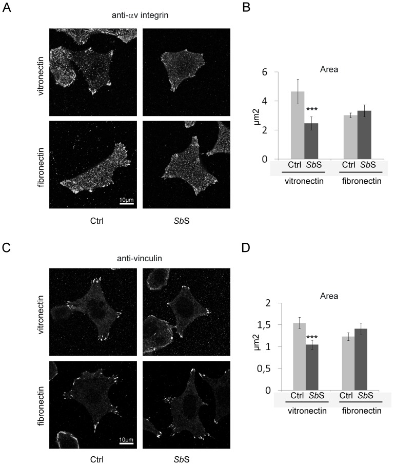
Activation of αvβ5 integrin leads to remodeling of the actin cytoskeleton through the formation and activation of a large signaling complex called focal adhesion. This adhesion complex contains enzymes and scaffolding molecules including FAK and vinculin [32]. Since SbS modulated HCT-8/E11 adhesion to Vn, we first checked whether SbS regulated αvβ5 integrin activation. We first analyzed the impact of SbS on the organization of adherence structures after plating cells on Vn. As depicted by Figure 7A , αv integrin subunit staining revealed that control cells plated on Vn, exhibited focal adhesion structures. However, αv integrin subunit staining in SbS treated cells displayed less focal adhesion structures. Analysis of the αv integrin subunit detected at the cell-substratum interface revealed that SbS markedly decreased the area of focal adhesion sites (Figure 7B ). The same results were obtained with vinculin (Figures 7C and 7D ). These results indicate that treatment with SbS is associated with structural modifications of focal adhesion structures. Interestingly this effect was not observed when Fn was used as a substrate (Figures 7A–B and 7C–D ).
Figure 7. SbS alters the organization of adherence structures when cells are plated on vitronectin.
HCT-8/E11 cells were plated on either fibronectin or vitronectin for 2 h and then fixed. αv integrin (A) or vinculin (C) were detected using specific Abs. Focal adhesion labeling was quantified by measuring the area of each focal adhesion for all cells (B and D). Data represent the mean+SD of 3 separate experiments. *** P<0.001.
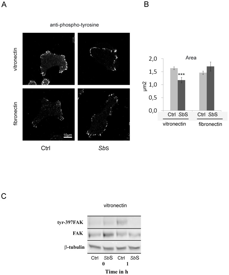
We next analyzed whether SbS the αvβ5-mediated signaling pathway. We first determined whether SbS affected protein tyrosine phosphorylation by immunolocalization. In untreated cells, the mAb PY20 directed against the phosphorylated tyrosine residue, mainly stained cell-ECM contact sites, namely focal adhesion structures (Figure 8A ). However, SbS treatment promoted a decrease in the area of the tyrosine phosphorylated focal adhesion sites (Figures 8A and 8B ). This suggests that SbS may negatively modulate the activity of focal adhesion structures. To confirm the impact of SbS on the αvβ5 signaling pathway, cells were either treated with SbS or not and plated on a Vn-coated surface for 1 h. The tyrosine phosphorylation status of FAK post-adhesion was determined by Western blot using an anti-Y397 FAK mAb. As depicted in figure 8C , adhesion of control cells to Vn promoted FAK tyrosine phosphorylation on residue 397. Interestingly, SbS decreased the tyrosine phosphorylation level of FAK, confirming that SbS may regulate αvβ5 integrin activation (Figure 8C ).
Figure 8. SbS alters the functionality of adherence structures.
(A) HCT-8/E11 cells were plated on either fibronectin or vitronectin for 2 h and then fixed. Tyrosine phosphorylated proteins were stained using a PY20 mAb. (B) Focal adhesion labeling was quantified by measuring the area of each focal adhesion for all cells. Data represent the mean+SD of 3 separate experiments. (C) HCT-8/E11 cells were allowed to adhere on vitronectin after SbS pretreatment. The phosphorylation of tyrosine residues in FAK (Tyr-397FAK) was determined after cell lysis at the indicated times of cell adhesion. Samples were analyzed by western blot analysis. Equal amounts of protein were analyzed and loading amounts were verified by probing the blot with anti-FAK (FAK) or anti- β-tubulin (β-tubulin) Abs. *** P<0.001.
SbS contains a heat-sensitive molecule, >9 kDa, that regulates both cell adhesion and migration
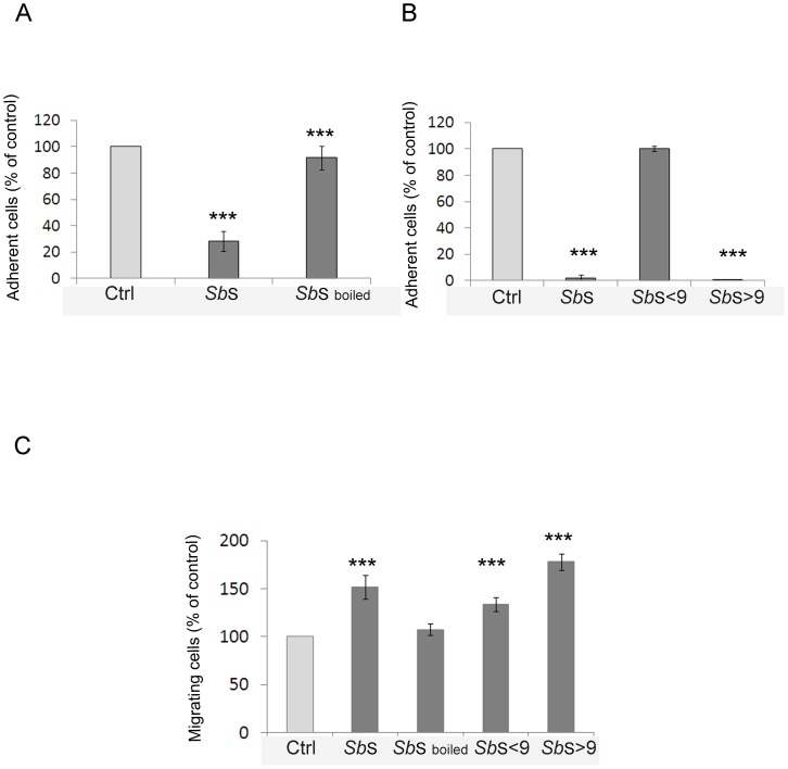
We next sought to better characterize the active component(s) in SbS that blocked cell adhesion to Vn and induced cell migration. We first determined the heat stability of the active component. As illustrated in figure 9A , boiling the SbS for 10 min abrogated the inhibitory effect of SbS on cell-Vn adhesion. Similar results as those obtained for cell adhesion were observed for cell migration (Figure 9C ), indicating that the blockade of both cell adhesion to Vn and induction of cell migration involved one or more proteins. We next examined the activity of SbS after passage through a 9 kDa cut-off filter. As illustrated in Figure 9B we found that the filtrate did not retain SbS activity in cell adhesion to Vn, clearly indicating that the active component(s) has a molecular weight higher than 9 kDa. Interestingly, although the filtrated fraction contained motogenic molecules, the flow-through filtration (<9 kDa) fraction was also able to increase cell migration. This indicates that SbS contains at least two molecules capable to modulate cell migration (Figure 9C ).
Figure 9. Sb secretes several molecules that differentially regulate both cell adhesion and migration.
(A) Isolated HCT-8/E11 cells were treated with or without SbS (SbS), boiled SbS (SbS boiled) and plated on 3.15 µg/ml vitronectin. Cell-vitronectin adhesion was evaluated as described in Methods. Results are expressed as the percentage of cell adhesion. Data represent the mean+SD of 3 separate experiments. (B) Isolated HCT-8/E11 cells were treated with or without SbS (SbS), or fractionated SbS (>9 kDa or <9 kDa) and plated on 3.15 µg/ml vitronectin. Cell-vitronectin adhesion was evaluated as described in Methods. Results are expressed as the percentage of cell adhesion. Data represent the mean+SD of 3 separate experiments. (C) HCT-8/E11 cell monolayers were wounded and incubated without (Ctrl) or with SbS (SbS), boiled SbS (SbS boiled) or fractionated Sb supernatant (>9 kDa or <9 kDa) for 5 h. Wound closure was determined as described in Methods. Results are expressed as the percentage of cell migration compared to control. Data represent the mean+SD of 2 separate experiments. *** P<0.001.
Discussion
Intestinal epithelial restitution, proliferation and differentiation are all requirements for wound healing, a process disrupted in infectious or inflammatory bowel diseases, such as ulcerative colitis and Crohn's disease [32]. Therefore, complete remission of such diseases requires both the cessation of inflammation and the repair of damaged epithelium. The development of novel therapies that accelerate the repair of intestinal epithelium has recently begun and various molecules are now being considered for clinical use. These include epidermal growth factor in combination with mesalamine [33], keratinocyte growth factor [34], and hepatocyte growth factor [35]. However, a better understanding of the biological effects of these molecules must be ascertained, not least to identify any undesired secondary effects, such as tumorigenesis. Clinical trials have suggested that Sb can be effective in the treatment of gastroenteritis and IBD [18] through the modulation of host cell signaling pathways implicated in the pro-inflammatory response. Moreover, we previously demonstrated both in vitro and in mice that Sb promoted intestinal restitution [15].
In the present study, we analyzed the effect of Sb on the capacity of intestinal epithelial cells to heal a wound. According to our findings, the impact of Sb on epithelial cells can be summarized as follows: (1) SbS contains heat-sensitive compounds that decreased αvβ5 integrin-mediated adhesion to Vn by competing with the integrin; (2) SbS-mediated changes in cell adhesion to Vn results in a reduction of the αvβ5 signaling pathway; (3) This perturbation is related to Sb-induced cell migration. According to these data, we postulated that SbS contains motogenic factor(s) that improved intestinal cell restitution by down-modulating the αvβ5 integrin-Vn interaction.
We demonstrated that SbS contains factors that blocked the cell-Vn interaction. Indeed, our adhesion assays showed that SbS competed with αv integrin subunit in binding to Vn. Indeed, (1) SbS blocked cell adhesion to Vn and (2) addition of SbS onto Vn prior to cell plating impaired cell adhesion. This blockade is selective since SbS did not block the cell interaction with Fn (this paper), laminin or type I collagen [15]. Moreover this phenomenon could be common to all intestinal epithelial cell lines since the same results were observed using HT29-D4 cells, another intestinal cell line. Vn associates with a wide variety of ligands including αv integrin, IGF-I family members, and urokinase plasminogen activator receptor [36] [11], [37]. The binding site(s) present in Vn for SbS factors remain to be elucidated. One might postulate that the tripeptide sequence RGD, involved in binding integrin to both Fn and Vn, does not play a role in this process since SbS did not block cell interaction with Fn. Further studies are required to explore whether sequences flanking the RGD peptide could interact with SbS , as they have been reported to be important for integrin selectivity [38]. On the other hand, other Vn domains could be involved in the interaction with compound(s) found in SbS. For example, Protein E from Hemophilus influenza competed with αv subunit containing integrins for binding to Vn by interacting with domains located at the heparin binding regions of Vn [39]. Moreover, the high molecular weight form of kinninogen confers a strong anti-adhesive function upon integrin-mediated cell interaction with Vn [40]. Nevertheless, we reported in this study that the anti-adhesive factor(s) found in SbS is a (are) heat-sensitive compound (s) with a molecular weight higher than 9 kDa.
It is now clear that Vn plays a crucial role in many biological processes including cell migration, adhesion, tissue repair and angiogenesis [41]. Interestingly, other studies have suggested potential roles of Vn in microbial colonization and serum resistance. According to this, recent findings unveiled that many bacterial species, including C. difficile and H. Pylori interact with Vn [32]. The functionality of these interactions in pathogenesis has not been fully elucidated, although Vn most likely functions as a bridge between bacteria and epithelial cells [32]. Therefore we could postulate that SbS contains compounds that block Vn-enterocyte interactions and then inhibits adhesion of pathogens to host cells. Further work is needed to explore this putative antimicrobial activity in more depth.
Activation of αvβ5 integrin leads to remodeling of the actin cytoskeleton through the formation and activation of signaling complexes called “adhesion complexes”. These multi-molecular complexes contain enzymes and scaffolding molecules including FAK and paxillin [42]. Activation of these signaling molecules, following αv integrin activation leads to the modulation of cell migration [42]. Several arguments suggest that SbS promotes dynamic changes in the αvβ5 integrin affinity for its ligand. Firstly, immunolocalization of both αv integrin subunit and paxillin showed that SbS decreased the area of focal adherence structures. Secondly, this SbS-induced reorganization of focal adherence structures was associated with both a decrease in FAK tyrosine phosphorylation on the Y397 residue upon cell adhesion to Vn and an increase in αvβ5 dependent migration. FAK is a key mediator of intracellular signaling by integrins and may serve as conduits for the transmission of the force necessary for cell migration and bidirectional signaling between the cell interior and its environment [43]. FAK activation leads to the stimulation of other signaling proteins such as paxillin, thereby activating various signaling pathways crucial in the regulation of cell adhesion and migration [43]. Moreover, signals that modify tyrosine phosphorylation may influence mucosal wound healing [44]. Therefore, we suggest that SbS regulates the strength of the αvβ5 integrin/Vn interaction and consequently modulates FAK tyrosine phosphorylation, leading to a change in αvβ5 integrin-dependent migration.
Changes in cell-ECM interactions occur spatially and temporally during intestinal wound healing. Several in vivo and in vitro studies in the gut indicate the requirement of the binding and interaction of ECM-specific integrins for this restitution, such as α3β1, α6β1, α6β4 laminin-binding integrins, α2β1 collagen-binding integrin or αv-subunit Fn-binding integrin [7], [45]. We recently provided data indicating that Sb exerts at least some of its motogenic effect through the activation of the collagen receptor α2β1 integrin [15]. Here we have reported that SbS also exerts some of its motogenic effect via down-modulation of the interaction between αvβ5 integrin and Vn. Indeed, (1) inhibition assays using anti-integrin mAbs demonstrated that αvβ5 integrins participate in the SbS-induced cell migration. Given the ligand-binding properties of these integrins, it appears likely that Vn supports this process. (2) Both cell adhesion and cell spreading to Vn, two processes required for cell migration, were altered by SbS. (3) Sb promoted the reorganization of αvβ5 integrins into adhesive structures localized at the leading edge of the cells. These are crucial to migration and are themselves regulated by SbS. Moreover, SbS was shown to promote the redistribution of the Vn receptor in the mouse intestine. This redistribution was observed for both epithelial and non-epithelial cells. In line with these data, some studies have indicated a crucial role of Vn in regulating cell migration during wound healing [46], [47].
Cell migration requires the dynamic interaction between cells and the substratum on which they are attached and over which they migrate [48]. Changes in the density of ligand, integrin repertoire, ligand-binding affinity and/or cytoskeletal associations are all key determinants of cell migration speed [48], [49]. In the present study, we showed, by flow cytometry analysis, that SbS did not alter the concentration of αvβ5integrin or, in previous work, α2β1integrin [15]. However, our data strongly suggest that SbS dynamically regulates the assembly and disassembly of adhesions that are essential for optimum cell migration. Indeed, to promote cell restitution, SbS activates the α2β1 integrin collagen receptor [15] whereas it down-regulates the αv integrin interactions with Vn (this work).
Sb acts as a shuttle that could liberate, during the intestinal transit, at least 1500 molecules that have not been totally characterized [50]. This large number of secreted peptidic and non-peptidic factors, including proteases, phosphatases and polyamines, may at least partially explain why Sb has pleiotropic effects on intestinal mucosa and also has therapeutic effects on such a wide variety of gastrointestinal disorders [14], [31], [51]. Some of the molecules, as yet unidentified, can interfere with host cell signaling pathways and are therefore able to modulate host cell behavior including intestinal mucosal inflammatory, secretory, and barrier functions [50]. Moreover, Sb produces other factors that reduce inflammation by blocking NF-κB and MAPK activation [26], [52] and enhancing PPAR-γ expression [53]. On the other hand, conditioned medium of Sb was shown both in vitro and in vivo to modulate host signaling pathways involved in the regulation of cell motility including the MAPK and FAK pathways [15], [26]. Although the motogenic molecule(s) secreted by Sb remain to be elucidated we have reported here that SbS contains two groups of heat-sensitive compounds capable to increase intestinal restitution: one group with a molecular weight lower than 9 kDa and another with a molecular weight higher than 9 kDa. We can postulate that this latest class of molecules corresponds to the same as those involved in the down-modulation of cell adhesion to Vn.
In conclusion, this report demonstrates that SbS contains various heat-sensitive motogenic factors that can improve intestinal restitution. These factors exerted their effect through multiple pathways, including the dynamic fine regulation of integrin-mediated adhesion to the ECM. This could be of major importance in diseases characterized by severe mucosal injury, as seen in IBD or infectious gastroenteritis.
Supporting Information
HCT-8/E11 cell monolayers were wounded as described in Methods , incubated with Sb supernatant (dilution 1/8), then placed in a temperature and CO2-controlled chamber mounted on a Nikon TE2000 inverted microscope. Images were captured every 5 minutes for a total observation period of 5 h, using a Cool SnapHQ camera (Princeton Instrument) through a 10× objective lens.
(MOV)
HCT-8/E11 cell monolayers were wounded as described in Methods and incubated with Sb S. Cell monolayers were further incubated with or without function-blocking anti- αv integrin mAb 1 h before wounding and during cell migration. Plates were placed in a temperature and CO2-controlled chamber mounted on a Nikon TE2000 inverted microscope. Images were captured every 5 minutes for a total observation time of 5 h using a Cool SnapHQ camera (Princeton Instrument) through a 10× objective lens.
(MOV)
Funding Statement
This work was financed in part by an institutional grant from Institut National du Cancer (InCa, Cancéropôle PACA). The funders had no role in study design, data collection and analysis, decision to publish, or preparation of the manuscript. No additional external funding received for this study.
References
- 1. Blikslager AT, Moeser AJ, Gookin JL, Jones SL, Odle J (2007) Restoration of barrier function in injured intestinal mucosa. Physiol Rev 87: 545–564. [DOI] [PubMed] [Google Scholar]
- 2. Mumy KL, Chen X, Kelly CP, McCormick BA (2008) Saccharomyces boulardii interferes with Shigella pathogenesis by postinvasion signaling events. Am J Physiol Gastrointest Liver Physiol 294: G599–609. [DOI] [PMC free article] [PubMed] [Google Scholar]
- 3. Martins FS, Dalmasso G, Arantes RM, Doye A, Lemichez E, et al. (2010) Interaction of Saccharomyces boulardii with Salmonella enterica serovar Typhimurium protects mice and modifies T84 cell response to the infection. PLoS ONE 5: e8925. [DOI] [PMC free article] [PubMed] [Google Scholar]
- 4. Sturm A, Dignass AU (2008) Epithelial restitution and wound healing in inflammatory bowel disease. World J Gastroenterol 14: 348–353. [DOI] [PMC free article] [PubMed] [Google Scholar]
- 5. Swanson PA 2nd, Kumar A, Samarin S, Vijay-Kumar M, Kundu K, et al. (2011) Enteric commensal bacteria potentiate epithelial restitution via reactive oxygen species-mediated inactivation of focal adhesion kinase phosphatases. Proc Natl Acad Sci U S A 108: 8803–8808. [DOI] [PMC free article] [PubMed] [Google Scholar]
- 6. Goke M, Kanai M, Lynch-Devaney K, Podolsky DK (1998) Rapid mitogen-activated protein kinase activation by transforming growth factor alpha in wounded rat intestinal epithelial cells. Gastroenterology 114: 697–705. [DOI] [PubMed] [Google Scholar]
- 7. Basson MD (2001) In vitro evidence for matrix regulation of intestinal epithelial biology during mucosal healing. Life Sci 69: 3005–3018. [DOI] [PubMed] [Google Scholar]
- 8. Zhang J, Owen CR, Sanders MA, Turner JR, Basson MD (2006) The motogenic effects of cyclic mechanical strain on intestinal epithelial monolayer wound closure are matrix dependent. Gastroenterology 131: 1179–1189. [DOI] [PubMed] [Google Scholar]
- 9. Basson MD, Modlin IM, Madri JA (1992) Human enterocyte (Caco-2) migration is modulated in vitro by extracellular matrix composition and epidermal growth factor. J Clin Invest 90: 15–23. [DOI] [PMC free article] [PubMed] [Google Scholar]
- 10. Basson MD, Rashid Z, Turowski GA, West AB, Emenaker NJ, et al. (1996) Restitution at the cellular level: regulation of the migrating phenotype. Yale J Biol Med 69: 119–129. [PMC free article] [PubMed] [Google Scholar]
- 11. Gilcrease MZ (2007) Integrin signaling in epithelial cells. Cancer Lett 247: 1–25. [DOI] [PubMed] [Google Scholar]
- 12. Lussier C, Basora N, Bouatrouss Y, Beaulieu JF (2000) Integrins as mediators of epithelial cell-matrix interactions in the human small intestinal mucosa. Microsc Res Tech 51: 169–178. [DOI] [PubMed] [Google Scholar]
- 13. Dignass AU (2001) Mechanisms and modulation of intestinal epithelial repair. Inflamm Bowel Dis 7: 68–77. [DOI] [PubMed] [Google Scholar]
- 14. Czerucka D, Piche T, Rampal P (2007) Review article: yeast as probiotics – Saccharomyces boulardii. Aliment Pharmacol Ther 26: 767–778. [DOI] [PubMed] [Google Scholar]
- 15. Canonici A, Siret C, Pellegrino E, Pontier-Bres R, Pouyet L, et al. (2011) Saccharomyces boulardii improves intestinal cell restitution through activation of the alpha2beta1 integrin collagen receptor. PLoS ONE 6: e18427. [DOI] [PMC free article] [PubMed] [Google Scholar]
- 16. Pothoulakis C (2009) Review article: anti-inflammatory mechanisms of action of Saccharomyces boulardii. Aliment Pharmacol Ther 30: 826–833. [DOI] [PMC free article] [PubMed] [Google Scholar]
- 17. Buts JP, De Keyser N (2010) Transduction pathways regulating the trophic effects of Saccharomyces boulardii in rat intestinal mucosa. Scand J Gastroenterol 45: 175–185. [DOI] [PubMed] [Google Scholar]
- 18. McFarland LV (2010) Systematic review and meta-analysis of Saccharomyces boulardii in adult patients. World J Gastroenterol 16: 2202–2222. [DOI] [PMC free article] [PubMed] [Google Scholar]
- 19. Plein K, Hotz J (1993) Therapeutic effects of Saccharomyces boulardii on mild residual symptoms in a stable phase of Crohn's disease with special respect to chronic diarrhea–a pilot study. Z Gastroenterol 31: 129–134. [PubMed] [Google Scholar]
- 20. Guslandi M, Giollo P, Testoni PA (2003) A pilot trial of Saccharomyces boulardii in ulcerative colitis. Eur J Gastroenterol Hepatol 15: 697–698. [DOI] [PubMed] [Google Scholar]
- 21. Girard P, Pansart Y, Coppe MC, Gillardin JM (2005) Saccharomyces boulardii inhibits water and electrolytes changes induced by castor oil in the rat colon. Dig Dis Sci 50: 2183–2190. [DOI] [PubMed] [Google Scholar]
- 22. Dalmasso G, Loubat A, Dahan S, Calle G, Rampal P, et al. (2006) Saccharomyces boulardii prevents TNF-alpha-induced apoptosis in EHEC-infected T84 cells. Res Microbiol 157: 456–465. [DOI] [PubMed] [Google Scholar]
- 23. Mammen JM, Matthews JB (2003) Mucosal repair in the gastrointestinal tract. Crit Care Med 31: S532–537. [DOI] [PubMed] [Google Scholar]
- 24. Andre F, Rigot V, Thimonier J, Montixi C, Parat F, et al. (1999) Integrins and E-cadherin cooperate with IGF-I to induce migration of epithelial colonic cells. Int J Cancer 83: 497–505. [DOI] [PubMed] [Google Scholar]
- 25. Vermeulen SJ, Bruyneel EA, Bracke ME, De Bruyne GK, Vennekens KM, et al. (1995) Transition from the noninvasive to the invasive phenotype and loss of alpha-catenin in human colon cancer cells. Cancer Res 55: 4722–4728. [PubMed] [Google Scholar]
- 26. Chen X, Kokkotou EG, Mustafa N, Bhaskar KR, Sougioultzis S, et al. (2006) Saccharomyces boulardii inhibits ERK1/2 mitogen-activated protein kinase activation both in vitro and in vivo and protects against Clostridium difficile toxin A-induced enteritis. J Biol Chem 281: 24449–24454. [DOI] [PubMed] [Google Scholar]
- 27. Canonici A, Steelant W, Rigot V, Khomitch-Baud A, Boutaghou-Cherid H, et al. (2008) Insulin-like growth factor-I receptor, E-cadherin and alpha v integrin form a dynamic complex under the control of alpha-catenin. Int J Cancer 122: 572–582. [DOI] [PubMed] [Google Scholar]
- 28. Gorvel JP, Ferrero A, Chambraud L, Rigal A, Bonicel J, et al. (1991) Expression of sucrase-isomaltase and dipeptidylpeptidase IV in human small intestine and colon. Gastroenterology 101: 618–625. [DOI] [PubMed] [Google Scholar]
- 29. Defilles C, Montero MP, Lissitzky JC, Rome S, Siret C, et al. (2011) alphav integrin processing interferes with the cross-talk between alphavbeta5/beta6 and alpha2beta1 integrins. Biol Cell 103: 519–529. [DOI] [PubMed] [Google Scholar]
- 30. Defilles C, Lissitzky JC, Montero MP, Andre F, Prevot C, et al. (2009) alphavbeta5/beta6 integrin suppression leads to a stimulation of alpha2beta1 dependent cell migration resistant to PI3K/Akt inhibition. Exp Cell Res 315: 1840–1849. [DOI] [PubMed] [Google Scholar]
- 31. Thomas S, Metzke D, Schmitz J, Dorffel Y, Baumgart DC (2011) Anti-inflammatory effects of Saccharomyces boulardii mediated by myeloid dendritic cells from patients with Crohn's disease and ulcerative colitis. Am J Physiol Gastrointest Liver Physiol 301: G1083–1092. [DOI] [PubMed] [Google Scholar]
- 32. Singh B, Su YC, Riesbeck K (2010) Vitronectin in bacterial pathogenesis: a host protein used in complement escape and cellular invasion. Mol Microbiol 78: 545–560. [DOI] [PubMed] [Google Scholar]
- 33. Sinha A, Nightingale J, West KP, Berlanga-Acosta J, Playford RJ (2003) Epidermal growth factor enemas with oral mesalamine for mild-to-moderate left-sided ulcerative colitis or proctitis. N Engl J Med 349: 350–357. [DOI] [PubMed] [Google Scholar]
- 34. Zeeh JM, Procaccino F, Hoffmann P, Aukerman SL, McRoberts JA, et al. (1996) Keratinocyte growth factor ameliorates mucosal injury in an experimental model of colitis in rats. Gastroenterology 110: 1077–1083. [DOI] [PubMed] [Google Scholar]
- 35. Ido A, Numata M, Kodama M, Tsubouchi H (2005) Mucosal repair and growth factors: recombinant human hepatocyte growth factor as an innovative therapy for inflammatory bowel disease. J Gastroenterol 40: 925–931. [DOI] [PubMed] [Google Scholar]
- 36. Chantret I, Rodolosse A, Barbat A, Dussaulx E, Brot-Laroche E, et al. (1994) Differential expression of sucrase-isomaltase in clones isolated from early and late passages of the cell line Caco-2: evidence for glucose-dependent negative regulation. J Cell Sci 107 (Pt 1) 213–225. [DOI] [PubMed] [Google Scholar]
- 37. Kricker JA, Towne CL, Firth SM, Herington AC, Upton Z (2003) Structural and functional evidence for the interaction of insulin-like growth factors (IGFs) and IGF binding proteins with vitronectin. Endocrinology 144: 2807–2815. [DOI] [PubMed] [Google Scholar]
- 38. Avraamides CJ, Garmy-Susini B, Varner JA (2008) Integrins in angiogenesis and lymphangiogenesis. Nat Rev Cancer 8: 604–617. [DOI] [PMC free article] [PubMed] [Google Scholar]
- 39. Hallstrom T, Blom AM, Zipfel PF, Riesbeck K (2009) Nontypeable Haemophilus influenzae protein E binds vitronectin and is important for serum resistance. J Immunol 183: 2593–2601. [DOI] [PubMed] [Google Scholar]
- 40. Chavakis T, Kanse SM, Lupu F, Hammes HP, Muller-Esterl W, et al. (2000) Different mechanisms define the antiadhesive function of high molecular weight kininogen in integrin- and urokinase receptor-dependent interactions. Blood 96: 514–522. [PubMed] [Google Scholar]
- 41. Preissner KT, Reuning U (2011) Vitronectin in vascular context: facets of a multitalented matricellular protein. Semin Thromb Hemost 37: 408–424. [DOI] [PubMed] [Google Scholar]
- 42. Banno A, Ginsberg MH (2008) Integrin activation. Biochem Soc Trans 36: 229–234. [DOI] [PMC free article] [PubMed] [Google Scholar]
- 43. Zaidel-Bar R, Itzkovitz S, Ma'ayan A, Iyengar R, Geiger B (2007) Functional atlas of the integrin adhesome. Nat Cell Biol 9: 858–867. [DOI] [PMC free article] [PubMed] [Google Scholar]
- 44. Owen KA, Abshire MY, Tilghman RW, Casanova JE, Bouton AH (2010) FAK regulates intestinal epithelial cell survival and proliferation during mucosal wound healing. PLoS ONE 6: e23123. [DOI] [PMC free article] [PubMed] [Google Scholar]
- 45. André F, Rigot V, Thimonier J, Montixi C, Parat F, et al. (1999) Integrins and E-cadherin cooperate with IGF-I to induce migration of epithelial colonic cells. Int J Cancer 83: 497–505. [DOI] [PubMed] [Google Scholar]
- 46. Adair JE, Stober V, Sobhany M, Zhuo L, Roberts JD, et al. (2009) Inter-alpha-trypsin inhibitor promotes bronchial epithelial repair after injury through vitronectin binding. J Biol Chem 284: 16922–16930. [DOI] [PMC free article] [PubMed] [Google Scholar]
- 47. Upton Z, Wallace HJ, Shooter GK, van Lonkhuyzen DR, Yeoh-Ellerton S, et al. (2011) Human pilot studies reveal the potential of a vitronectin: growth factor complex as a treatment for chronic wounds. Int Wound J 8: 522–532. [DOI] [PMC free article] [PubMed] [Google Scholar]
- 48. Huttenlocher A, Horwitz AR (2011) Integrins in cell migration. Cold Spring Harb Perspect Biol 3: a005074. [DOI] [PMC free article] [PubMed] [Google Scholar]
- 49. Palecek SP, Loftus JC, Ginsberg MH, Lauffenburger DA, Horwitz AF (1997) Integrin-ligand binding properties govern cell migration speed through cell-substratum adhesiveness. Nature 385: 537–540. [DOI] [PubMed] [Google Scholar]
- 50. Buts JP (2009) Twenty-five years of research on Saccharomyces boulardii trophic effects: updates and perspectives. Dig Dis Sci 54: 15–18. [DOI] [PubMed] [Google Scholar]
- 51. Zanello G, Meurens F, Berri M, Salmon H (2009) Saccharomyces boulardii effects on gastrointestinal diseases. Curr Issues Mol Biol 11: 47–58. [PubMed] [Google Scholar]
- 52. Sougioultzis S, Simeonidis S, Bhaskar KR, Chen X, Anton PM, et al. (2006) Saccharomyces boulardii produces a soluble anti-inflammatory factor that inhibits NF-kappaB-mediated IL-8 gene expression. Biochem Biophys Res Commun 343: 69–76. [DOI] [PubMed] [Google Scholar]
- 53. Lee SK, Kim HJ, Chi SG, Jang JY, Nam KD, et al. (2005) [Saccharomyces boulardii activates expression of peroxisome proliferator-activated receptor-gamma in HT-29 cells]. Korean J Gastroenterol 45: 328–334. [PubMed] [Google Scholar]
Associated Data
This section collects any data citations, data availability statements, or supplementary materials included in this article.
Supplementary Materials
HCT-8/E11 cell monolayers were wounded as described in Methods , incubated with Sb supernatant (dilution 1/8), then placed in a temperature and CO2-controlled chamber mounted on a Nikon TE2000 inverted microscope. Images were captured every 5 minutes for a total observation period of 5 h, using a Cool SnapHQ camera (Princeton Instrument) through a 10× objective lens.
(MOV)
HCT-8/E11 cell monolayers were wounded as described in Methods and incubated with Sb S. Cell monolayers were further incubated with or without function-blocking anti- αv integrin mAb 1 h before wounding and during cell migration. Plates were placed in a temperature and CO2-controlled chamber mounted on a Nikon TE2000 inverted microscope. Images were captured every 5 minutes for a total observation time of 5 h using a Cool SnapHQ camera (Princeton Instrument) through a 10× objective lens.
(MOV)


