Abstract
Posterior reversible encephalopathy syndrome (PRES) is often associated with hypertension, however recent advances in the understanding of this condition have shown that endothelial dysfunction is responsible for much of the pathogenesis and the condition can occur in the absence of hypertension. This case describes a 32-year-old lady with untreated HIV infection who developed PRES at a normal blood pressure and without opportunistic infection or other conditions known to precipitate PRES. HIV, particularly when untreated, is associated with endothelial dysfunction and this may have been sufficient to cause PRES in this patient. To our knowledge this is the first case to describe PRES in HIV without uncontrolled hypertension, sepsis or other precipitating cause.
Background
Posterior reversible encephalopathy syndrome (PRES) is characterised by a potentially reversible vasogenic oedema of the occipital lobes with a characteristic MRI appearance1 resulting in a variety of clinical manifestations including headache, cortical blindness, altered mental status and seizures.2
PRES is reported in six HIV-positive individuals who had known risk factors for the disease making it difficult to discern its relationship with HIV. Here, we report a case without such risk factors in which the most notable association of PRES was with untreated HIV infection. Recent advances in the understanding of PRES implicate endothelial dysfunction, something often associated with HIV. This case suggests that HIV infection itself may precipitate PRES without other risk factors.
Case presentation
A 32-year-old lady was diagnosed with HIV in 2004. She received antiretroviral treatment during a pregnancy in 2007 with zidovudine, lamivudine and lopinovir which was stopped postpartum. This pregnancy was complicated by a pulmonary embolus for which she was anticoagulated for 6 months. No cause for thrombophilia was identified, other than HIV infection and pregnancy. One year later she developed an uncomplicated community acquired pneumonia but has otherwise been clinically well.
One afternoon, while watching television at home, she developed an acute onset of severe frontal throbbing headache associated with nausea and photophobia. Her vision simultaneously deteriorated over the course of a few hours. Ophthalmological investigations revealed a right inferior quadrantinopia with acuity of 6/60 in the remaining field of vision. The anterior eye and retina were normal.
She was 9 weeks gestation in her second pregnancy at time of presentation. She had no pre-existing visual problems and a recent opticians report records visual acuity of 6/6 in both eyes and normal fields. There was no history of fever or neck stiffness and she was systemically well. There was no history of vasoactive or other drug abuse.
Investigations
CD4 T lymphocyte count was 290 cells/mm3 and HIV viral load 63 288 copies/ml. Blood tests showed a mildly elevated erythrocyte sedimentation rate, D-dimer and fibrinogen levels consistent with early pregnancy, but routine blood tests were otherwise normal or negative. The C reactive protein was 11 and blood cultures were sterile.
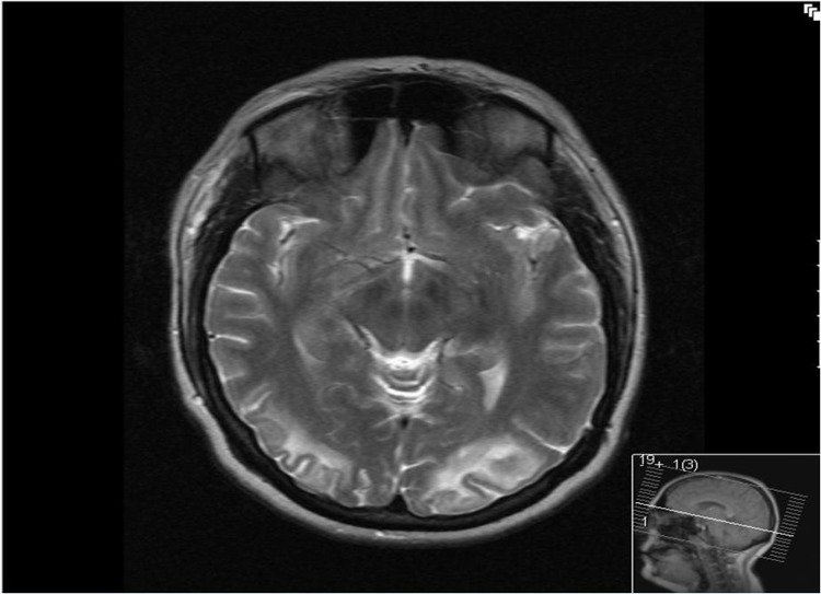
Contrast enhanced CT of the brain demonstrated low density in the white matter of both occipital lobes with relative sparing of the cortex and no enhancement post intravenous contrast. MRI demonstrated high T2 signal in the same distribution (figure 1). There was high signal on diffusion weighted MRI which was matched on the apparent diffusion coefficient map. MR venogram and MR angiogram were normal.
Figure 1.
T2 weighted MRI demonstrating high signal in bilateral occipital lobes consistent with posterior reversible encephalopathy syndrome.
Cerebrospinal fluid (CSF) was acellular with a normal protein and glucose ratio. CSF viral PCRs were negative including John Cunningham virus, cytomegalovirus, Epstein–Barr, varicella zoster, enterovirus and herpes simplex. CSF syphilis serology and cryptococcal antigen were also negative.
There were no apparent pregnancy related complications. Ultrasound scan showed a 9 week fetus with normal development. Twenty four hour blood pressure recording was normal with a mean of 128/76 mm Hg and a peak of 140/80 mm Hg. At presentation, there was significant proteinuria with a urine protein creatinine ratio (UPCR) of 188 mg/mmol. Antiretroviral therapy was started with zidovudine, lamivudine and lopinovir following which UPCR decreased to normal limits within 2 weeks.
Differential diagnosis
In this case, the MRI angiogram and CSF were normal against the diagnosis of cerebral vasculitis or an acute demyelinating encephalopathy. The occipital distribution of the brain lesions, the absence of gadolinium enhancement, and the clinical presentation were in keeping with PRES. The nature of the headache and persisting symptoms were in keeping with an overlap with the reversible cerebral vasoconstriction syndrome (Call–Fleming syndrome) which occurs in around 10% of cases. Further examination of the CSF did not suggest progressive multifocal leucoencephalopathy.
Outcome and follow-up
The initial headache subsided over 12 h but she went on to have four or five episodes of recurrence with a similar severe headache associated with photophobia in the following few weeks. Follow-up MRI showed resolution of the T2 MRI changes, but with a degree of occipital lobe atrophy bilaterally. The visual loss and field deficit have improved only slightly and she remains significantly impaired with acuity of 6/60 in both eyes. Although PRES is typically reversible, persisting damage is described as a result of severe hypoperfusion.3
Discussion
A comprehensive hypothesis for PRES was suggested by Bartynski.4 He notes that while PRES is associated with hypertension the systemic blood pressure can be normal, as in our case, suggesting systemic hypertension is not the primary aetiology of PRES. Various radiological techniques have shown hypoperfusion in the brain of PRES cases, a finding against systemic hypertension causing a break through hyperperfusion and tissue injury as a general mechanism. Such a mechanism could however, account for PRES in cases where the blood pressure exceeds the limits of auto-regulation in the brain. Conversely, Bartynski proposes that the intrinsic vascular tone in PRES cases is unstable due to endothelial dysfunction. Vasoconstriction and hypoperfusion of organs that arises in this context he proposes could cause a secondary autoregulatory rise in blood pressure.
Bartynski notes that a generalised endothelial dysfunction is a sequelae of severe activation of the immune system and postulates that it is the basis of PRES. PRES is associated with the systemic inflammatory response syndrome, multi-organ failure and Gram-positive sepsis,5 preeclampsia/eclampsia,6 autoimmune disease, solid organ and bone marrow transplantation – here with graft versus host disease, graft rejection or opportunistic infection,7 and also with use of cancer chemotherapies or cyclosporine suggesting a contribution of drug specific endothelial toxicity.
Evidence to support the specific role of T cells and innate cells in this is that histological evidence of endothelial activation; expression of adhesion molecules and vascular endothelial growth factor in cerebral blood vessels is reported in a PRES case due to tacrolimus following cardiac transplantation.8 Further findings are of reactive astrocytes and microglial cells, intravascular, transmural, and perivascular CD4 and CD8 T-cell T-lymphocytes. Of note, histological changes do not include invasion of B cells or neutrophils and focal myeloid and lymphoid cell accumulations in blood vessels, nor focal encephalitis, leptomeningitis, or demyleination, rather oedema in keeping with capillary leakage syndrome.
There have been six case reports describing PRES in HIV positive individuals. PRES could have been caused by malignant hypertension in three cases (blood pressure >180/110),9 10 in two of these cases the patients took antiretroviral therapy and had undetectable HIV viral loads, and in further cases PRES was associated with hypercalcaemia11 or with severe systemic inflammation (thrombotic thrombocytopenia purpura)12 or with opportunistic infection (Blastomyces dermatitides).13 Therefore, our case is distinctive because the patient did not have evidence of risk factors known to be associated with PRES raising the possibility that HIV infection constitutes the risk for PRES.
We do not believe that pregnancy contributed to an increased risk for PRES. Although PRES is often seen in late pregnancy or the postpartum period in association with preeclampsia/eclampsia,6 it is not described in early or uncomplicated pregnancy and its onset at 6 weeks gestation here may have been coincidental. Renal dysfunction can precipitate PRES, however in this case microalbuminuria occurred without renal impairment and with normal serum urea and creatinine levels. This degree of impairment is unlikely to be sufficient to precipitate PRES, but may have comtributed. Twenty four-hour blood pressure was normal and all previous routine blood pressure measurements had been within normal limits. There was no evidence of pre-existing hypertentsion, although we cannot rule out short lasting increases in blood pressure.
HIV infected individuals have defects in intrinsic regulation of vascular tone demonstrated by abnormal flow mediated brachial artery dilation.14 This is consistent with the view that HIV has effects on endothelial function due to generalised immune activation, a well-known sequelae of HIV infection. Additionally, HIV proteins have multiple effects on endothelial cell function.15 16
In addition, immune activation constitutes a risk for cardiovascular and renal end organ disease in HIV-infected individuals. Endothelial inflammation is suspected to cause renal disease in HIV infected individuals. Our patient presented with proteinuria which resolved with HIV treatment. Although causality is not proven, it appears such immune activation and endothelial dysfunction may be sufficient to cause PRES in our case.
Learning points.
PRES can occur in the absence of hypertension and its pathogenesis is related to endothelial dysfunction.
HIV, particularly when untreated, can cause endothelial dysfunction due to generalised immune activation.
In this case untreated HIV infection alone may have been sufficient to precipitate PRES.
Footnotes
Competing interests: None.
Patient consent: Obtained.
References
- 1.Bartynski WS. Posterior reversible encephalopathy syndrome, part 1: fundamental imaging and clinical features. AJNR Am J Neuroradiol 2008;29:1036–42. [DOI] [PMC free article] [PubMed] [Google Scholar]
- 2.Ducros A, Bousser MG. Reversible cerebral vasoconstriction syndrome. Pract Neurol 2009;9:256–67. [DOI] [PubMed] [Google Scholar]
- 3.Calabrese LH, Dodick DW, Schwedt TJ, et al. Narrative review: reversible cerebral vasoconstriction syndromes. Ann Intern Med 2007;146:34–44. [DOI] [PubMed] [Google Scholar]
- 4.Bartynski WS. Posterior reversible encephalopathy syndrome, part 2: controversies surrounding pathophysiology of vasogenic edema. AJNR Am J Neuroradiol 2008;29:1043–9. [DOI] [PMC free article] [PubMed] [Google Scholar]
- 5.Bartynski WS, Boardman JF, Zeigler ZR, et al. Posterior reversible encephalopathy syndrome in infection, sepsis and shock. Am J Neuroradiol 2006;27:2179–90. [PMC free article] [PubMed] [Google Scholar]
- 6.Roth C, Ferbert A. Posterior reversible encephalopathy syndrome: is there a difference between pregnant and non-pregnant patients? Eur Neurol 2009;62:142–8. [DOI] [PubMed] [Google Scholar]
- 7.Bartynski WS, Tan HP, Boardman JF, et al. Posterior reversible encephalopathy syndrome after solid organ transplantation. Am J Neuroradiol 2008;29:924–30. [DOI] [PMC free article] [PubMed] [Google Scholar]
- 8.Horbinski C, Bartynski WS, Carson-Walter E, et al. Reversible encephalopathy after cardiac transplantation: histologic evidence of endothelial activation, T-cell specific trafficking, and vascular endothelial growth factor expression. AJNR Am J Neuroradiol 2009;30:588–90. [DOI] [PMC free article] [PubMed] [Google Scholar]
- 9.Ridolfo AL, Resta F, Milazzo L, et al. Reversible posterior leukoencephalopathy syndrome in 2 HIV-infected patients receiving antiretroviral therapy. Clin Infect Dis 2008;46:e19–22. [DOI] [PubMed] [Google Scholar]
- 10.Giner V, Fernández C, Esteban MJ, et al. Reversible posterior leukoencephalopathy secondary to indinavir-induced hypertensive crisis: a case report. Am J Hypertens 2002;15:465–7. [DOI] [PubMed] [Google Scholar]
- 11.Choudhary M, Rose F. Posterior reversible encephalopathic syndrome due to severe hypercalcemia in AIDS. Scand J Infect Dis 2005;37:524–6. [DOI] [PubMed] [Google Scholar]
- 12.Sylvester SL, Diaz LA, Jr, Port JD, et al. Reversible posterior leukoencephalopathy in an HIV-infected patient with thrombotic thrombocytopenic purpura. Scand J Infect Dis 2002;34:706–9. [DOI] [PubMed] [Google Scholar]
- 13.Saeed MU, Dacuycuy MA, Kennedy DJ. Posterior reversible encephalopathy syndrome in HIV patients: case report and review of the literature. AIDS 2007;21:781–2. [DOI] [PubMed] [Google Scholar]
- 14.Torriani FJ, Komarow L, Parker RA, et al. ; ACTG 5152s Study Team. Endothelial function in human immunodeficiency virus-infected antiretroviral-naive subjects before and after starting potent antiretroviral therapy: The ACTG (AIDS Clinical Trials Group) Study 5152s. J Am Coll Cardiol 2008;52:569–76. [DOI] [PMC free article] [PubMed] [Google Scholar]
- 15.Chaudhuri A, Duan F, Morsey B, et al. HIV-1 activates proinflammatory and interferon-inducible genes in human brain microvascular endothelial cells: putative mechanisms of blood-brain barrier dysfunction. J Cereb Blood Flow Metab 2008;28:697–711. [DOI] [PubMed] [Google Scholar]
- 16.Alessandri G, Fiorentini S, Licenziati S, et al. CD8(+)CD28(-) T lymphocytes from HIV-1-infected patients secrete factors that induce endothelial cell proliferation and acquisition of Kaposi’s sarcoma cell features. J Interferon Cytokine Res 2003;23:523–31. [DOI] [PubMed] [Google Scholar]


