

Abstract
A 28-year-old offshore worker attended accident and emergency department with a tender benign-feeling lump inferior to the left testis. He was previously investigated abroad with an ultrasound scan showing a homogenous mass posterior to the left testis. Subsequent CT was unremarkable. As there was no clinical suspicion of malignancy, a scrotal exploration was performed. During scrotal exploration, the left testicular mass appeared to be a supernumerary testis, which shared the same tunica albuginea. Histology has confirmed the diagnosis. Polyorchidism is an extremely rare congenital anomaly, and can be associated with hydrocele, testicular torsion or rarely malignancy. Leung has classified polyorchidism in four types. This case has been described as type 2; the supernumerary testis shares the epididymis and the vas deferens of the other testis. Treatment can either be conservative or surgical excision. However, if the supernumerary testis is asymptomatic, with negative tumour markers and radiological findings, surgery can be avoided.
Background
This is a rare disorder, with only about 100 cases reported in the literatures.1 Good understanding of this anomaly can allow clinician to make better clinical decision.
Case presentation
The patient presented with a left tender small lump inferior to the left testis.
Investigations
Ultrasound showed a homogenous mass posterior to the left testis.
CT staging scan did not show any distant metastasis.
Differential diagnosis
Testicular carcinoma (figure 1).
Figure 1.
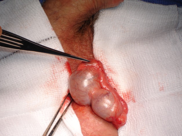
A supernumerary testis, sharing the same tunica albuginea and epididymis.
Treatment
Conservative or surgical excision.
Outcome and follow-up
No further follow-up required if no neoplasia has been confirmed on excisional biopsy.
Discussion
This is a rare anomaly, with only about 100 cases reported in the literatures. One published case has revealed the diagnosis can be made radiologically, hence conservative management.
Learning points.
-
▶
Polyorchidism is a rare anomaly. Four types of variants have been described according to its relation to epididymis and vas deferens.
-
▶
Diagnosis can confidently be made with tumour markers and radiological findings.
-
▶
Thorough history taking and clinical examination can avoid surgery.
Footnotes
Competing interests: None.
Patient consent: Obtained.
References
- 1.Kharrazi SMH, Rahmani MR, Sakipour M, et al. Polyorchidism: A Case Report and Review of Literature. Urol J (Tehran) 2006;3:180–3. [PubMed] [Google Scholar]