Abstract
Menopause in women occurs at mid-life. Chimpanzees, in contrast, continue to display cycles of menstrual bleeding and genital swelling, suggestive of ovulation, until near their maximum life span of about 60 years. Because ovulation was not confirmed hormonally, however, the age at which chimpanzees experience menopause has remained uncertain. In the present study, we provide hormonal data from urine samples collected from 30 female chimpanzees, of which 9 were old (>30 years), including 2 above the age of 50 years. Eight old chimpanzees showed clear endocrine evidence of ovulation, as well as cycles of genital swelling that correlated closely with measured endocrine changes. Endocrine evidence thus confirms prior observations (cyclic anogenital swelling) that menopause is a late-life event in the chimpanzee. We also unexpectedly discovered an idiopathic anovulation in some young and middle-aged chimpanzees; this merits further study. Because our results on old chimpanzees validate the use of anogenital swelling as a surrogate index of ovulation, we were able to combine data on swelling and urinary hormones to provide the first estimates of age-specific rates of menopause in chimpanzees. We conclude that menopause occurs near 50 years of age in chimpanzees as it does in women. Our finding identifies a basic difference between the human and chimpanzee aging processes: female chimpanzees can remain reproductively viable for a greater proportion of their life span than women. Thus, while menopause marks the end of the chimpanzee’s life span, women may thrive for decades more.
Keywords: Menopause, Sexual swelling, Reproduction, Aging, Estrogen, Progesterone
Introduction
Females of all primate species experience clinical menopause, defined as the permanent termination of ovulation (Walker and Herndon 2008). A question of greater interest to evolutionary biologists, however, is when in the life cycle menopause occurs. In other words: do any non-human primates (NHPs) undergo a human-like midlife menopause, or do they experience reproductive cessation late in life when other physiological systems are also failing? Because the chimpanzee is our closest living living evolutionary relative, the question of the timing of menopause in this species is of particular importance. Yet until recently, there has been relatively little information concerning its reproductive aging. One reason for this is the scarcity of specimens that live long enough to undergo the cessation of normal ovulatory cycles. An early study of chimpanzee reproductive aging (Graham 1979) monitored menstrual cycles by observing genital swelling and menstrual bleeding in 10 chimpanzees above the age of 35 years. These apes were studied until natural death occurred at ages between 38 and 48 years, which in 1979 was thought to represent the upper limit of age in captive chimpanzees. All of the chimpanzees in the study had “some evidence of reproductive cyclicity in the last year of life.” The 48-year-old subject, for example, died of a cerebral hemorrhage during the luteal phase of her cycle, as indicated by a well-developed corpus luteum in her ovary. In a subsequent study (Gould et al. 1981), ovarian and gonadotrophic hormone levels in two old (49 and 50 years) chimpanzees (Pan troglodytes) and one old (exact age unknown) bonobo (Pan paniscus) were studied. While the bonobo was menopausal, the two chimpanzees showed evidence of continued ovulation, albeit in an altered, perimenopause-like pattern. When findings of a workshop convened by the National Institute on Aging (NIA) were published more than two decades later, these two studies, comprising a total of 12 old chimpanzees, were found to be the only reports on chimpanzee menopause (Bellino and Wise 2003). The authors summarized the state of knowledge at that time by concluding that menstrual cycles continue until the end of natural life in this ape.
Three years after the NIA workshop, a new study of captive chimpanzees concluded that chimpanzees reach “perimenopause at 30 to 35y and menopause at 40y” (Videan et al. 2006). Although this study included only 3 animals above the age of 40 years and inferred menopause from serum hormone levels collected biannually, this new conclusion was adopted by the editors of a review volume on primate reproductive aging published 2 years later (Erwin and Hof 2008). The same data on early menopause were republished in that volume (Videan et al. 2008), giving the incorrect impression that evidence of early menopause in chimpanzees was continuing to accumulate.
Since 2006, however, a number of findings have confirmed Graham’s observation (Graham 1979) that menstrual cycles in chimpanzees can persist into very old age. In our analysis of archival records on 89 female chimpanzees, whose cycling and menstruation had been recorded as part of a breeding program, we found that menstrual cycles continued in the 20 apes that lived beyond the age of 39 years (Lacreuse et al. 2008). Three apes continued to cycle in their 6th decade, and menopause (defined as survival for at least 1 year in good health without menstrual cycles) was observed in only one female at about the age of 56 years. Our observations are consistent with recent data on production of live offspring in older female chimpanzees. For example, although fertility in wild chimpanzees declined steadily through adulthood, they gave birth as late as their 50th year—essentially up until the maximum life span in these populations (Nishida et al. 2003; Emery Thompson et al. 2007). Captive chimpanzees have also been documented to give birth to healthy infants at the advanced maternal age of 49 (Puschmann and Federer 2008) and 56 years (Ross 2009). Nevertheless, histological analysis of ovarian follicles in chimpanzees ranging in age from 0 to 47 years indicated an age-related ovarian decline similar to that seen in women (Jones et al. 2007). To date, however, no study has provided comprehensive evidence to support or reject the notion of menopause occurring in chimpanzees. In summary, many studies strongly suggest that chimpanzee fertility can continue into their 40s and 50s.
The present study was designed to further our knowledge of ovarian cyclicity across the life span by examining menstrual bleeding, genital swelling and urinary reproductive hormones in a group of chimpanzees representing the entire life span of this species. Measures of serum anti-Müllerian hormone (AMH), an index of ovarian reserve in other NHPs, were also obtained (Appt et al. 2009).
Materials and methods
Subjects and environment
Subjects were adult female chimpanzees (P. troglodytes) from the colony of the Yerkes National Primate Research Center (YNPRC) of Emory University; they comprised a subset of a group of 39 chimpanzees included in a comprehensive NIA-funded project examining several aspects of aging in this species. The apes were selected to provide a sample covering the adult age range of the species. In our colony, the maximum age attained by a chimpanzee was 59 years, and the mean survival of females is about 25 years (Herndon et al. 1999). The apes are housed in small groups of compatible individuals and are trained to cooperate with clinical examinations and scientific study. Housing, husbandry, and the experimental procedures described in this report are approved by the Institutional Animal Care and Use Committee of Emory University in accordance with the Animal Welfare Act and the US Department of Health and Human Services “Guide for the Care and Use of Laboratory Animals.” The YNPRC is accredited by the Association for Assessment and Accreditation of Laboratory Animal Care International. As a part of the normal care of the chimpanzees, they are given annual physical examinations, which include health assessments, collection of blood samples for hematology and clinical chemistry, and determinations of body weight.
Menstrual bleeding and genital swelling
External signs of ovarian cycles in chimpanzees, including anogenital swelling and menstrual bleeding, have been recorded for many years in our colony (Graham 1979). The magnitude of swelling is rated on a 5-point scale (0 to 4) and the presence of menstrual blood on the perineum of the animal or on cage surfaces is noted, as previously described (Lacreuse et al. 2008).
Urine collection
Urine samples were collected by means of non-invasive techniques from 30 chimpanzees, ranging in age from 13 to 54 years at the time of study. Briefly, chimpanzees were desensitized to a wand (a 1-in. diameter PVC pipe with a cap on one end and an elbow attached to the other end) that was used to collect urine. Subsequent to desensitization of the wand, positive reinforcement training was used to teach the chimpanzees to approach the front of their cage and provide urine on command. Urine samples were collected twice a week (on average) over the course of 2 to 3 months during the spring and summer of 2009 and 2010, and were immediately frozen at −20°C and stored until assayed. Although this sampling regimen may not detect the ovulatory peak in estradiol and gonadotropins, it can identify the luteal phase by elevation in urinary progestin levels and thus identify the follicular phase and provide an estimate of the day of ovulation (Kaplan et al. 2002; Pope et al. 1986; Shimizu et al. 2003; Wilson et al. 1982). Furthermore, in the absence of increases in urinary progestins, anovulatory periods can be inferred.
Blood collection and upper body mass index
Blood was collected from 30 chimpanzees for AMH assay from the inguinal vein following anesthesia with Telazol (5 mg/kg). These samples were collected opportunistically from chimpanzees during annual health examinations or brain imaging sessions (as part of another project). Because AMH is an index of ovarian stores, its levels are not influenced by the stage of the menstrual cycle. Blood samples were centrifuged (3,000 rpm for 15 min) and serum was frozen immediately at −20°C and stored until assayed. When possible, animals were also weighed and measured for calculation of UMBI (weight in kilograms divided by the square of the crown-to-rump length in meters).
Hormone assays
All urine samples were analyzed in duplicate for estrone-3-glucuronide (E1G; Immunometrics; London, UK) and pregnanediol-3α-glucuronide (PdG; Immunometrics; London, UK). Creatinine was measured by a colorimetric assay based on a modified Jaffee reaction. Frozen urine samples were thawed and centrifuged (3,000 rpm for 5 min). The samples were diluted according to the assay protocol provided by the manufacturer so that hormone concentrations of applied samples fell in the ranges of the standards provided by the assays. Briefly, the urine supernatant (20 μl) was diluted 1:100 in assay buffer for the E1G assay and diluted 1:200 in assay buffer for the PdG assay. To compensate for variations in urine volume and concentration, creatinine concentration was also measured in a subset of urine samples from each ape. Specifically, creatinine was measured in all samples with a significant elevation of E1G or PdG, as well as in a random subset of the samples with low and unvarying steroid levels. Creatinine levels from this random subset were used as estimates for those samples for which creatinine was not measured. In order to determine the urinary concentration of creatinine, 20 μl of urine was diluted 1:40 in deionized water and assayed according to the protocol provided by the assay manufacturer (Quidel Corporation; San Diego, CA).
The optical density of plastic cuvettes (VWR; Suwanee, GA) used to measure E1G and PdG were read from a Model 690 spectrophotometer (Turner; Mountain View, CA) set at 550 nm. E1G and PdG concentrations were determined by fitting the absorbance to the standard curve constructed with curve-fitting software (RIASMART; Packard Instrument Company). A MultiWash 3 automatic plate washer (Tricontinent; Green Valley, CA) was used to wash plates thoroughly with wash solution between each step during the creatinine assay. Plates were read on a SpectraMax 340 PC 384 (Molecular Devices; Sunnyvale, CA) set to read at 490 nm. The creatinine concentration was calculated by fitting the absorbance to the standard curve using SoftMax Pro v5.4 (Molecular Devices; Sunnyvale, CA). Urinary hormone concentrations are expressed as ng/mg of creatinine.
Serum samples were analyzed in duplicate for AMH (Beckman Coulter) by enzyme-linked immunosorbent assays according to the protocol provided by the manufacturer. On the day of the assay, frozen serum samples were thawed and centrifuged (3,000 rpm for 5 min) immediately prior to beginning the assay. The inter-assay and intra-assay coefficients of variation (CVs) are 14.0% and 8.9%, respectively, for the PdG assay, 15.53% and 5.63% for E1G, 4.52% and 0.8% for creatinine, and 8.47% and 4.45% for the AMH assay.
Data analysis
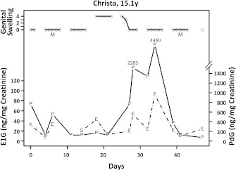
Each chimpanzee’s urinary data for E1G and PdG (corrected for creatinine levels), genital swelling, and observed menses for a period of about 50 days were plotted in the form shown in Fig. 1. PdG levels greater that 1,000 ng/mg creatinine were taken as definitive evidence of ovulation (Graham et al. 1972; Shimizu 2005). This increase corresponds to the mid-cycle decline on perineal swelling and typically continues to rise before declining to levels less than 1,000 ng/mg creatinine typical of the follicular phase or anovulatory periods. Each chimpanzee’s record was evaluated to determine whether ovulation occurred, and whether there were cyclic patterns of genital swelling and menstrual bleeding.
Fig. 1.
Urinary E1G (E, broken line) and PdG (P, solid line) over a 50-day period of observation of a young, ovulating chimpanzee (Christa). Genital swelling (circle) is depicted near the top of the graph and is based upon a 5-point scale. A solid line connects observations on consecutive days. Menstrual bleeding (M) is indicated when observed
We used Fisher’s exact test to compare frequencies of ovulation between groups, and analysis of variance followed by t tests to compare body weight and upper body mass index among the groups. Tukey’s t test was used to correct for the underestimation of alpha as a result of multiple comparisons. The variance of AMH levels was found to correlate with age. This correlation was removed by log-transforming the AMH levels. The relationship between log-transformed AMH and age was examined by means of the Pearson product–moment correlation. Logistic regression analyses were used to model the log-odds of ovulation vs. non-ovulation (expressed as a binary variable) as a function of several variables that were judged to be potential predictors of ovulation.
Standard methods of survival analysis were employed to produce Kaplan–Meier estimates and standard errors of age-specific prevalence of menopause (Lee 1992).
Results
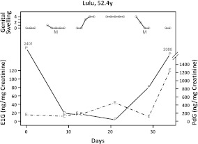
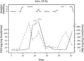
Several different patterns of cyclicity were observed. Figure 1 depicts one menstrual cycle from a young female (Christa, 15.1 years) and shows characteristics that may be considered typical of the species. The circles near the top of the chart show genital swelling, rated on a 5-point scale; a solid line connects consecutive days of observation. Swelling is rated at 0 for Days 0 to 1, and remains at 0 through Day 15, with two interruptions of observation during weekends. Swelling then rises to a maximal level of 4, where it remains until Day 25 of the cycle. The “M’s” show the days of observed menstrual bleeding. The left and right vertical axes correspond to E1G and PdG levels, as depicted in the lower two traces. The PdG peak, indicative of ovulation, occurs just after the detumescence of the genital area. Figure 2 displays a very similar pattern of swelling, menstruation and hormone levels in an old chimpanzee (Lulu, 52.4 years). Figure 3 depicts another hormone cycle from the same old female, about 1 year later. Monthly patterns of genital swelling appeared regularly throughout the intervening months. Note, however, that menstrual bleeding was not detectible (“M’s” are absent) in this second cycle, despite clear hormonal evidence of ovulation. Menstrual bleeding was, however, evident in some subsequent cycles for which urine samples were not collected.
Fig. 2.
An old, ovulating chimpanzee (Lulu). For abbreviations, see Fig. 1
Fig. 3.
An old, ovulating chimpanzee (Lulu), about 1 year after previous figure. For abbreviations, see Fig. 1
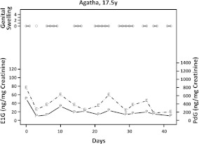
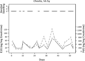
Figure 4 displays a 42-day period in another young female (Agatha, 17.5 years). There is no evidence of cyclic swelling or menses; the hormonal profile clearly shows that this ape did not ovulate. The absence of ovarian activity in this apparently healthy young female was unexpected (see below). Figure 5 shows a similar infertile cycle in an old chimpanzee (Cheeta, 53.5 years).
Fig. 4.
A young, non-ovulating chimpanzee (Agatha). For abbreviations, see Fig. 1
Fig. 5.
An old, non-ovulating chimpanzee (Cheeta). For abbreviations, see Fig. 1
The basic patterns of hormonal activity in young, middle-aged, and old chimpanzees are listed in Table 1. This table also indicates whether a cyclic pattern of genital swelling was observed and provides AMH levels. While AMH showed a tendency to decline with age, this tendency did not reach statistical significance.
Table 1.
The 30 chimpanzees studied
| Chimpanzee | Age | OV | SWELLa | AMHb | ||
|---|---|---|---|---|---|---|
| Young | n = 11 | Samantha | 12.9 | yes | yes | 0.160 |
| Azalea | 13.4 | no | irr | 0.725 | ||
| Christa | 15.1 | yes | yes | 0.373 | ||
| Jamie | 15.2 | yes | yes | 0.740 | ||
| Abby | 15.2 | no | irr | 1.651 | ||
| Fiona | 15.6 | no | irr | 1.301 | ||
| Julie | 15.9 | no | irr | 0.160 | ||
| Liza | 16.3 | no | irr | 0.160 | ||
| Sylvia | 17.0 | no | no | 1.769 | ||
| Agatha | 17.5 | no | no | 0.734 | ||
| Frannie | 17.7 | yes | yes | 0.160 | ||
| Average | 15.6 | 36% c | 82% | 0.721 | ||
| SEM | 0.5 | 15% | 12% | 0.183 | ||
| Middle aged | n = 10 | Jewelle | 18.4 | yes | yes | 0.340 |
| Evelyne | 18.9 | no | no | 0.160 | ||
| Edwina | 19.1 | no | yes | – | ||
| Rebecca | 19.2 | no | yes | 0.160 | ||
| Callie | 19.4 | yes | yes | 0.275 | ||
| Elvira | 21.7 | yes | yes | 0.506 | ||
| Rowena | 21.9 | yes | yes | 0.160 | ||
| Suwannee | 25.4 | yes | yes | 0.357 | ||
| Shirley | 25.5 | yes | no | 1.994 | ||
| Brodie | 26.5 | yes | yes | 0.861 | ||
| Average | 21.6 | 70% c | 80% | 0.535 | ||
| SEM | 1.0 | 15% | 13% | 0.187 | ||
| Lena | 33.1 | yes | yes | – | ||
| Old | n = 9 | Cissie | 33.6 | yes | yes | 0.160 |
| Jacqueline | 33.7 | yes | yes | 0.694 | ||
| Marietta | 37.0 | yes | yes | 0.669 | ||
| Barbara | 37.6 | yes | yes | 0.541 | ||
| Lil’One | 41.5 | yes | yes | 0.160 | ||
| Foxy | 42.6 | yes | yes | 0.160 | ||
| Lulu | 53.5 | yes | yes | 0.160 | ||
| Cheeta | 53.5 | no | no | 0.160 | ||
| Average | 40.6 | 89%c | 89% | 0.338 | ||
| SEM | 2.7 | 11% | 11% | 0.083 |
Shown are age (years), presence or absence of ovulation (OV) based on cyclic changes in urinary PdG, genital swelling (SWELL), and serum level of Anti-Müllerian Hormone (AMH ng/mL, a putative indicator of ovarian store). Averages and standard error of the mean (SEM) are shown for each age group
ayes means cyclic swelling; no means no swelling; irr means irregular swelling
bLower detection limit was 0.16 mg/ml
c% of animals ovulating in the Old group is significantly higher than the % in the Young group. The Middle-aged group does not differ from the other two
Table 1 shows that that only 4/11 (36%) of the young chimpanzees ovulated, as indicated by levels of PdG. The idiopathic anovulation seen in 7/11 young chimpanzees is described in more detail below. Of the middle-aged chimpanzees 7/10 (70%) ovulated, as did 8/9 (88%) of the old apes.
Age-specific prevalence of menopause in chimpanzees over 40 years of age
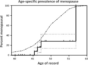
The perfect correlation of anogenital swelling with ovulatory levels of urinary PdG within the old age group validates our prior assumption that continued cyclic genital swelling in aged apes is indicative of continued ovulation. Therefore we examined the swelling records of the 20 chimpanzees from our previous study (Lacreuse et al. 2008) that were 40 years or older to determine whether any had converted to menopausal status in the interval, as defined by the absence of ovulation for a period of 12 months (Burger 1999). We used the definition of menopause proposed for women by Burger (1999); i.e., 12 months without menstrual cycles. While the application of the 12-month criterion to chimpanzees is somewhat arbitrary, we chose it rather than the average interbirth interval, used by others (e.g., Nishida et al. 2003) because the latter criterion does not seem appropriate for captive apes with controlled breeding. Table 2 lists the chimpanzees included in this extended analysis, and indicates the name of each chimpanzee and “age of record,” and whether the chimpanzee exhibited menopause (“1”) or not (“0”). The age of record in the study is defined as the latest age at which each chimpanzee was at risk for menopause (i.e., was not yet menopausal). Age of record was thus the age of menopause or, if menopause did not occur, the age at death. The data in Table 2 are represented as a Kaplan–Meier plot showing the age-specific prevalence of menopause in Fig. 6. For comparison, the percentage of women past their final menstrual cycle is presented as a dashed line, which represents the graphically determined median of the 4 populations of women presented by Hawkes et al. (2009). In the terminology of survival analysis, menopause is considered the event of interest, and data from animals that died before menopause were considered “right-censored.” That is, we know that they cycled at least as long as the age of record, but we cannot determine when or if they would have become menopausal. From the plot of these data in Fig. 6, it can be seen that no chimpanzee was menopausal before 45 years, and about 16% (±10%) of chimpanzees were menopausal by 46 years, and 28% (±14%) were menopausal by 47 years. The Kaplan–Meier curve reaches a level of 100% at 57 years of age when the oldest ape studied became menopausal. While these percentages are numerically lower than the percentages of women that are menopausal at equivalent ages, the human average falls within the 95% confidence interval of the chimpanzee curve (not shown in Fig. 6).
Table 2.
Summary of cycling history of female chimpanzees above the age of 40 years in the present study and in that of Lacreuse et al. 2008
| Chimpanzee | Age of record | Menopausal status | Comments |
|---|---|---|---|
| Pati | 40.00 | 0 | Age at last pregnancy |
| Vera | 40.95 | 0 | Age at death |
| Lil’Onea | 41.50 | 0 | Age at last observation |
| Jeanie | 41.95 | 0 | Age at death |
| Wenka | 42.00 | 0 | Age at the time of diagnosis of enlarged uterus |
| Foxya | 42.60 | 0 | Age at last observation |
| Reba | 44.16 | 0 | Age at death |
| Beleka | 44.21 | 0 | Age at death |
| Garbo | 45.30 | 0 | Age at which observations ceased (animal was donated to another facility) |
| Anna | 45.32 | 0 | Age at death |
| Jenda | 45.37 | 1 | Age at last anogenital swelling cycle |
| Sparkle | 45.89 | 0 | Age at death |
| Martha | 46.23 | 1 | Age at last anogenital swelling |
| Banana | 46.84 | 0 | Age at death |
| Soda | 47.04 | 0 | Age at death |
| Boka | 47.05 | 0 | Age at last observation |
| Cheetaa | 47.24 | 1 | Age at last anogenital swelling cycle |
| Wendy | 48.36 | 0 | Age at death |
| Cheri | 48.67 | 0 | Age at death |
| Lulua | 53.54 | 0 | Age at last observation |
| Flora | 55.28 | 0 | Age at death |
| Bula | 56.07 | 0 | Age at death |
| Gamma | 56.94 | 1 | Age at last anogenital swelling cycle |
Age of record indicates age at last anogenital swelling or age last observed. A status of “1” indicates that the chimpanzee was menopausal at age of record; “0” indicates that animal was not menopausal.
aOvulation confirmed via urinary hormone analysis or that menopausal status was inferred by persistently low levels of hormonal metabolites normally associated with ovulation
Fig. 6.
The solid line is a Kaplan–Meier (K-M) plot of the age-specific prevalence of menopause in 24 chimpanzees above the age of 40 years. The horizontal dotted lines show the standard error of the K-M estimate. The curved dashed line shows the prevalence of menopause for women, drawn after Hawkes et al. (2009). Chimpanzees include those in the present study as well as those in Lacreuse et al. (2008a)
Idiopathic anovulation in young chimpanzees
While the absence of ovulation in one old chimpanzee (Cheeta, 53.5 years) appears to be explained by menopause, the anovulation of 7 young and 3 middle-aged chimpanzees must be considered to be abnormal. The idiopathic nature of this phenomenon is emphasized by the finding that the old animals were actually more likely to display ovulation than were the young (Fisher’s exact test, p = 0.03). An analysis of this apparent infertility is beyond the scope of this paper and will be presented in a subsequent report. The high prevalence of this phenomenon underscores the importance of verifying prior reproductive viability of chimpanzee subjects in future studies of menopause.
Discussion
Here we report that 8 of 9 old female chimpanzees (33.1 to 53.5 years) continue to display ovarian cycles characterized by cyclic anogenital swelling and, in most cases, marked by visible menses. Urinary levels of PdG, a urinary progestin, confirmed the occurrence of ovulation in each of these females, including 3 beyond the age of 40 years. One chimpanzee in this age group, Cheeta, had no ovulatory cycles. Cheeta, previously reported to have regular cycles up to the age of 48 years (Lacreuse et al. 2008), had no apparent health problems and thus meets the definition of menopause which requires the absence of cycles for at least 12 months, in the absence of any underlying pathology (Burger 1999). The present findings are consistent with several previous reports stating that reproductive cycles continue in the chimpanzee into the 5th and 6th decade of life (Gould et al. 1981; Graham 1979; Lacreuse et al. 2008) but contradict reports suggesting that menopause occurs by 40 years of age (Atsalis and Videan 2009; Videan et al. 2006, 2008).
AMH showed a trend toward decline with age. Although this trend did not reach statistical significance in the present analysis, studies of this hormone in humans (Ledger 2010; Shebl et al. 2011) and cynomolgus monkeys (Appt et al. 2009) suggest that serum levels of AMH are a good index of the number of oocytes present in the ovary. Further study with a larger number of chimpanzees may confirm this pattern in these apes as well. Table 1 shows that AMH levels below the limit of detection are not inconsistent with confirmed ovulation. Indeed, of the 12 apes with undetectable AMH, 8 ovulated, including 5 in the aged group. This is consistent with the recent finding that AMH levels do not predict the chances of pregnancy in women (Tremellen and Kolo 2010).
One argument for the idea that menopause occurs 10 to 15 years before the disappearance of anogenital swelling cycles is that continuation of cycles of anogenital swelling may not be an accurate gauge of reproductive status and may instead serve to maintain affiliative relationships within the social group (Videan et al. 2006). The present results refute this claim by revealing that swelling cycles occurred in all eight of the old chimpanzees that ovulated but did not occur in the single old animal that did not ovulate. Indeed, anogenital swelling has an underlying physiology precisely regulated by fluctuations in ovarian steroids (Dahl et al. 1991; Deschner et al. 2004; Emery Thompson 2005; Graham et al. 1972; McArthur et al. 1981; Nadler et al. 1985). This high degree of association between anogenital swelling and ovulation reinforces our previous conclusion that ovulation in chimpanzees continues into advanced old age.
Because there are only a few hundred aged chimpanzees available for study (Erwin et al. 2002), conclusions about their biology often rely on small samples. But given that we have now observed menstruation beyond 40 years of age in 100% of the 25 individual chimpanzees in this age category assessed in the current study and in previous work (Lacreuse et al. 2008), we are confident in our claim that menopause typically occurs well beyond the age of 40 years. Even the two chimpanzees that eventually became menopausal (8% of those observed) continued cycling until advanced ages. One of these, Gamma, ceased cycling at about 57 years and was the only confirmed case of menopause in our prior report. Cheeta ceased cycling at 48 years but was not considered menopausal because at that time she did not meet the criterion of a 12-month interval without menstrual cycles; she has since met this criterion. Our findings on reproductive aging in captivity are entirely consistent with the report that chimpanzees in the wild continue to produce viable offspring throughout the life span, into the 6th decade of life, although fertility declines after about 30 years of age (Emery Thompson et al. 2007). Live births have occurred at the advanced maternal ages of 42, 49, and 56 years, respectively, in our colony at the YNPRC (in 1997, unpublished observations), at a zoo in Basel, Switzerland (Puschmann and Federer 2008), and at a zoo in Manhattan, Kansas, USA (Ross 2009).
An unexpected finding of the present study was the absence of ovulation in 7 of 11 (64%) young and 3 of 10 (30%) middle-aged chimpanzees. Although this pattern was not seen in old animals, the phenomenon has important implications for studies of menopause in that it emphasizes the need for longitudinal studies. Future cross-sectional studies of aged chimpanzees could incorrectly conclude that any non-ovulating subjects are menopausal unless their ovulatory history at earlier ages is carefully considered. We are unaware of any other report of spontaneous anovulation in non-aged chimpanzees and have not previously observed this phenomenon in our own colony. Although idiopathic infertility is seen in women, the rates are lower (8.4% between 14 and 24 years, and 14.1% between 25 and 35 years; Chandra et al. 2005) than those we observed. We plan a separate publication in which we will present a detailed analysis of this phenomenon.
The primary purpose of this study was to measure urinary metabolites of ovarian hormones to determine whether ovarian cycles and ovulation continue into old age in chimpanzees, or whether they terminate in the mid to late 40s, as has been suggested by a recent study based primarily upon evaluation of baseline gonadotrophin levels (Videan et al. 2006; Videan et al. 2008). That study relied upon an FSH-based criterion for menopause that was derived from human data and not yet validated in chimpanzees. The present study, in contrast, monitored ovarian cycles by means of urinary PdG level, a direct endocrine product of ovulation. Our finding identifies a basic difference between the human and chimpanzee aging process: female chimpanzees can remain reproductively viable for a greater proportion of their life span than women. By providing endocrine confirmation of Graham’s conclusion that “[r]eproductive cyclicity continues in the chimpanzee to approximately the same absolute age as in women” (Graham 1979), we lend support to the proposition that the age at menopause has not changed significantly since chimpanzees and humans diverged from a common ancestor and that longevity beyond reproductive age evolved in the human line without a change in the age at menopause. One possible mechanism for increased human longevity is the “grandmother effect,” as proposed by Hawkes (2003). The grandmother hypothesis has important implications for understanding human aging, as it identifies the human aging phenotype as a derived trait (Herndon 2010). Many argue that life beyond menopause in women is a relatively modern phenomenon, because average life expectancy has risen above the age of 50 years only in the past century or so. This is not the case, however. The fact that women in hunter–gatherer societies have been documented to live past the age of menopause suggests that post-reproductive infertility was common in ancestral humans as well (Hawkes and Smith 2010). Furthermore, although there have been increases in human life expectancy for all ages (e.g., Finch and Crimmins 2004), Oeppen and Vaupel note that until the mid-twentieth century, “most of the gain in life expectancy was due to large reductions in death rates at younger ages” (Oeppen and Vaupel 2002).
Because the chimpanzee is our closest biological relative, it is of great importance to understand the biological similarities and differences between the two species. The chimpanzee life history follows a similar course to our own until the age of menopause. The chimpanzee’s life ends as reproductive function ends, while a woman may thrive for many more years.
Acknowledgments
We thank Kimberly Neu, Caroline Griffis, and Andrea Franklin for recording daily observations of anogenital swelling and for urine collection, Doris Jane Langford for the assistance with manuscript preparation, Drs. Agnès Lacreuse and Johannes Tigges for comments on the manuscript, and the Animal Resources Division of the Yerkes National Primate Research Center of Emory University for its assistance in the extraction of data for this study. This study was supported through the following grants: NIH grants P51RR000165 and P01AG026423.
References
- Appt SE, Clarkson TB, Chen H, Adams MR, Christian PJ, Hoyer PB, Wilson ME, Kaplan JR. Serum antimüllerian hormone predicts ovarian reserve in a monkey model. Menopause. 2009;16(3):597–601. doi: 10.1097/gme.0b013e3181906fb6. [DOI] [PMC free article] [PubMed] [Google Scholar]
- Atsalis S, Videan E. Reproductive aging in captive and wild common chimpanzees: factors influencing the rate of follicular depletion. Am J Primatol. 2009;71(4):271–282. doi: 10.1002/ajp.20650. [DOI] [PubMed] [Google Scholar]
- Bellino FL, Wise PM. Nonhuman primate models of menopause workshop. Biol Reprod. 2003;68(1):10–18. doi: 10.1095/biolreprod.102.005215. [DOI] [PubMed] [Google Scholar]
- Burger HG. The endocrinology of the menopause. J Steroid Biochem Mol Biol. 1999;69(1–6):31–35. doi: 10.1016/S0960-0760(98)00145-9. [DOI] [PubMed] [Google Scholar]
- Chandra A, Martinez GM, Mosher WD, Abma JC, Jones J (2005) Fertility, family planning, and reproductive health of U.S. women: data from the 2002 National Survey of Family Growth. National Center for Health Statistics. Vital Health Stat 23(25):1–160 [PubMed]
- Dahl J, Nadler RD, Collins DC. Monitoring the ovarian cycles of Pan troglodytes and P. paniscus: a comparative approach. Am J Primatol. 1991;24:195–209. doi: 10.1002/ajp.1350240306. [DOI] [PubMed] [Google Scholar]
- Deschner T, Heistermann M, Hodges K, Boesch C. Female sexual swelling size, timing of ovulation, and male behavior in wild West African chimpanzees. Horm Behav. 2004;46(2):204–215. doi: 10.1016/j.yhbeh.2004.03.013. [DOI] [PubMed] [Google Scholar]
- Emery Thompson M. Reproductive endocrinology of wild female chimpanzees (Pan troglodytes schweinfurthii): methodological considerations and the role of hormones in sex and conception. Am J Primatol. 2005;67:137–158. doi: 10.1002/ajp.20174. [DOI] [PubMed] [Google Scholar]
- Emery Thompson M, Jones JH, Pusey AE, Brewer-Marsden S, Goodall J, Marsden D, Matsuzawa T, Nishida T, Reynolds V, Sugiyama Y, Wrangham RW. Aging and fertility patterns in wild chimpanzees provide insights into the evolution of menopause. Curr Biol. 2007;17:2150–2156. doi: 10.1016/j.cub.2007.11.033. [DOI] [PMC free article] [PubMed] [Google Scholar]
- Erwin JM, Hof PR. Menopause and reproductive senescence in comparative context. In: Atsalis S, Margulis S, Hof P, editors. Primate reproductive aging: cross-taxon perspectives. Interdisciplinary top gerontology. Basel, Switzerland: Karger; 2008. pp. 4–16. [Google Scholar]
- Erwin JM, Hof PR, Ely JJ, Perl DP. One gerontology: advancing understanding of aging through studies of great apes and other primates. In: Erwin JM, Hof PR, editors. Aging in nonhuman primates. Interdiscipl Top Gerontol. Basel: Karger; 2002. pp. 1–21. [Google Scholar]
- Finch CE, Crimmins EM. Inflammatory exposure and historical changes in human life-spans. Science. 2004;305(5691):1736–9. doi: 10.1126/science.1092556. [DOI] [PubMed] [Google Scholar]
- Gould KG, Flint M, Graham CE. Chimpanzee reproductive senescence: a possible model for evolution of the menopause. Maturitas. 1981;3(2):157–166. doi: 10.1016/0378-5122(81)90007-4. [DOI] [PubMed] [Google Scholar]
- Graham CE. Reproductive function in aged female chimpanzees. Am J Phys Anthropol. 1979;50:291–300. doi: 10.1002/ajpa.1330500302. [DOI] [PubMed] [Google Scholar]
- Graham CE, Collins DC, Robinson H, Preedy JR. Urinary levels of estrogens and pregnanediol and plasma levels of progesterone during the menstrual cycle of the chimpanzee; relationship to the sexual swelling. Endocrinology. 1972;91(1):13–24. doi: 10.1210/endo-91-1-13. [DOI] [PubMed] [Google Scholar]
- Hawkes K. Grandmothers and the evolution of human longevity. Am J Hum Biol. 2003;15(3):380–400. doi: 10.1002/ajhb.10156. [DOI] [PubMed] [Google Scholar]
- Hawkes K, Smith KR. Do women stop early? Similarities in fertility decline in humans and chimpanzees. Ann NY Acad Sci. 2010;1204:43–53. doi: 10.1111/j.1749-6632.2010.05527.x. [DOI] [PMC free article] [PubMed] [Google Scholar]
- Hawkes K, Smith KR, Robson SL. Mortality and fertility rates in humans and chimpanzees: how within-species variation complicates cross-species comparisons. Am J Hum Biol. 2009;21:578–586. doi: 10.1002/ajhb.20890. [DOI] [PMC free article] [PubMed] [Google Scholar]
- Herndon JG. The grandmother effect: implications for studies on aging and cognition. Gerontology. 2010;56(1):73–79. doi: 10.1159/000236045. [DOI] [PMC free article] [PubMed] [Google Scholar]
- Herndon JG, Tigges J, Anderson DC, Klumpp SA, McClure HM. Brain weight throughout the life span of the chimpanzee. J Comp Neurol. 1999;409(4):567–572. doi: 10.1002/(SICI)1096-9861(19990712)409:4<567::AID-CNE4>3.0.CO;2-J. [DOI] [PubMed] [Google Scholar]
- Jones K, Walker L, Anderson DC, Lacreuse A, Robson S, Hawkes K. Depletion of ovarian follicles with age in chimpanzees: similarities to humans. Biol Reprod. 2007;77(2):247–251. doi: 10.1095/biolreprod.106.059634. [DOI] [PubMed] [Google Scholar]
- Kaplan JR, Manuck SB, Anthony MS, Clarkson TB. Premenopausal social status and hormone exposure predict postmenopausal atherosclerosis in female monkeys. Obstet Gynecol. 2002;99(3):381–388. doi: 10.1016/S0029-7844(01)01659-3. [DOI] [PubMed] [Google Scholar]
- Lacreuse A, Chennareddi L, Gould K, Hawkes K, Wijayawardana S, Chen J, Easley K, Herndon JG. Menstrual cycles continue into advanced old age in the common chimpanzee (Pan troglodytes) Biol Reprod. 2008;79(3):407–412. doi: 10.1095/biolreprod.108.068494. [DOI] [PMC free article] [PubMed] [Google Scholar]
- Ledger WL. Clinical utility of measurement of anti-Müllerian hormone in reproductive endocrinology. J Clin Endocrinol Metab. 2010;95(12):5144–5154. doi: 10.1210/jc.2010-0701. [DOI] [PubMed] [Google Scholar]
- Lee ET. Statistical methods for survival data analysis. New York: Wiley; 1992. [Google Scholar]
- McArthur JW, Collins DC, Preedy JRK, Graham CE. The interrelationships between sex skin swelling and the urinary excretion of LH, estrone, and pregnanediol by the cycling female chimpanzee. Am J Primatol. 1981;1:265–279. doi: 10.1002/ajp.1350010303. [DOI] [PubMed] [Google Scholar]
- Nadler RD, Graham CE, Gosselin R, Collins DC. Serum levels of gonadotropins and gonadal steroids, including testosterone, during the menstrual cycle of the chimpanzee (Pan troglodytes) Am J Primatol. 1985;9:273–284. doi: 10.1002/ajp.1350090404. [DOI] [PubMed] [Google Scholar]
- Nishida T, Corp N, Hamai M, Hasegawa T, Hiraiwa-Hasegawa M, Hosaka K, Hunt KD, Itoh N, Kawanaka K, Matsumoto-Oda A, Mitani JC, Nakamura M, Nori-koshi K, Sakamaki T, Turner L, Uehara S, Zamma K. Demography, female life history, and reproductive profiles among the chimpanzees of Mahale. Am J Primatol. 2003;59:99–121. doi: 10.1002/ajp.10068. [DOI] [PubMed] [Google Scholar]
- Oeppen J, Vaupel JW. Demography. Broken limits to life expectancy. Science. 2002;296(5570):1029–1031. doi: 10.1126/science.1069675. [DOI] [PubMed] [Google Scholar]
- Pope NS, Gordon TP, Wilson ME. Age, social rank and lactational status influence ovulatory patterns in seasonally breeding rhesus monkeys. Biol Reprod. 1986;35(2):353–359. doi: 10.1095/biolreprod35.2.353. [DOI] [PubMed] [Google Scholar]
- Puschmann W, Federer E. Ein neuer Fertilitätsnachweis bei einer hoch betagten Schimpansin und Anmerkungen zum Höchstalter von Pan troglodytes. Der zoologische Garten. 2008;77:182–185. doi: 10.1016/j.zoolgart.2007.10.007. [DOI] [Google Scholar]
- Ross SR. North American Regional Chimpanzee Studbook (Pan troglodytes) Chicago, IL, USA: Lincoln Park Zoo; 2009. [Google Scholar]
- Shebl O, Ebner T, Sir A, Schreier-Lechner E, Mayer RB, Tews G, Sommergruber M. Age-related distribution of basal serum AMH level in women of reproductive age and a presumably healthy cohort. Fertil Steril. 2011;95(2):832–834. doi: 10.1016/j.fertnstert.2010.09.012. [DOI] [PubMed] [Google Scholar]
- Shimizu K. Studies on reproductive endocrinology in non-human primates: application of non-invasive methods. J Reprod Dev. 2005;51(1):1–13. doi: 10.1262/jrd.51.1. [DOI] [PubMed] [Google Scholar]
- Shimizu K, Douke C, Fujita S, Matsuzawa T, Tomonaga M, Tanaka M, Matsubayashi K, Hayashi K. Urinary steroids, FSH and CG measurements for monitoring the ovarian cycle and pregnancy in the chimpanzee. J Med Primatol. 2003;32:15–22. doi: 10.1034/j.1600-0684.2003.01039.x. [DOI] [PubMed] [Google Scholar]
- Tremellen K, Kolo M. Serum anti-Mullerian hormone is a useful measure of quantitative ovarian reserve but does not predict the chances of live-birth pregnancy. Aust N Z J Obstet Gynaecol. 2010;50(6):568–572. doi: 10.1111/j.1479-828X.2010.01232.x. [DOI] [PubMed] [Google Scholar]
- Videan E, Fritz J, Heward CB, Murphy J. The effects of aging on hormone and reproductive cycles in female chimpanzees (Pan troglodytes) Com Med. 2006;56(4):291–299. [PubMed] [Google Scholar]
- Videan E, Fritz J, Heward CB, Murphy J. Reproductive aging in female chimpanzees (Pan troglodytes) Interdiscip Top Gerontol. 2008;36:103–118. doi: 10.1159/000137688. [DOI] [PubMed] [Google Scholar]
- Walker ML, Herndon JG. Menopause in nonhuman primates? Biol Reprod. 2008;79(3):398–406. doi: 10.1095/biolreprod.108.068536. [DOI] [PMC free article] [PubMed] [Google Scholar]
- Wilson ME, Gordon TP, Collins DC. Serum 17 beta-estradiol and progesterone associated with mating behavior during early pregnancy in female rhesus monkeys. Horm Behav. 1982;16(1):94–106. doi: 10.1016/0018-506X(82)90010-1. [DOI] [PubMed] [Google Scholar]


