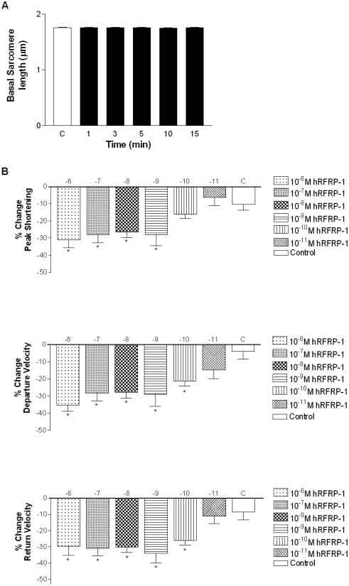
Fig. 1.
Basal sarcomere length and percent change in peak shortening, departure velocity, and return velocity in response to 15 minute perfusion with 10−6 M to 10−11 M hRFRP-1 at 37°C and paced at 0.2 Hz in isolated adult rat cardiac myocytes (Table 1). Fig. 1A. The resting sarcomere lengths were 1.76 ± 0.01 μm (baseline), 1.76 ± 0.01 μm (1 minute), 1.76 ± 0.01 μm (3 minutes), 1.76 ± 0.01 μm (5 minutes), 1.75 ± 0.01 μm (10 minutes), and 1.75 ± 0.01 μm (15 minutes). Fig. 1B. Percent change in peak shortening, departure velocity, and return velocity during 15 minutes perfusion with peptide. Values for each concentration (y-axis; hRFRP-1 (log [ ])) were compared to media control (C) with 1-way ANOVA followed by a Dunnett's Multiple Comparison Test with p < 0.05 considered statistically significant (*; Table 1). The best-fit EC50 values were calculated to be 5×10⁻¹⁰ M (peak shortening), 5×10⁻¹¹ M (departure velocity), and 5×10⁻¹¹M (return velocity). Recordings were made from n = 7-20, 1-day and 2-day myocytes isolated from n = 1-4 hearts.

