Abstract
Myelosuppression and drug resistance are common adverse effects in cancer patients with chemotherapy, and those severely limit the therapeutic efficacy and lead treatment failure. It is unclear by which cellular mechanism anticancer drugs suppress bone marrow, while drug-resistant tumors survive. We report that due to the difference of glucosylceramide synthase (GCS), catalyzing ceramide glycosylation, doxorubicin (Dox) eliminates bone marrow stem cells (BMSCs) and expands breast cancer stem cells (BCSCs). It was found that Dox decreased the numbers of BMSCs (ABCG2+) and the sphere formation in a dose-dependent fashion in isolated bone marrow cells. In tumor-bearing mice, Dox treatments (5 mg/kg, 6 days) decreased the numbers of BMSCs and white blood cells; conversely, those treatments increased the numbers of BCSCs (CD24−/CD44+/ESA+) more than threefold in the same mice. Furthermore, therapeutic-dose of Dox (1 mg/kg/week, 42 days) decreased the numbers of BMSCs while it increased BCSCs in vivo. Breast cancer cells, rather than bone marrow cells, highly expressed GCS, which was induced by Dox and correlated with BCSC pluripotency. These results indicate that Dox may have opposite effects, suppressing BMSCs versus expanding BCSCs, and GCS is one determinant of the differentiated responsiveness of bone marrow and cancer cells.
Keywords: myelosuppression, doxorubicin, glucosylceramide synthase, breast cancer stem cells, bone marrow
1. Introduction
Most chemotherapeutic agents target proliferative cells, which include cancer cells as well as normal adult stem cells in regenerating tissues such as bone marrow (Wang et al., 2006, Nurgalieva et al., 2010). Thus, these anti-proliferative effects, without tumor specificity, unavoidably cause myelosuppression or bone marrow suppression, while anticancer agents eradicate tumors. Myelosuppression is a dose-limiting toxicity for most chemotherapeutic regents (Repetto, 2009, Nurgalieva et al., 2010, Daniel and Crawford, 2006). The risk of aplastic anemia, a common syndrome of myelosuppression is strongly associated with regimens of cyclophosphamide/doxorubicin/fluorouracil, platinum/taxane, and cyclophosphamide/methotrexate/fluorouracil (Nurgalieva et al., 2010). There is a dose-response relationship between the use of taxane or platinum and myelosuppression, and the increasing myelosuppression risk is consistently higher in patients using alkylating agents or anthracycline-based regimens (Nurgalieva et al., 2010, Kroger et al., 1999). Drug-induced myelosuppression not only limits the treatments with cytostatic agents, but also is a risk factor for poor prognosis, as it substantially diminishes immunity and other systems against malignancy (Richardson and Johnson, 1997, Busch et al., 1990, Nurgalieva et al., 2010). Bone marrow transplantation and the supplements of erythropoietin (Epoetin), granulocyte colony-stimulating factor (G-CSF, Neupogen) and interleukin-11 (Oprelvekin) have been demonstrated recovering bone marrow and significantly improvement chemotherapy outcome (Bartsch and Steger, 2009, Seidman, 2006, Moore and Crom, 2006, Janni et al., 2001, Timmer-Bonte et al., 2005, Hood, 2003, Carey, 2003). An understanding of cellular mechanisms underlying drug-induced myelosuppression may guide development of chemotherapeutics with high efficacy and little bone marrow toxicity.
Chemotherapeutic agents can induce drug-resistant tumors, which may result in treatment failure (Gonzalez-Angulo et al., 2007, Szakacs et al., 2006). Many anticancer drugs including anthracyclines can amplify the expression of multidrug resistance 1 (MDR1), breast cancer resistant protein (BCRP), Bcl-2 and glucosylceramide synthase (GCS) to result in drug resistance in cancer cells (Clarke et al., 1992, Doyle et al., 1998, Gasparini et al., 1995, Liu et al., 1999, Liu et al., 2008). Emerging evidence supports that cancer stem cells (CSCs) might be an important mechanism of drug resistance. Chemotherapy kills most differentiated cancer cells in tumors, but surviving CSCs may cause disseminated metastasis or recurrence of aggressive tumors after treatment failure (Dean et al., 2005, Maugeri-Sacca et al., 2011). Recent studies show that a prolonged selection with doxorubicin (Dox) enriches CSCs in breast cancer cells (Calcagno et al., 2010); the CSCs detected as side population by flow cytometry are increased in paclitaxel-resistant ovarian cancer cell lines (Kobayashi et al., 2011). An increase of BCSCs after systemic chemotherapy is a poor prognostic factor for patients with breast cancers (Lee et al., 2011).
The present study simultaneously assessed the effects of Dox on bone marrow and CSCs, probing cellular mechanisms underlying myelosuppression and drug resistance generated during the course of chemotherapy.
2. Materials & Methods
2.1 Preparation of bone marrow cells
Bone marrow cells (BMCs) were extracted from the femurs of mice under sterile condition, as described previously with minor modification (Zhou et al., 2001). Athymic nude mice (Foxn1nu/Foxn1+, 4–5 weeks, female) were purchased from Harlan (Indianapolis, IN) and maintained in the vivarium at University of Louisiana at Monroe (ULM). The animal study was approved by the IACUC of ULM and was handled in strict accordance with good animal practice as defined by NIH guidelines. After dissection, the femurs were rinsed with phosphate-buffered saline solution (PBS, pH 7.4, 10 mM, containing 200 units/ml penicillin, 200 μg/ml streptomycin, 1168 mg/liter L-glutamine) and then immersed with 1 ml of RPMI-1640 medium containing 10% fetus bovine serum (FBS), 200 units/ml penicillin, 200 μg/ml streptomycin, 1168 mg/liter L-glutamine and 200 μg/ml fungizone for 30 minutes. After crushing delicately with scissors, the BMC suspension was filtered through a 40 μm mesh to remove bone pieces and other debris. BMC suspension (~6 × 10⁶ cells/mice) was used for further experiments. The FBS was purchased from HyClone (Waltham, MA) and other materials for cell culture were from Invitrogen (Carlsbad, CA).
2.2 Treatments of BMCs with Dox and paclitaxel
BMCs, isolated from mice (Foxn1nu/Foxn1+, 6–7 weeks, female), were cultured in 60-mm dishes (3 × 10⁶ cells/dish) with 10% FBS RPMI-1640 medium containing Dox (0–1000 nM) or paclitaxel (0–500 nM) for 6 days, and the medium with agents was refreshed at day 4. After treatments, BMCs were counted by using hemocytometer. The ABCG2+ BMCs were analyzed by using flow cytometry, following the incubation of BMCs with anti-ABCG2 antibody. Dox hydrochloride and paclitaxel were purchased from Sigma-Aldrich (St. Louis, MO).
2.3 Treatments of mice with Dox
The dose-response effects of Dox on bone marrow stem cells (BMSCs) and breast cancer stem cells (BCSCs) were examined in mice with orthotopic breast tumors after 6 days treatments. The suspensions of human MCF-7/Dox breast cancer cells (3–5 passages, 1 × 10⁶ cells in 20 μl per mouse) were inoculated into the second left mammary gland of nude mice (Foxn1nu/Foxn1+, 4~5 weeks, female), those were implanted with 17β-estradiol tablet (0.72 mg, 90 days release; Innovative Research of American, Sarasota, FL) to generate orthotopic breast tumors (Patwardhan et al., 2009, Liu et al., 2010). Once tumors reached ~5 mm in diameter, mice were randomly divided into treatment and control groups (4 mice per group). Dox was administered by intraperitoneal injection (0–5 mg/kg, i.p., every three days; Dox); and a mixed-backbone oligonucleotide (MBO-asGCS) silencing of human GCS was injected intratumorally (1 mg/kg every three days, twice) (Patwardhan et al., 2009, Liu et al., 2010). After 6 days of treatments, the tumor tissues were dissected to quantify BCSCs, blood cells (~200 μl) were collected by retro-orbital puncture using EDTA-coated capillaries. The blood cells were analyzed by flow cytometry in clinical laboratory (St. Francis Medical Center, Monroe, LA, USA). Human MCF-7/Dox cells were gift from Dr. Kapil Mehta (M.D. Anderson Cancer Center, Houston, TX) (Mehta, 1994, Herman et al., 2006). MBO-asGCS (20-mer) was synthesized and purified by reverse-phase high-performance liquid chromatography and desalting by Integrated DNA Technologies (Coralville, IA) (Patwardhan et al., 2009).
The long-term effects of Dox on BMSCs and BCSCs were examined in mice treated with therapeutic dose. Once the orthotopic breast tumors (MCF-7/Dox) reached ~2 mm in diameter, mice were randomly divided into treatment and control groups (5 mice per group). Dox (1 mg/kg) at a dose used for patient treatment was administered (i.p, once a week; Dox); MBO-asGCS was administered (4 mg/kg, i.p. every three days) alone (asGCS) or in combination with Dox (Dox+asGCS). After 42 days treatments, the bone marrow and tumor tissues were collected for further analyses.
2.4 Flow cytometry analyses of BMSCs and BCSCs
These analyses were performed as described previously (Gupta et al., 2011). After washing with PBS, the extracted BMCs were resuspended in RPMI-1640 medium (106 cells/100 μl) and incubated with the Alexa Fluro®647 conjugated anti-ABCG2 antibody (5 μl/106 cells; clone 5D3 from Biolegend, San Diego, CA) for 30 minutes at 4 °C. Unbound antibody was washed off with medium and centrifugation. The cell pellets were resuspended in 1 ml of PBS and analyzed on a BD FACSCalibur flow cytometer with BD CellQuest Pro program (BD Bioscience, San Jose, CA). To identify ABCG2+ cells that were enclosed in the rectangle box of histogram, each sample incubated with RPMI medium containing BSA was analyzed as negative control, respectively.
To analyze BCSCs, tumors (~60 mg per each) were immediately resected from mice under sterile condition and dispersed in RPMI-1640 medium with collagenase IV (500 units/ml), at 37°C for 120 min with shaking (20 rpm), as described with minor modification (Gupta et al., 2010, Al-Hajj et al., 2003). After filtration through a 70-μm cell strainer, tumor cells were washed twice with PBS and cultured in 10 % FBS RPMI-1640 medium overnight. After trypsinization and washing, cells (~1 × 10⁷) were incubated with Anti-CD24 antibody in 1 ml of buffer 1 (PBS with 0.1% BSA and 2 mM EDTA, pH 7.4) at 4°C for 10 min. Cells were washed with PBS and centrifugation and incubated with Dynabeads (1 × 10⁷ beads per ml, Invitrogen) at 4°C for 20 min. The CD24− cells were collected after depletion of CD24+ cells with MagCellect magnet (R&D systems, Minneapolis, MN), and cultured overnight. After trypsinization and washing with PBS, cells (106 cells/100 μl) were incubated with FITC conjugated anti-CD44, Alexa Fluo®647 conjugated anti-ESA antibodies (5 μl/106 cells) for 30 minutes at 4 °C. Cells were resuspended in 1 ml PBS and analyzed on BD FACSCalibur following washing and centrifugation. To identify CD44+/ESA+ cells that were enclosed in the rectangle box of histogram, each sample incubated with RPMI medium containing BSA was analyzed as negative control, respectively.
2.5 Histochemistry of BMCs
Smear slides of BMCs were prepared as described previously (Gupta et al., 2010, Patwardhan et al., 2010). Briefly, approximately 5 μl suspension of extracted BMCs with adjusted density (~200 cells/μl) was dispersed in a monolayer on standard microscope slide, and fixed with heating and methanol. Cells were blocked with 5% goat serum in blocking buffer (Vector Laboratories, Burlingame, CA) for 20 min and incubated with anti-ABCG2 monoclonal antibody (1:100) (Santa Cruz Biotechnology, Santa Cruz, CA) in the blocking buffer at 4°C, overnight. The slides were then incubated with Alexa Fluor®647 conjugated anti-mouse IgG (BioLegend, San Diego, CA). After rinsing, the slides were mounted with Vectashield medium containing DAPI (4′,6′-diamidino-2-phenylindole) (Vector Laboratories). The fluorescence images were captured using LSM Pascal confocal microscope (Carl Zeiss Microimaging Inc., Thornwood, NY).
For Giemsa staining, the fixed smear slides were incubated with KaryoMax Giemsa stain improved R66 solution (Invitrogen) at room temperature for approximately 2 min. Following wash with deionized water, cells were photomicrographed under Nikon Eclipse TS-100 microscope equipped with digital camera.
2.6 Sphere formation of BMCs
Sphere formation was performed as described previously with minor modification (Shiota et al., 2007, Giarratana et al., 2005). Briefly, after extraction, BMCs (20,000 cells/well) were plated in ultralow attachment 24-well plates (Corning, Lowell, MA) with DMEM-F12 (1:1) medium containing insulin (5 μg/ml), human basic fibroblast growth factor (10 ng/ml), human epidermal growth factor (20 ng/ml) and 0.4% bovine serum albumin (BSA). The cells were treated with Dox (0–1,000 nM) for 6-days, and the medium was refreshed at the day 4. The cells of spheres were counted using hemocytometer following trypsinization. Spheres on smear slides were incubated with anti-ABCG2 antibody to recognize ABCG2+ BMSCs, as described above in the 2.5 section.
2.7 Western blot analysis
Cells were lysed using NP40 cell lysis buffer (Biosource, Camarillo, CA, USA) and proteins were measured using a bicinchoninic acid (BCA) protein assay kit (Pierce, Rockford, IL, USA). Equal amount of detergent-soluble proteins (50 μg/lane) were resolved using 4–20% gradient SDS-PAGE (Invitrogen). After transferring, blots were blocked in 5% fat-free milk in PBS, and incubated with primary antibodies against GCS (1:700 dilution), ABCG2 (1:200 dilution), Sca-1 (1:500 dilution), Thy-1 (1:500 dilution) and α-tubulin (1:500 dilution) overnight at 4 °C, and then with respective horseradish peroxide-conjugated secondary antibodies (1:5000 dilution). SuperSignal® West Femto Maximum Sensitivity Substrate (Thermo scientific, Rockford, IL) was employed for detection (Liu et al., 1999, Liu et al., 2010). Rabbit anti-Thy-1 polyclonal, rat anti-Sca-1/Ly-6A monoclonal and mouse anti-β-actin monoclonal antibodies were purchased from Santa Cruz Biotechnology (Santa Cruz, CA). Mouse anti-α-tubulin monoclonal antibody was purchased from Sigma-Aldrich (St. Louis, MO), and Mouse anti-GAPDH monoclonal antibody was from Invitrogen.
2.8 GCS enzymatic assay
GCS activity was performed as described previously (Gupta et al., 2010, Liu et al., 2010). BMCs or MCF-7/Dox cells were grown 24 hr in 35-mm dishes (5 × 10⁶ cells/dish) in 10% FBS RPMI-1640 medium and switched to 1% BSA RPMI-1640 medium containing 5 μM NBD C6-ceramide complexed to BSA (Invitrogen). After 2 hr incubation at 37°C, lipids were extracted, and resolved on partisil high performance thin-layer chromatograph (HPTLC) plates with fluorescent indicator (Whatman, Florham Park, NJ) in a solvent system containing chloroform/methanol/3.5N ammonium hydroxide (85:15:1, v/v/v) as described previously (Gupta et al., 2010). NBD C6-glucosylceramide (GC) and NBD C6-ceramide (Cer) were identified using AlphaImager HP imaging system (Alpha Innotech, San Leandro, CA) and quantified on a Synergy HT multi-detection microplate reader (BioTek). For quantification, calibration curves were established after TLC separation of NBD C6-ceramide (Invitrogen) and NBD C6-glucosylceramide (N-hexanol-NBD-glucosylceramide; Matreya, Pleasant Gap, PA). GCS enzyme activity was presented as GC/Cer, C6-glucosylceramide fluorescence to C6-ceramide fluorescence.
2.9 Statistical analysis
All cell experiments in triplicate were repeated twice. Data were analyzed by using Prism version 4 (GraphPad software, San Diego, CA) and presented as mean ± SD. Two-tailed student’s t tests were used to compare the continuous variables between groups and Fisher’s extract test was used to compare the proportion between groups. All p<0.001 was considered statistically significant.
3. Results
3.1 Dox decreased ABCG2+ BMSC in ex vivo
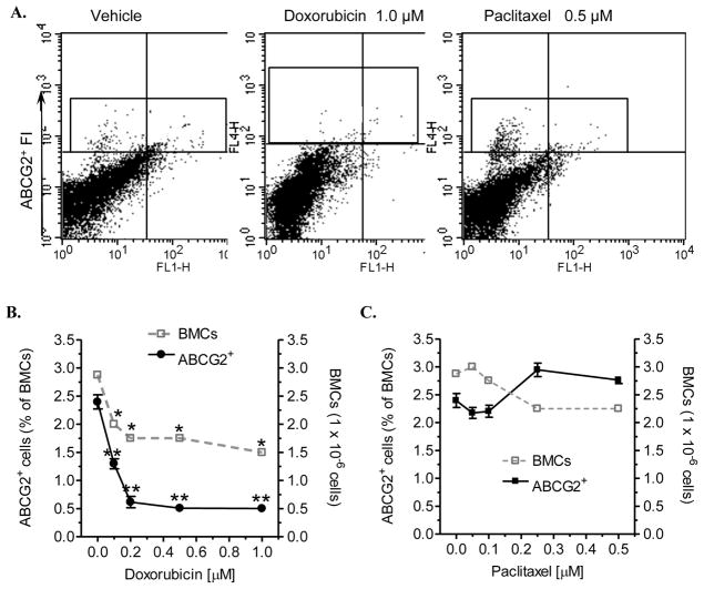
We employed flow cytometry analyzing ABCG2+ BMCs to assess myelosuppression. ABCG2 protein (also known as BCRP), which expresses in a wide variety of stem cells, effluxes the fluorescent dye Hoechst 33342, determining the side population (SP) phenotype (Goodell et al., 1996, Zhou et al., 2001). Bone marrow SP cells are enriched for hematopoietic stem cells (HSCs) as well as mesenchymal stem cells, and ABCG2 has been used as a marker to purify HSCs (Zhou et al., 2002, Zhou et al., 2001, Scharenberg et al., 2002). To test whether analysis of ABCG2+ BMCs can assess bone marrow responsiveness, we primarily analyzed BMCs exposed to Dox and paclitaxel in ex vivo. Compared with the negative staining control, we were able to define ABCG2+ cells and detected the alterations of ABCG2+ BMSCs in BMCs treated with Dox and paclitaxel (Fig. 1A). Dox treatments (6 days) significantly decreased BMC numbers to 61% (1.75 × 10⁶ vs. 2.87 × 10⁶ cells; p<0.001) at 0.2 μM of Dox (Fig. 1B). Furthermore, Dox treatments substantially decreased ABCG2+ cells, as those cells reduced to 25% (0.61% vs. 2.39% BMC; p<0.001) (Fig. 1B) at 0.2 μM of Dox. As shown in Fig 1C, in the same conditions, paclitaxel displayed less myelosuppressive effects. Paclitaxel did not significantly decrease the numbers of BMCs or ABCG2+ BMSCs (Fig. 1C). These data are consistent with other reports that Dox is a myelosuppressive agent (Richardson and Johnson, 1997, Busch et al., 1990) and furthermore indicate ABCG2+ BMSCs were more sensitive to myelotoxic agents such as Dox.
Fig. 1.
Toxicities of Dox and paclitaxel in BMCs. After extraction, BMCs (3 × 10⁶ cells per 60-mm dish) were incubated with Dox and paclitaxel in medium for 6 days. BMCs were counted with hemocytometer and BMSCs (ABCG2+) were analyzed by using flow cytometry. (A) Two-dimension fluorescence histograms of BMC. ABCG2+ cells were enclosed in the rectangle box (was identified as compared with the negative staining, no showed) on the top quadrants. FI, fluorescence intensity. (B) Effects of Dox on BMCs and ABCG2+ cells. *, p<0.001 compared with vehicle control of BMCs; **, p<0.001 compared with vehicle control of ABCG2+ cells. (C) Effects of paclitaxel on BMCs and ABCG2+ cells. The changes of BMCs (open square with dotted line) and ABCG2+ cells (solid circle and line) were indicated in right Y-axis and left Y-axis, respectively.
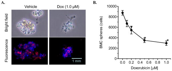
We further examined Dox effects on BMSCs in sphere formation to corroborate above observation. Dox treatment (1.0 μM) substantially decreased the sizes (bright filed) and numbers of bone marrow spheres, due to significantly reduced ABCG2+ BMSCs (red fluorescence), as compared with vehicle control (Fig. 2A). Dox significantly decreased spheres in dose-dependent fashion, and it reduced BMC spheres to 34% (2,958 vs. 8,767 cells, p<0.001) at the 1.0 μM concentration (Fig. 2B). These suggest that quantification of ABCG2+ BMSCs is a direct approach to assess myelosuppression sensitively.
Fig. 2.
The effect of Dox on sphere formation of BMSCs in ex vivo. BMCs were cultured in ultralow attachment dishes with stem cell medium containing Dox for 6 days. (A) Microphotograph of BMSC spheres (× 200 magnification). Red, Alexa Fluor@ 647-ABCG2; blue, nuclear counterstaining with DAPI. (B) Dox effects on sphere formation. *, p<0.001 compared with vehicle control.
3.2 ABCG2+ BMSCs, but not BMCs, were declined in immediate response to Dox treatment in vivo
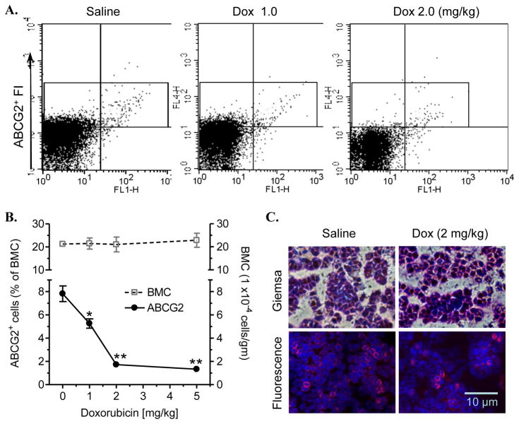
We examined the adverse effect of Dox on bone marrow in vivo. After 6-days Dox treatments, BMCs were extracted from mice. ABCG2+ BMSCs were identified and enclosed in the rectangle of histogram, as compared with each negative control (Fig. 3A). It was found that Dox treatments significantly decreased the numbers of ABCG2+ cells (Fig. 3A). Dox reduced the percentages of ABCG2+ cells in a dose-dependent fashion (1–5 mg/kg), and the ABCG2+ cells were reduced to 21% (1.7 % vs. 7.8% of total BMCs; p<0.001) at the dose of 2.0 μM, although these treatments has few effects on reducing BMC numbers (Fig. 3B). Consistently, bone marrow of mice treated Dox displayed the same cell densities as vehicle control in smear slides with Giemsa staining (Fig. 3B). However, the numbers of ABCG2+ cells (red fluorescence) in Dox treatment (2 mg/kg) were significantly decreased, as compared with in vehicle control (8 cells vs. 26 cells in the represented slides) (Fig. 3C). Furthermore, peripheral blood cells, particularly white blood cells significantly reduced to 39% and 36% at the Dox doses of 2.0 mg/kg and 5.0 mg/kg (Table 1). Those data indicate that Dox eliminate ABCG2+ BMSCs and cause myelosuppression, and quantification of ABCG2+ BMCs by flow cytometry can be applied to detect myelosuppression early.
Fig. 3.
Acute myelotoxicity of Dox in mice. BMC were extracted from mice after 6 days Dox administration (i.p.). (A) The fluorescence histograms of BMCs. ABCG2+ cells were enclosed in the rectangle box on the top quadrants of each one. FI, fluorescence intensity. (B) Effects of Dox on BMCs and ABCG2+ cells. BMCs were normalized against body weight (gram) of each individual (4 mice/group). *, p<0.01 and **, p<0.001 compared with vehicle control. The changes of BMCs (open square and dotted line) and ABCG2+ cells (solid circle and line) were indicated in the right Y-axis and left Y-axis, respectively. (C) ABCG2+ cells in bone marrow. Red, Alexa Fluor®647-ABCG2; blue, nuclear counterstaining with DAPI.
Table 1.
Peripheral blood cell profiles of mice exposed to doxorubicin.
| Lineage | 0 | 1.0 (mg/kg) | 2.0 (mg/kg) | 5.0 (mg/kg) |
|---|---|---|---|---|
| WBC, ×103/μl | 8.30±0.51 | 9.23±0.62 | 3.29*±0.43 | 2.99*±0.32 |
| neutrophil (%) | 9.4 | 11.7 | 13.7 | 17.6 |
| monocyte (%) | 15.5 | 24.5 | 12.4 | 13.6 |
| lymphocyte (%) | 73.6 | 62.2 | 61.2 | 67.8 |
| RBC, ×106/μl | 9.70±0.45 | 10.4±0.64 | 7.16±0.38 | 7.48±0.34 |
| Platelet, ×103/μl | 444±20 | 481±19 | 580±29 | 450±34 |
WBC, white blood cells; RBC, red blood cells.
3.3 Dox increased BCSCs in tumor-bearing mice
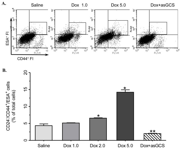
We examined the effects of Dox on BCSCs in mice with orthotopic breast tumors. After 35 days inoculation of human MCF-7/Dox cells, tumor-bearing mice (~5 mm in diameters) were treated with Dox (1–5 mg/kg, i.p.) for 6 days. The BCSCs were isolated and quantified by the CD24−/CD44+/ESA+ markers that have been broadly used to identify human BCSCs (Al-Hajj et al., 2003, Fillmore and Kuperwasser, 2008, Gupta et al., 2011). After CD24 negative separation, the CD44+/ESA+ cells were identified in the rectangle of histogram (top-right quadrant), as compared with each negative control (Fig. 4A). Surprisingly, Dox treatments significantly increased the numbers of CD44+/ESA+ cells, as compared with in saline group; MBO-asGCS treatment, which suppressed GCS expression, decreased CD44+/ESA+ cells, as compared with Dox treatments (Fig. 4A). It was found that the numbers of BCSCs (CD24−/CD44+/ESA+ cells) increased with the doses of Dox (1–5 mg/kg); at the doses of 2 mg/kg and 5 mg/kg of Dox treatments, BCSCs were increased to 150% (6.56% vs. 4.35% total cells; p<0.001) and 326% (14.22% vs. 4.35% total cells; p<0.001), respectively, as compared with saline group (Fig. 4B). Interestingly, it was also found that MBO-asGCS treatment (4 mg/kg, i.p., every three days) decreased the BCSCs to 39% (2.02% vs. 5.14% of total cells; p<0.001) in the combination treatment group, as compared with Dox alone treatments (1.0 mg/kg or 5 mg/kg). These data suggest that Dox can enrich CSCs while it eliminates BMSCs.
Fig. 4.
Acute effects of Dox on BCSCs in tumor-bearing mice. Cancer cells were extracted from tumors of mice (4 mice/group) after 6-days treatments. Dox, Dox treatments (1.0–5.0 mg/kg, i.p); Dox+asGCS, combination treatment of Dox (1 mg/kg, i.p) with MBO-asGCS (1 mg/kg intratumoral injection, every three days). The CD24− cells from negative magnetic separation were incubated with fluorescence conjugated anti-CD44 and anti-ESA antibodies, and analyzed by using flow cytometry. (A) The 2-D fluorescence histograms of CD24− cells. CD44+/ESA+ cells that represented BCSCs were enclosed in the rectangle box (was identified as compared with the negative staining) on the top-right quadrant of each one. FI, fluorescence intensity. (B) Dox increased BCSCs in tumors. BCSCs were represented as percentages of CD24−/CD44+/ESA+ cells to total cancer cells. *, p<0.001 compared with saline group; ** p<0.001 compared with Dox 1.0–5.0 groups.
3.4 Opposite effects of Dox on BMSCs and BCSCs after a long-term and therapeutic dose treatment
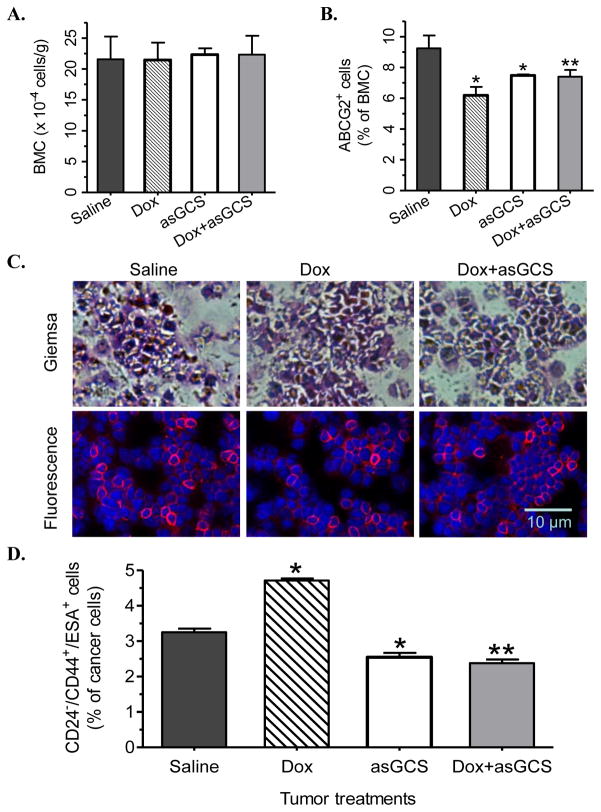
We observed adverse effects of Dox in tumor-bearing mice treated with therapeutic dose of Dox. Breast tumors (MCF/7/Dox, ~2 mm in diameters) were treated with Dox (1 mg/kg. i.p. once a week) that was close to the administration used for cancer patients, and did not display severely acute toxicities in mice (Patwardhan et al., 2009). There were no abnormal clinical syndromes or body weight alterations observed among these groups. Dox and MBO-asGCS (asGCS) alone or combination had fewer effects on the numbers of total BMCs, as compared to saline (Fig. 5A). It was found that Dox treatments significantly reduced the BMSCs (ABCG2+ cells) to 67% (6.18% vs. 9.24% of BMCs; p<0.001) in Dox group, and to 80% (7.40% vs. 9.24% of BMCs; p<0.01) in Dox+asGCS group, respectively, as compared with in saline group (Fig. 5B). The numbers of BMSCs were 120% (7.46% vs. 6.16% of BMCs and 7.40% vs. 6.18% of BMCs; p<0.01) in MBO-asGCS group (asGCS) and in the combination (Dox+asGCS) groups, as compared with Dox alone (Fig. 5B). Bone marrow of these groups displayed almost the same cell densities in smear slides after Giemsa staining (Fig. 5C, top panel); however, the numbers of ABCG2+ cells (red fluorescence) were significantly decreased (16 vs. 26 cells/filed) in Dox group, as compared with in saline group (Fig. 5C bottom panel).
Fig. 5.
Opposite effects of Dox in tumor-bearing mice. Mice with orthotopic breast tumors treated with Dox (1 mg/kg, i.p., per week; Dox), MBO-asGCS (4 mg/kg, i.p., every three days; asGCS) alone and MBO-asGCS combined with Dox (Dox+asGCS) for 42 days. (A) Dox effect on BMCs. BMCs were counted and normalized against body weight. (B) Dox effect on ABCG2+ bone marrow cells. *, p<0.01 compared with saline group; **, p<0.001 compared with Dox group. (C) ABCG2+ cells in bone marrow. Red, Alexa Fluor@ 647-ABCG2; blue, nuclear counterstaining with DAPI. (D) Dox effects on BCSCs of tumors. BCSCs (CD24−/CD44+/ESA+) were analyzed by flow cytometry and were represented as percentages of BCSCs of total tumor cells. *, p<0.01 compared with saline group; **, p<0.001 compared with Dox group.
Furthermore, we assessed the alterations of BCSCs of tumors in the same groups of mice. It was found that Dox treatment significantly increased the numbers of BCSCs to 145% (4.71% vs. 3.25% of tumor cells, p<0.001) in Dox group (Dox; 1 mg/kg, 42 days), as compared with saline (Fig. 5D). Conversely, the BCSCs were decreased to 73% (2.54 vs. 3.25% of tumor cells and 2.38 vs. 3.25 % tumor cells, p<0.01) in MBO-asGCS group (asGCS, 4 mg/kg) and in the combination group (Dox+asGCS), respectively, as compared with saline (Fig. 5D). MBO-asGCS treatment significantly decreased the BCSCs to 50% (2.38 vs. 4.71% cancer cells; p<0.001) in combination group (Dox+asGCS), as compared with Dox alone (Dox) (Fig. 5D).
3.5 GCS determined the cellular effects of Dox on BMSCs and BCSCs
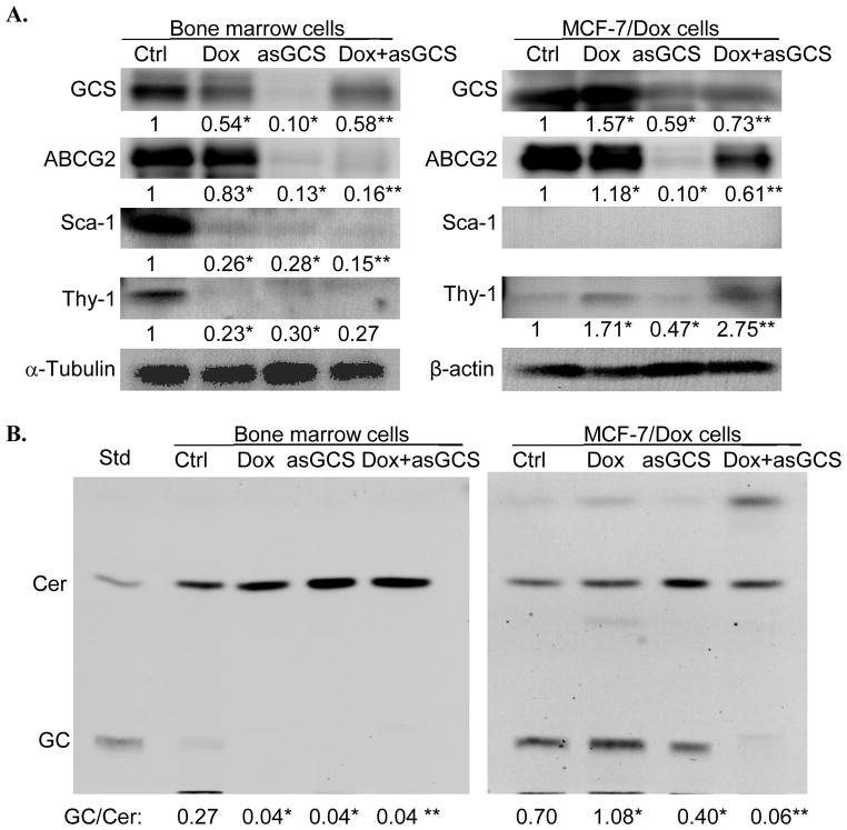
GCS, catalyzing ceramide glycosylation, converts apoptotic ceramide to glucosylceramide and confers cancer cells resistance to Dox; conversely, suppression of GCS sensitizes cells to anticancer drugs (Liu et al., 1999, Liu et al., 2001, Patwardhan et al., 2009). To elucidate the opposite effects of Dox on BMSCs and BCSCs observed above, we assessed GCS expression and its responsiveness to Dox in bone marrow and cancer cells. As shown in Fig. 6, the GCS enzyme activity as well protein level was lower in murine BMCs, approximately 40% of MCF-7/Dox cancer cells. Dox treatments (0.5 μM, 6 days) significantly increased GCS protein as well as enzyme to approximately 150% (1.08 vs. 0.70 GC/Cer; p<0.001) in MCF-7/Dox cells, as compared with vehicle control; conversely, Dox reduced GCS enzyme activity to approximately 20% (0.04 vs. 0.27 GC/Cer; p<0.001), as well as GCS protein level to 50% in bone marrow cells. Silencing of GCS significantly reduced GCS enzyme and protein levels to 60% (0.40 vs. 0.70 GC/Cer) in MCF-7/Dox cells treated with MBO-asGCS alone, as compared with vehicle control. MBO-asGCS treatment substantially reduced GCS activity (by 18-fold, 0.06 vs. 1.08 GC/Cer) and protein level (by 2-fold, 0.73 vs. 1.57) in MCF-7/Dox cells treated with combination, as compared with Dox group (Fig. 6). It was also aware that ABCG2 protein levels of MCF-7/Dox cells were significantly decreased with GCS silencing in MCF-7/Dox cells. The protein levels of ABCG2 and Sca-1 (another marker of bone marrow hematopoietic stem cells) (Weilbaecher et al., 1991, Yilmaz et al., 2006) were decreased in BMCs treated with Dox, MBO-asGCS, and both combination (Fig. 6A).
Fig. 6.
The protein levels and enzyme activities of GCS in BMCs and cancer cells. BMCs extracted and MCF-7/Dox cells were treated with Dox (0.5 μM; Dox), MBO-asGCS (100 nM; asGCS) alone and combined with Dox (Dox+asGCS) for 6 days. Ctrl, vehicle control. (A) Western blotting. Equal amounts of detergent-soluble protein (50 μg/lane) were resolved on 4–20% SDS-PAGE and immunoblotted with individual antibody. The protein levels of GCS or others were normalized against α-tubulin in bone marrow or β-actin in MCF-7/Dox cells, and presented under each individual blot after measurement of optical density. *, p<0.001 compared with vehicle control; **, p<0.001 compared with Dox treatment. (B) Thin-layer chromatography of GCS enzymatic reactions. After incubation with NBD C6-ceramide, cellular lipids were extracted and resolved by thin-layer chromatography. GC/Cer, the ratio of NBD C6-glucosylceramide (GC) to NBD C6-ceramide (Cer). *, p<0.001 compared with vehicle control; **, p<0.001 compared with Dox alone.
4. Discussion
We examined the myelosuppressive effect of Dox by assessing BMSCs in ex vivo and in tumor-bearing mice. Flow cytometry can directly and quantitatively determine the alterations of BMSCs with ABCG2+ phenotype and distinguishes the adverse effects of drugs such as Dox on BMCs. In contrast, the total BMCs could sensitively represent myelotoxicity under these conditions (Fig. 1). Assessment of BMSCs has succeeded in detection of mouse myelotoxicity under chemotherapy (Fig. 3, 5). Early recognition of drug-induced myelosuppression is critical for patients with chemotherapy, as adjustment of therapeutic agents and addition of supportive therapy prior to severe syndromes can significantly improve the clinical outcome (Carey, 2003, Daniel and Crawford, 2006). Different from current examinations for myelosuppression, the flow cytometry of ABCG2 directly detects BMSCs that are responsible for the regeneration of blood cells and other tissues. Peripheral cytological alterations including complete blood cells or reticulocytes and platelets, which are simultaneous with anemia, leucopenia and thrombocytopenia, cannot represent early sign of bone marrow cytotoxicity. The fall of leukocytes after chemotherapy is more sensitive than platelets or erythrocytes, and reaches its nadir about 6-days (Jimenez et al., 1992). In the present study, we find the significant decreases of BMSCs accompanied with leukocytes decreased in mice after 6 days of Dox treatments (Fig. 3, Table 1). Additionally, the decreased BMSCs are detected in mice exposed to therapeutic dose of Dox, even though there are no other abnormal clinical signs including loss of body weight or alterations of activity observed in these mice (Fig. 5). Another feature of this assay is that the BMSCs of bone marrow can be stably remained in culture condition. We still can find more than 95% of BMSCs after 48 hr culture of bone marrow in 10% FBS RPMI-1640 medium at 37°C, as compared to samples measured immediately after extraction. This would offer a time frame to analyze BMSCs by flow cytometry in clinical setting.
Interestingly, we find that anticancer drug such as Dox has opposite effects on normal stem cells versus CSCs in vivo. Dox significantly increase BCSC numbers of mice either in 6-days or 42-days treatments (Fig. 4, Fig. 5), even the BMSC numbers are reduced under the same conditions in the same individuals (Fig. 3, Fig. 5). Early reports show that BCSCs are one reason of drug resistance in cancer cells and in patient tumors (Fillmore and Kuperwasser, 2008, Shafee et al., 2008, Tanei et al., 2009). BCSCs are substantially higher in human MCF-7 breast cancer cells after stepwise-exposure to Dox (Calcagno et al., 2010). In cancer patients, BCSCs (CD44+/CD24− or ALDH1+) are significantly increased after chemotherapy of Dox plus docetaxel or cyclophosphamide (Lee et al., 2011). In present study, 6-days Dox treatments increase BCSCs in dose-dependent fashion and a therapeutic-dose of Dox enhances BCSCs significantly (Fig. 4, Fig. 5). In both conditions, the percentages of BCSCs and the absolute numbers of BCSCs are increased in tumors treated with Dox. Lagadec et al. recently report that radiation induces BCSC phenotype in differentiated breast cancer cells to increase BCSC numbers (Lagadec et al., 2012). Whether Dox enriches BCSCs via dysdiferention of the progenies or differentiated tumor cells or via increase of symmetric cell division of BCSCs need to be examined in further studies.
Our study, for the first time, indicates that GCS is crucial for the stemness of adult stem cells and cancer stem cells. GCS converts ceramide to glucosylceramide, and is a limiting-enzyme regulating cellular ceramide levels and the synthesis of glycosphingolipids (GSLs) (Hakomori, 2008, Patwardhan and Liu, 2011). Early studies demonstrate that GCS confers cancer drug resistance, and is overexpressed in metastatic breast cancers (Liu et al., 1999, Liu et al., 2001, Liu et al., 2011). Globopentosylceramide (Gb5) and monosialyl Gb5 (MSGb5), synthesized by a series of enzymes following ceramide glycosylation, are stage specific embryonic antigen 3 (SSEA-3) and SSEA-4 commonly used to characterize human embryonic stem cells (Thomson et al., 1998, Klimanskaya et al., 2006). The disappearance of Gb5 and MSGb5 is associated with the differentiation of normal stem cells (Liang et al., 2010, Brimble et al., 2007). It is detected in the present study that GCS protein level and enzyme activity in MCF-7/Dox cancer cells are significantly higher than bone marrow cells (Fig. 6). Exposure to Dox upregulates GCS expression and its activity in cancer cells, rather than in BMCs (Fig. 6). Silencing of GCS by MBO-asGCS significantly reduces GCS activity in cancer cells (Fig. 6) and decreases BCSCs in tumor-bearing mice (Fig. 4, Fig. 5). Conversely, silencing GCS increases BMSCs of mice, even those mice exposed to Dox (Fig. 5B). It is unclear whether the species difference between MCF-7/Dox and murine bone marrow plays any role in regulating the opposite effects of Dox on GCS; at least GCS homology is extremely high between human and mouse (98%) (Ichikawa and Hirabayashi, 1998), and MBO-asGCS silences GCS expression in normal or cancer cell lines of human and mouse (Patwardhan et al., 2009) (Fig. 6). Our previous works show that overexpression of GCS enhances globo-series GSLs (Gb3) and activate cSrc/β-catenin signaling, inducing drug resistance by upregulation of MDR1 expression (Liu et al., 2010, Patwardhan and Liu, 2011). Whether cSrc/β-catenin reprograms cancer cells to induce CSCs should be investigated in further study. Collectively, this study demonstrates that GCS, as modulator of stem cell pluripotency, determines the differentiated responsiveness of bone marrow and cancer cells to anticancer drug, Dox.
Acknowledgments
This work was supported by the National Center for Research Resources (5P20RR016456-11) and the National Institute of General Medical Sciences (8 P20 GM103424-11) from the National Institutes of Health (Y.Y.L), and the Mizutani Foundation for Glycoscience, Japan (Y.Y.L.). The authors thank Dr. Brian Rowan (Tulane University School of Medicine, New Orleans, LA) for his advices on cancer stem cell separation, Dr. K. Briski and Mr. A. Gujar (Basic Pharmaceutical Sciences, University of Louisiana at Monroe) for help in confocal microscopy.
Footnotes
Publisher's Disclaimer: This is a PDF file of an unedited manuscript that has been accepted for publication. As a service to our customers we are providing this early version of the manuscript. The manuscript will undergo copyediting, typesetting, and review of the resulting proof before it is published in its final citable form. Please note that during the production process errors may be discovered which could affect the content, and all legal disclaimers that apply to the journal pertain.
References
- Al-Hajj M, Wicha MS, Benito-Hernandez A, Morrison SJ, Clarke MF. Prospective identification of tumorigenic breast cancer cells. Proc Natl Acad Sci U S A. 2003;100:3983–3988. doi: 10.1073/pnas.0530291100. [DOI] [PMC free article] [PubMed] [Google Scholar]
- Bartsch R, Steger GG. The Role of Supportive Therapy in the Era of Modern Adjuvant Treatment - Current and Future Tools. Breast Care (Basel) 2009;4:167–176. doi: 10.1159/000223360. [DOI] [PMC free article] [PubMed] [Google Scholar]
- Brimble SN, Sherrer ES, Uhl EW, Wang E, Kelly S, Merrill AH, Jr, Robins AJ, Schulz TC. The cell surface glycosphingolipids SSEA-3 and SSEA-4 are not essential for human ESC pluripotency. Stem Cells. 2007;25:54–62. doi: 10.1634/stemcells.2006-0232. [DOI] [PubMed] [Google Scholar]
- Busch FW, Schmittele U, Ehninger G. Toxicity of novel anthracycline derivatives towards normal myeloid bone marrow progenitor cells (CFU-GM) is not increased by verapamil. Blut. 1990;60:219–222. doi: 10.1007/BF01728787. [DOI] [PubMed] [Google Scholar]
- Calcagno AM, Salcido CD, Gillet JP, Wu CP, Fostel JM, Mumau MD, Gottesman MM, Varticovski L, Ambudkar SV. Prolonged drug selection of breast cancer cells and enrichment of cancer stem cell characteristics. J Natl Cancer Inst. 2010;102:1637–1652. doi: 10.1093/jnci/djq361. [DOI] [PMC free article] [PubMed] [Google Scholar]
- Carey PJ. Drug-induced myelosuppression : diagnosis and management. Drug Saf. 2003;26:691–706. doi: 10.2165/00002018-200326100-00003. [DOI] [PubMed] [Google Scholar]
- Clarke R, Currier S, Kaplan O, Lovelace E, Boulay V, Gottesman MM, Dickson RB. Effect of P-glycoprotein expression on sensitivity to hormones in MCF-7 human breast cancer cells. J Natl Cancer Inst. 1992;84:1506–1512. doi: 10.1093/jnci/84.19.1506. [DOI] [PubMed] [Google Scholar]
- Daniel D, Crawford J. Myelotoxicity from chemotherapy. Semin Oncol. 2006;33:74–85. doi: 10.1053/j.seminoncol.2005.11.003. [DOI] [PubMed] [Google Scholar]
- Dean M, Fojo T, Bates S. Tumour stem cells and drug resistance. Nat Rev Cancer. 2005;5:275–284. doi: 10.1038/nrc1590. [DOI] [PubMed] [Google Scholar]
- Doyle LA, Yang W, Abruzzo LV, Krogmann T, Gao Y, Rishi AK, Ross DD. A multidrug resistance transporter from human MCF-7 breast cancer cells. Proc Natl Acad Sci U S A. 1998;95:15665–15670. doi: 10.1073/pnas.95.26.15665. [DOI] [PMC free article] [PubMed] [Google Scholar]
- Fillmore CM, Kuperwasser C. Human breast cancer cell lines contain stem-like cells that self-renew, give rise to phenotypically diverse progeny and survive chemotherapy. Breast Cancer Res. 2008;10:R25. doi: 10.1186/bcr1982. [DOI] [PMC free article] [PubMed] [Google Scholar]
- Gasparini G, Barbareschi M, Doglioni C, Palma PD, Mauri FA, Boracchi P, Bevilacqua P, Caffo O, Morelli L, Verderio P, et al. Expression of bcl-2 protein predicts efficacy of adjuvant treatments in operable node-positive breast cancer. Clin Cancer Res. 1995;1:189–198. [PubMed] [Google Scholar]
- Giarratana MC, Kobari L, Lapillonne H, Chalmers D, Kiger L, Cynober T, Marden MC, Wajcman H, Douay L. Ex vivo generation of fully mature human red blood cells from hematopoietic stem cells. Nat Biotechnol. 2005;23:69–74. doi: 10.1038/nbt1047. [DOI] [PubMed] [Google Scholar]
- Gonzalez-Angulo AM, Morales-Vasquez F, Hortobagyi GN. Overview of resistance to systemic therapy in patients with breast cancer. Adv Exp Med Biol. 2007;608:1–22. doi: 10.1007/978-0-387-74039-3_1. [DOI] [PubMed] [Google Scholar]
- Goodell MA, Brose K, Paradis G, Conner AS, Mulligan RC. Isolation and functional properties of murine hematopoietic stem cells that are replicating in vivo. J Exp Med. 1996;183:1797–1806. doi: 10.1084/jem.183.4.1797. [DOI] [PMC free article] [PubMed] [Google Scholar]
- Gupta V, Patwardhan GA, Zhang QJ, Cabot MC, Jazwinski SM, Liu YY. Direct quantitative determination of ceramide glycosylation in vivo: a new approach to evaluate cellular enzyme activity of glucosylceramide synthase. J Lipid Res. 2010;51:866–874. doi: 10.1194/jlr.D002949. [DOI] [PMC free article] [PubMed] [Google Scholar]
- Gupta V, Zhang QJ, Liu YY. Evaluation of anticancer agents using flow cytometry analysis of cancer stem cells. Methods Mol Biol. 2011;716:179–191. doi: 10.1007/978-1-61779-012-6_11. [DOI] [PMC free article] [PubMed] [Google Scholar]
- Hakomori SI. Structure and function of glycosphingolipids and sphingolipids: recollections and future trends. Biochim Biophys Acta. 2008;1780:325–346. doi: 10.1016/j.bbagen.2007.08.015. [DOI] [PMC free article] [PubMed] [Google Scholar]
- Herman JF, Mangala LS, Mehta K. Implications of increased tissue transglutaminase (TG2) expression in drug-resistant breast cancer (MCF-7) cells. Oncogene. 2006;25:3049–3058. doi: 10.1038/sj.onc.1209324. [DOI] [PubMed] [Google Scholar]
- Hood LE. Chemotherapy in the elderly: supportive measures for chemotherapy-induced myelotoxicity. Clin J Oncol Nurs. 2003;7:185–190. doi: 10.1188/03.CJON.185-190. [DOI] [PubMed] [Google Scholar]
- Ichikawa S, Hirabayashi Y. Glucosylceramide synthase and glycosphingolipid synthesis. Trends Cell Biol. 1998;8:198–202. doi: 10.1016/s0962-8924(98)01249-5. [DOI] [PubMed] [Google Scholar]
- Janni W, Rjosk D, Strobl B, Bergauer F, Linka F, Dimpfl T, Schindlbeck C, Rack B, Kaestner R, Sommer H. Chemotherapy-associated myelosuppression in gynecological oncology. Gynakol Geburtshilfliche Rundsch. 2001;41:166–173. doi: 10.1159/000049471. [DOI] [PubMed] [Google Scholar]
- Jimenez JJ, Huang HS, Hindahl M, Pearson FC, Yunis AA. Protection from chemotherapy-induced neutropenia by ImuVert. Am J Med Sci. 1992;303:83–85. doi: 10.1097/00000441-199202000-00003. [DOI] [PubMed] [Google Scholar]
- Klimanskaya I, Chung Y, Becker S, Lu SJ, Lanza R. Human embryonic stem cell lines derived from single blastomeres. Nature. 2006;444:481–485. doi: 10.1038/nature05142. [DOI] [PubMed] [Google Scholar]
- Kobayashi Y, Seino K, Hosonuma S, Ohara T, Itamochi H, Isonishi S, Kita T, Wada H, Kojo S, Kiguchi K. Side population is increased in paclitaxel-resistant ovarian cancer cell lines regardless of resistance to cisplatin. Gynecol Oncol. 2011;121:390–394. doi: 10.1016/j.ygyno.2010.12.366. [DOI] [PubMed] [Google Scholar]
- Kroger N, Achterrath W, Hegewisch-Becker S, Mross K, Zander AR. Current options in treatment of anthracycline-resistant breast cancer. Cancer Treat Rev. 1999;25:279–291. doi: 10.1053/ctrv.1999.0137. [DOI] [PubMed] [Google Scholar]
- Lagadec C, Vlashi E, Della Donna L, Dekmezian C, Pajonk F. Radiation-induced reprogramming of breast cancer cells. Stem Cells. 2012;30:833–844. doi: 10.1002/stem.1058. [DOI] [PMC free article] [PubMed] [Google Scholar]
- Lee HE, Kim JH, Kim YJ, Choi SY, Kim SW, Kang E, Chung IY, Kim IA, Kim EJ, Choi Y, Ryu HS, Park SY. An increase in cancer stem cell population after primary systemic therapy is a poor prognostic factor in breast cancer. Br J Cancer. 2011;104:1730–1738. doi: 10.1038/bjc.2011.159. [DOI] [PMC free article] [PubMed] [Google Scholar]
- Liang YJ, Kuo HH, Lin CH, Chen YY, Yang BC, Cheng YY, Yu AL, Khoo KH, Yu J. Switching of the core structures of glycosphingolipids from globo-and lacto- to ganglio-series upon human embryonic stem cell differentiation. Proc Natl Acad Sci U S A. 2010;107:22564–22569. doi: 10.1073/pnas.1007290108. [DOI] [PMC free article] [PubMed] [Google Scholar]
- Liu YY, Gupta V, Patwardhan GA, Bhinge K, Zhao Y, Bao J, Mehendale H, Cabot MC, Li YT, Jazwinski SM. Glucosylceramide synthase upregulates MDR1 expression in the regulation of cancer drug resistance through cSrc and beta-catenin signaling. Mol Cancer. 2010;9:145. doi: 10.1186/1476-4598-9-145. [DOI] [PMC free article] [PubMed] [Google Scholar]
- Liu YY, Han TY, Giuliano AE, Cabot MC. Expression of glucosylceramide synthase, converting ceramide to glucosylceramide, confers adriamycin resistance in human breast cancer cells. J Biol Chem. 1999;274:1140–1146. doi: 10.1074/jbc.274.2.1140. [DOI] [PubMed] [Google Scholar]
- Liu YY, Han TY, Giuliano AE, Cabot MC. Ceramide glycosylation potentiates cellular multidrug resistance. FASEB J. 2001;15:719–730. doi: 10.1096/fj.00-0223com. [DOI] [PubMed] [Google Scholar]
- Liu YY, Patwardhan GA, Xie P, Gu X, Giuliano AE, Cabot MC. Glucosylceramide synthase, a factor in modulating drug resistance, is overexpressed in metastatic breast carcinoma. Int J Oncol. 2011;39:425–431. doi: 10.3892/ijo.2011.1052. [DOI] [PMC free article] [PubMed] [Google Scholar]
- Liu YY, Yu JY, Yin D, Patwardhan GA, Gupta V, Hirabayashi Y, Holleran WM, Giuliano AE, Jazwinski SM, Gouaze-Andersson V, Consoli DP, Cabot MC. A role for ceramide in driving cancer cell resistance to doxorubicin. FASEB J. 2008;22:2541–2551. doi: 10.1096/fj.07-092981. [DOI] [PubMed] [Google Scholar]
- Maugeri-Sacca M, Vigneri P, De Maria R. Cancer stem cells and chemosensitivity. Clin Cancer Res. 2011;17:4942–4947. doi: 10.1158/1078-0432.CCR-10-2538. [DOI] [PubMed] [Google Scholar]
- Mehta K. High levels of transglutaminase expression in doxorubicin-resistant human breast carcinoma cells. Int J Cancer. 1994;58:400–406. doi: 10.1002/ijc.2910580316. [DOI] [PubMed] [Google Scholar]
- Moore K, Crom D. Hematopoietic support with moderately myelosuppressive chemotherapy regimens: a nursing perspective. Clin J Oncol Nurs. 2006;10:383–388. doi: 10.1188/06.CJON.383-388. [DOI] [PubMed] [Google Scholar]
- Nurgalieva Z, Liu CC, Du XL. Chemotherapy use and risk of bone marrow suppression in a large population-based cohort of older women with breast and ovarian cancer. Med Oncol. 2010 doi: 10.1007/s12032-010-9512-5. [DOI] [PubMed] [Google Scholar]
- Patwardhan G, Gupta V, Huang J, Gu X, Liu YY. Direct assessment of P-glycoprotein efflux to determine tumor response to chemotherapy. Biochem Pharmacol. 2010;80:72–79. doi: 10.1016/j.bcp.2010.03.010. [DOI] [PMC free article] [PubMed] [Google Scholar]
- Patwardhan GA, Liu YY. Sphingolipids and expression regulation of genes in cancer. Prog Lipid Res. 2011;50:104–114. doi: 10.1016/j.plipres.2010.10.003. [DOI] [PMC free article] [PubMed] [Google Scholar]
- Patwardhan GA, Zhang QJ, Yin D, Gupta V, Bao J, Senkal CE, Ogretmen B, Cabot MC, Shah GV, Sylvester PW, Jazwinski SM, Liu YY. A new mixed-backbone oligonucleotide against glucosylceramide synthase sensitizes multidrug-resistant tumors to apoptosis. PLoS One. 2009;4:e6938. doi: 10.1371/journal.pone.0006938. [DOI] [PMC free article] [PubMed] [Google Scholar]
- Repetto L. Incidence and clinical impact of chemotherapy induced myelotoxicity in cancer patients: an observational retrospective survey. Crit Rev Oncol Hematol. 2009;72:170–179. doi: 10.1016/j.critrevonc.2009.03.004. [DOI] [PubMed] [Google Scholar]
- Richardson DS, Johnson SA. Anthracyclines in haematology: preclinical studies, toxicity and delivery systems. Blood Rev. 1997;11:201–223. doi: 10.1016/s0268-960x(97)90020-5. [DOI] [PubMed] [Google Scholar]
- Scharenberg CW, Harkey MA, Torok-Storb B. The ABCG2 transporter is an efficient Hoechst 33342 efflux pump and is preferentially expressed by immature human hematopoietic progenitors. Blood. 2002;99:507–512. doi: 10.1182/blood.v99.2.507. [DOI] [PubMed] [Google Scholar]
- Seidman AD. Systemic treatment of breast cancer. Two decades of progress. Oncology (Williston Park) 2006;20:983–990. discussion 991–982, 997–988. [PubMed] [Google Scholar]
- Shafee N, Smith CR, Wei S, Kim Y, Mills GB, Hortobagyi GN, Stanbridge EJ, Lee EY. Cancer stem cells contribute to cisplatin resistance in Brca1/p53-mediated mouse mammary tumors. Cancer Res. 2008;68:3243–3250. doi: 10.1158/0008-5472.CAN-07-5480. [DOI] [PMC free article] [PubMed] [Google Scholar]
- Shiota M, Heike T, Haruyama M, Baba S, Tsuchiya A, Fujino H, Kobayashi H, Kato T, Umeda K, Yoshimoto M, Nakahata T. Isolation and characterization of bone marrow-derived mesenchymal progenitor cells with myogenic and neuronal properties. Exp Cell Res. 2007;313:1008–1023. doi: 10.1016/j.yexcr.2006.12.017. [DOI] [PubMed] [Google Scholar]
- Szakacs G, Paterson JK, Ludwig JA, Booth-Genthe C, Gottesman MM. Targeting multidrug resistance in cancer. Nat Rev Drug Discov. 2006;5:219–234. doi: 10.1038/nrd1984. [DOI] [PubMed] [Google Scholar]
- Tanei T, Morimoto K, Shimazu K, Kim SJ, Tanji Y, Taguchi T, Tamaki Y, Noguchi S. Association of breast cancer stem cells identified by aldehyde dehydrogenase 1 expression with resistance to sequential Paclitaxel and epirubicin-based chemotherapy for breast cancers. Clin Cancer Res. 2009;15:4234–4241. doi: 10.1158/1078-0432.CCR-08-1479. [DOI] [PubMed] [Google Scholar]
- Thomson JA, Itskovitz-Eldor J, Shapiro SS, Waknitz MA, Swiergiel JJ, Marshall VS, Jones JM. Embryonic stem cell lines derived from human blastocysts. Science. 1998;282:1145–1147. doi: 10.1126/science.282.5391.1145. [DOI] [PubMed] [Google Scholar]
- Timmer-Bonte JN, de Boo TM, Smit HJ, Biesma B, Wilschut FA, Cheragwandi SA, Termeer A, Hensing CA, Akkermans J, Adang EM, Bootsma GP, Tjan-Heijnen VC. Prevention of chemotherapy-induced febrile neutropenia by prophylactic antibiotics plus or minus granulocyte colony-stimulating factor in small-cell lung cancer: a Dutch Randomized Phase III Study. J Clin Oncol. 2005;23:7974–7984. doi: 10.1200/JCO.2004.00.7955. [DOI] [PubMed] [Google Scholar]
- Wang Y, Probin V, Zhou D. Cancer therapy-induced residual bone marrow injury-Mechanisms of induction and implication for therapy. Curr Cancer Ther Rev. 2006;2:271–279. doi: 10.2174/157339406777934717. [DOI] [PMC free article] [PubMed] [Google Scholar]
- Weilbaecher K, Weissman I, Blume K, Heimfeld S. Culture of phenotypically defined hematopoietic stem cells and other progenitors at limiting dilution on Dexter monolayers. Blood. 1991;78:945–952. [PubMed] [Google Scholar]
- Yilmaz OH, Kiel MJ, Morrison SJ. SLAM family markers are conserved among hematopoietic stem cells from old and reconstituted mice and markedly increase their purity. Blood. 2006;107:924–930. doi: 10.1182/blood-2005-05-2140. [DOI] [PMC free article] [PubMed] [Google Scholar]
- Zhou S, Morris JJ, Barnes Y, Lan L, Schuetz JD, Sorrentino BP. Bcrp1 gene expression is required for normal numbers of side population stem cells in mice, and confers relative protection to mitoxantrone in hematopoietic cells in vivo. Proc Natl Acad Sci U S A. 2002;99:12339–12344. doi: 10.1073/pnas.192276999. [DOI] [PMC free article] [PubMed] [Google Scholar]
- Zhou S, Schuetz JD, Bunting KD, Colapietro AM, Sampath J, Morris JJ, Lagutina I, Grosveld GC, Osawa M, Nakauchi H, Sorrentino BP. The ABC transporter Bcrp1/ABCG2 is expressed in a wide variety of stem cells and is a molecular determinant of the side-population phenotype. Nat Med. 2001;7:1028–1034. doi: 10.1038/nm0901-1028. [DOI] [PubMed] [Google Scholar]


