Abstract
There are a number of genetic tools available for studying Francisella tularensis, the etiological agent of tularemia; however, there is no effective inducible or repressible gene expression system. Here, we describe inducible and repressible gene expression systems for F. tularensis based on the Tet repressor, TetR. For the inducible system, a tet operator sequence was cloned into a modified F. tularensis groESL promoter sequence and carried in a plasmid that constitutively expressed TetR. To monitor regulation the luminescence operon, luxCDABE, was cloned under the hybrid Francisella tetracycline-regulated promoter (FTRp), and transcription was initiated with addition of anhydrotetracycline (ATc), which binds TetR and alleviates TetR association with tetO. Expression levels measured by luminescence correlated with ATc inducer concentrations ranging from 20 to 250 ng ml−1. In the absence of ATc, luminescence was below the level of detection. The inducible system was also functional during the infection of J774A.1 macrophages, as determined by both luminescence and rescue of a mutant strain with an intracellular growth defect. The repressible system consists of FTRp regulated by a reverse TetR mutant (revTetR), TetR r1.7. Using this system with the lux reporter, the addition of ATc resulted in decreased luminescence, while in the absence of ATc the level of luminescence was not significantly different from that of a construct lacking TetR r1.7. Utilizing both systems, the essentiality of SecA, the protein translocase ATPase, was confirmed, establishing that they can effectively regulate gene expression. These two systems will be invaluable in exploring F. tularensis protein function.
INTRODUCTION
Regulated expression systems are important tools for the manipulation of gene transcription for the study of organismal biology. Currently, there are no suitable genetic control systems for Francisella tularensis, a Gram-negative bacterium which is the etiological agent of tularemia (13). A conditional expression system was developed for F. tularensis that relies on the F. tularensis glucose-repressible promoter, FGRp (26). However, this system relies on a ubiquitous carbon source and is not flexible. Hence, we developed regulated gene expression systems for F. tularensis based upon tetracycline that can be used for both induction and repression.
Tetracycline-regulated systems have become a useful tool in analyzing gene function in prokaryotes (6). Such systems, derived from Tn10, involve a constitutively expressed tetracycline repressor, TetR, which binds the tetracycline operator, tetO, of the tetracycline promoter, tetAp, blocking transcription. TetR is released from tetO in the presence of tetracycline, allowing transcription from tetAp. The tetracycline analog anhydrotetracyline (ATc) is significantly less toxic and binds more avidly to TetR, making it a useful chemical for modulating TetR-mediated gene expression in bacteria (21). The TetR system has been used with a diverse group of bacteria, including other respiratory pathogens, such as Yersinia pestis, Brucella abortus, Coxiella burnetii, and Mycobacterium tuberculosis (4, 14, 27, 37). In addition, a reverse mutant derivative, revTetR, acts as a corepressor binding tetAo only when associated with ATc and can thus function to silence gene expression (34). Here, we describe TetR and revTetR plasmid systems for the regulation of one or more target genes in Francisella tularensis.
MATERIALS AND METHODS
Bacterial strains, transformation, and cell culture.
Escherichia coli strains (Table 1) were grown in Luria-Bertani (LB) broth (BD Biosciences) or on LB agar. F. tularensis strains (Table 1) were grown at 37°C on chocolate agar (25 g brain heart infusion liter−1, 10 μg hemoglobin ml−1, 15 g agarose liter−1) supplemented with 1% IsoVitaleX (Becton-Dickson) or in Chamberlain's defined medium (CDM) (10) When necessary, kanamycin (Km; Sigma-Aldrich) was used at 50 μg ml−1 for Escherichia coli and 10 μg ml−1 for F. tularensis. Hygromycin B (Hyg; Roche Applied Science) was used at 200 μg ml−1 for both species. Sucrose was used at a final concentration of 10% (wt/vol). Anhydrotetracycline (ATc; Sigma-Aldrich) was used at the concentrations stated.
Table 1.
Bacterial strains and plasmids
| Strain or plasmid | Description | Reference or source |
|---|---|---|
| Strains | ||
| E. coli | ||
| DH10B | F− mcrA Δ(mcrBC-hsdRMS-mrr) [φ80dΔlacZΔM15] ΔlacX74 deoR recA1 endA1 araD139 Δ(ara, leu)7697 galU galKλ− rpsL nupG | Invitrogen |
| S17-1 λpir | Tpr Smr hsdR pro recARP4-2-Tc::Mu-Km::Tn7 λpir | 12 |
| NK7402 | trpC83::Tn10, IN(rrnD-rrnE)1 source of tetR | CGSC strain 6688 |
| F. tularensis | ||
| LVS | F. tularensis subsp. holarctica live vaccine strain | CDC |
| LVSΔripA | LVSΔripA | 18 |
| LE149/TetOn | LVSΔsecA pEDL54 in trans | This work |
| LE150/TetOff | LVSΔsecA pEDL58 in trans | This work |
| Plasmids | ||
| pcDNA3-mOrange2 | Apr, Neor; source of mOrange2 | 36 |
| pJH1 | Hygr; source of oriT | 25 |
| pMP812 | Kmr; SacB suicide vector | 28 |
| pNR55 | Kmr; source of TetR r1.7 | 33 |
| pXB173-lux | Kmr; E. coli-F. tularensis shuttle vector containing the luxCDABE operon | 7 |
| pEDL17 | Hygr; FTRp-mOrange2, rpsLp-tetR | This work |
| pEDL20 | Hygr; FTRp-ripA, rpsLp-tetR | This work |
| pEDL40 | Hygr; FTRp-luxCDABE, rpsLp-tetR | This work |
| pEDL41 | Hygr; FTRp-luxCDABE with deletion of tetR | This work |
| pEDL47 | Hygr; FTRp-luxCDABE, rpsLp-tetR r1.7 | This work |
| pEDL50 | Kmr; pMP812 SacB suicide vector with oriT from pJH1 | This work |
| pEDL54 | Hygr; FTRp-secA, rpsLp-tetR | This work |
| pEDL55 | pEDL50 with LVS ΔsecA region | This work |
| pEDL58 | Hygr; FTRp-secA, rpsLp-tetR r1.7 | This work |
E. coli-F. tularensis shuttle vectors were introduced into F. tularensis strains via electroporation as described previously (30). Transformants were selected on chocolate agar supplemented with the appropriate antibiotics.
J774A.1 (ATCC TIB-67) is a mouse macrophage-like cell line that was cultured in Dulbecco's minimal essential medium with 4.5 g liter−1 glucose, 2 mM l-glutamine, and 10% heat-inactivated fetal bovine serum. Cell lines were maintained at 37°C in 5% CO2.
secA mutagenesis and allelic exchange.
A DNA fragment containing secA was obtained from F. tularensis LVS genomic DNA (GenBank accession no. AM233362.1) using PCR and cloned into the SacB/oriT vector pEDL50, which was used as a template for PCR to generate an in-frame deletion of 2,703 bp within secA. There is 761 bp of DNA upstream and 836 bp downstream of the ΔsecA1 allele in the deletion construct, pEDL55.
Conjugation and allelic exchange were performed similarly to the previously described method (24), except pEDL55 was mobilized into F. tularensis using E. coli S17-1 λpir and primary recombinants were selected on chocolate plates supplemented with polymyxin B at 200 μg ml−1 and kanamycin at 10 μg ml−1.
DNA manipulation.
Recombinant DNA methods were performed essentially as described previously (2). DNA fragments were isolated using agarose gel electrophoresis and QIAquick spin columns (Qiagen Inc.). Oligonucleotides were synthesized by Invitrogen Life Technologies. All restriction endonucleases were from New England BioLabs (NEB). DNA ligations were performed with the Fast-Link DNA ligation kit (Epicentre). PCRs were performed with Phusion high-fidelity DNA polymerase (NEB) according to the manufacturer's recommendations.
Plasmid construction.
Plasmids pertinent to this study are listed in Table 1. Detailed descriptions of the construction of the plasmids are available upon request.
Broth culture luminescence assay and SecA depletion assay.
Bacteria were grown with shaking at 37°C in 96-well, flat, clear-bottomed black polystyrene plates (Corning) in CDM in an Infinite 200 microplate reader (Tecan) with luminescence and absorbance (optical density at 600 nm [OD600]) monitored every 15 min. The SecA depletion assay was performed similarly, except growth was in 96-well, flat, clear polystyrene plates (Corning) and only the absorbance (OD600) monitored.
Intracellular induction and ripA growth rescue.
To determine the rate of intracellular induction by ATc, F. tularensis LVS strains harboring luminescent FTRp constructs were cultured to mid-exponential phase in CDM and then added to J774A.1 cells at a multiplicity of infection (MOI) of 100. Wells were washed 2 h postinfection, and 50 μg ml−1 gentamicin was added to inhibit any remaining extracellular bacteria. As peak luminescence of an LVS J774A.1 gentamicin protection assay is ∼24 to 30 h postinfection, the strains were induced with ATc at 23 h and placed in an Infinite 200 Pro microplate reader (Tecan) equipped with a gas control module set at 5% CO2, and luminescence was monitored every 15 min.
We used the gentamicin protection assay described above for the ripA intracellular growth rescue experiments, except that ATc was added at 12 h postinfection and CFU were enumerated at 12 and 36 h postinfection. Assays were performed in sextuplicate, and statistical significance was determined using unpaired t tests on the fold change of recovered CFU to compare the differences in growth between rescued strains and the ripA deletion strains.
RESULTS AND DISCUSSION
As Francisella tularensis does not recognize the Tn10 tetA promoter (data not shown), we chose to modify the well-characterized Francisella groESL promoter (15, 22), which drives strong gene expression in vitro and in vivo (7, 30). groESLp was modified by inserting a tetO sequence immediately downstream of the predicted transcriptional start site, creating the Francisella tetracycline-regulated promoter (FTRp) (Fig. 1). An MluI restriction site was added to facilitate cloning open reading frames and also to allow for changing the 5′-untranslated region (UTR) or start codon (e.g., a GTG start has been found to decrease expression in F. tularensis [3]). The tetR gene encoding the repressor TetR was placed under the constitutively active Francisella rpsL promoter and cloned divergent from FTRp (28).
Fig 1.
Francisella tetracycline-regulated promoter. FTRp has both sigma 32 and sigma 70 recognition, delineated by the underlined −35 and −10 sequences. The asterisk signifies the transcriptional start. Following the 5′ UTR is the tet operator, tetO, as well as a MluI site to facilitate cloning. ATG represents the translational start.
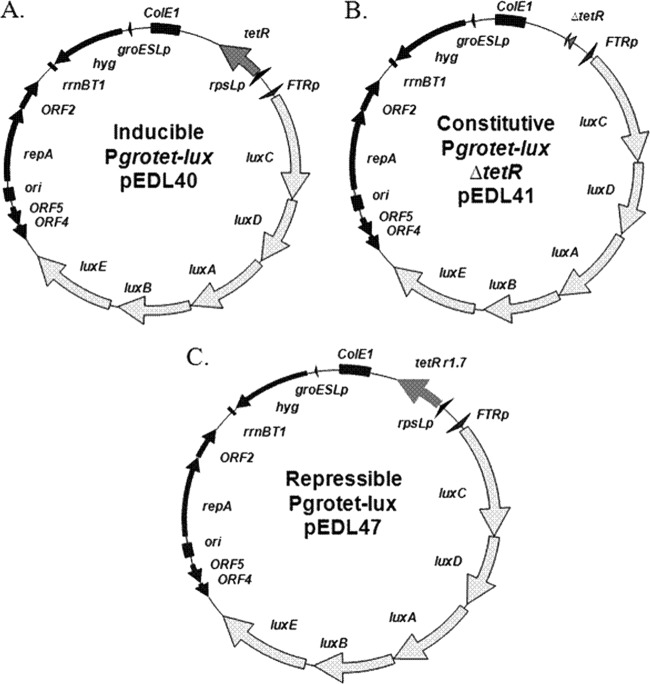
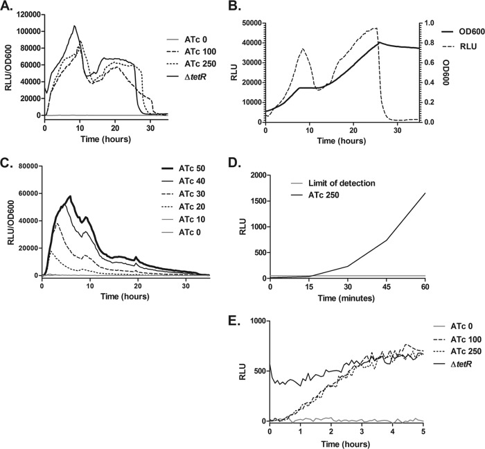
We used the bacterial luminescence operon luxCDABE from Photorhabdus luminescens, which has been shown to function in F. tularensis (7), to assess our ability to regulate transcription of the hybrid promoter. We created two separate constructs: pEDL40 (Fig. 2A), an E. coli-F. tularensis shuttle vector bearing FTRp driving the lux operon and an rpsLp-tetR cassette, and pEDL41 (Fig. 2B), which is pEDL40 with tetR deleted, allowing for constitutive FTRp-luxCDABE expression. As the groESL promoter is strong, we specifically chose the low-copy-number, hygromycin-resistant vector platform (29). The plasmids were introduced into F. tularensis LVS, and growth curves in CDM were performed in the presence of ATc at 0, 100, or 250 ng ml−1 while monitoring luminescence and absorbance (OD600) (Fig. 3A). The constitutive ΔtetR strain was not supplemented; however, we found that ATc had no effect on luminescence except at high levels (>250 ng ml−1 ATc), becoming increasingly bacteriostatic and causing an overall reduction in the relative light unit (RLU)/OD600 ratio (data not shown). No luminescence was detected at 0 ng ml−1, suggesting the system is effectively silenced by TetR, while luminescence with concentrations of 100 and 250 ng ml−1 was similar to that of the constitutive promoter, suggesting that at these levels the system was fully induced. LVS exhibits diauxie in CDM, which leads to the two peaks in the luminescence curve (Fig. 3B). The system was titrated with concentrations of 20 to 50 ng ml−1 ATc, and as the concentration increased so did the luminescence, although concentrations of less than 20 ng ml−1 were below the level of luminescence detection (Fig. 3C). Thus, by luminescence the TetR system has a useable range of 20 to 250 ng ml−1 ATc in broth culture. To determine the speed of induction, a mid-log culture was induced with 250 ng ml−1 ATc and measured over time (Fig. 3D). Luminescence exceeded the limit of detection within 18 min, showing that induction of transcription and subsequent translation is rapid.
Fig 2.
Maps of the FTRp-luxCDABE vectors. (A) The inducible vector, pEDL40, is a stable Hygr E. coli-F. tularensis shuttle vector that contains the hybrid Francisella FTR promoter driving the luminescence operon, luxCDABE. The tetR gene, constitutively expressed from the Francisella rpsL promoter, is divergently transcribed. (B) The vector pEDL41 is pEDL40 with the tetR gene deleted. This allows the constitutive expression of the lux operon from FTRp. (C) The repressible vector, pEDL47, is a Hygr, stable E. coli-F. tularensis shuttle vector that contains FTRp-luxCDABE. The TetR r1.7 gene (a revTetR), constitutively expressed from the Francisella rpsL promoter, is divergently transcribed.
Fig 3.
TetR system. (A) Growth curves were performed in Chamberlain's defined medium (CDM) with LVS bearing plasmid pEDL40 in the presence of ATc at concentrations of 0, 100, and 250 ng ml−1 and LVS bearing pEDL41 ΔtetR as the constitutive control. The absorbance at OD600 and luminescence were monitored every 15 min for 36 h. To determine the relative light produced per bacterium, the luminescence was divided by the absorbance and is represented as RLU/OD600. (B) A growth curve was performed in CDM with LVS bearing pEDL41. The absorbance at OD600 and luminescence were monitored every 15 min for 36 h. The RLU and OD600 were plotted on separate y axes. (C) Growth curves were performed in CDM utilizing ATc concentrations from 0 to 50 ng ml−1 in 10-ng ml−1 increments, and luminescence per bacterium was monitored as described above. (D) A mid-log culture of LVS/pEDL40 was induced with 250 ng ml−1 ATc to test the induction time. Luminescence was monitored every 15 min to ascertain the time it took to increase above 50 RLU, which represents the limit of detection. (E) A gentamicin protection assay was performed with LVS bearing plasmid pEDL40, with LVS bearing pEDL41 ΔtetR as the constitutive control. Induction was at 23 h postinfection with ATc concentrations of 0, 100, and 250 ng ml−1. Luminescence was measured every 15 min for 5 h after induction.
These results confirm that the system is useful in broth culture; however, as F. tularensis is a facultative intracellular bacterium, we wanted to determine if induction of the TetR system was possible in cell culture. A J774A.1 mouse macrophage-like cell line was infected with LVS bearing pEDL40 or pEDL41. Since peak luminescence in a J774A.1 infection occurs at ∼24 to 30 h postinfection (data not shown), ATc was added at 23 h postinfection at 0, 100, or 250 ng ml−1. With addition of ATc at 100 or 250 ng ml−1, the luminescence was above background within 45 min and reached the level of the constitutive promoter within 3 h (Fig. 3E). The bacteriostatic concentration of ATc in cell culture was found to be greater than 500 ng ml−1.
We wanted to establish if the TetR system was suitable for rescuing a mutant defective for intracellular replication. RipA is a cytoplasmic transmembrane protein essential for Francisella tularensis intracellular growth in host macrophage cells and for growth at neutral pH in the host cell's cytoplasmic environment (19, 38). The F. tularensis ΔripA mutant can infect host macrophages and escape the phagosome at the same frequency as the wild type (WT) but fails to replicate inside host cells, and it is impaired in its ability to colonize and disseminate in a mouse model of tularemia (18). A gentamicin protection assay was performed with LVS bearing the TetR control plasmid pEDL17, in which FTRp drives production of a red fluorescent protein (RFP), mOrange2 (36), LVSΔripA bearing pEDL17, and LVSΔripA containing the plasmid pEDL20 with ripA under the control of FTRp. J774A.1 macrophages were infected, and 100 ng ml−1 ATc was added at 12 h postinfection to determine if the LVSΔripA strain could be rescued after such a long period of stasis. As expected, the control strain LVS/pEDL17 replicated inside host macrophages, increasing in numbers by 3- to 5-fold between 12 and 36 h postinfection, while the ripA/pEDL17 strain failed to replicate inside macrophages. Furthermore, the addition of 100 ng ml−1 ATc did not change the intracellular growth of LVS or the ripA deletion mutant (Fig. 4). In contrast, the ΔripA/pEDL20 strain was rescued for growth when 100 ng ml−1 ATc was added to the cells, increasing in viable counts by 50-fold in the subsequent 24 h (P < 0.0001) (Fig. 4). The rescue of ΔripA/pEDL20 by addition of ATc confirmed that the system works intracellularly and suggests that although the ripA deletion strain is unable to replicate, it is still viable at 12 h postinfection.
Fig 4.
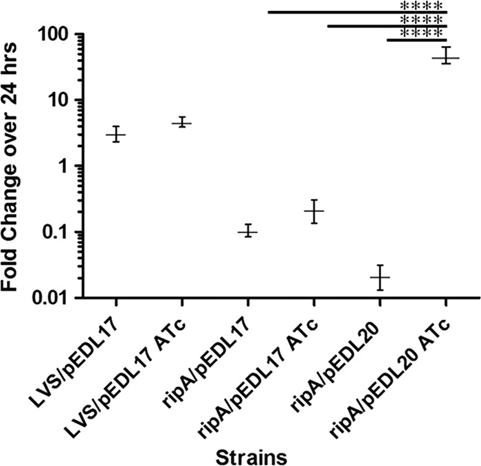
TetR system rescues LVSΔripA intracellular growth in J774A.1 macrophages. Fold change in growth was measured after 24 h during a gentamicin protection assay with LVS bearing the TetR control plasmid pEDL17, LVSΔripA bearing pEDL17, and LVSΔripA containing pEDL20 with Pgrotet-ripA. All strains were added to J774A.1 cells at a multiplicity of infection of 100. Gentamicin was added at 2 h postinfection. At 12 h, 100 ng ml−1 ATc was added to wells. Numbers of CFU were enumerated at 12 and 36 h postinfection. Data are represented as average fold changes in growth (n = 6) between 12 and 36 h postinfection. Error bars represent standard deviations. ****, P < 0.0001.
To create a repressible TetR system in Francisella, we chose a highly efficient revTetR, TetR r1.7 (23, 34). We replaced the tetR gene in pEDL40 with the TetR r1.7 gene to create pEDL47 (Fig. 2C). The plasmid pEDL47 was introduced into LVS, and growth curves were performed in the presence of ATc at 0, 100, or 250 ng ml−1 (Fig. 5). There was no significant difference between strains treated with 0 ng ml−1 ATc and the constitutive ΔtetR strain, which suggests that there is no binding of TetR r1.7 to tetAo in the absence of ATc. Both the 100 and 250 ng ml−1 ATc levels inhibited transcription of the operon, and while it took more than 20 h to reach baseline, it was significantly different from 0 ng ml−1 ATc. This slow decline in luminescence is likely due to the time it takes for depletion of the Lux proteins after transcriptional silencing by TetR r1.7.
Fig 5.
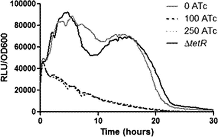
revTetR system. Growth curves were performed with LVS bearing plasmid pEDL47 in the presence of ATc concentrations of 0, 100, and 250 ng ml−1 and LVS bearing pEDL41 ΔtetR as the constitutive control. The absorbance at OD600 and luminescence were monitored every 15 min for 36 h. To determine the relative light produced per bacterium, the luminescence was divided by the absorbance.
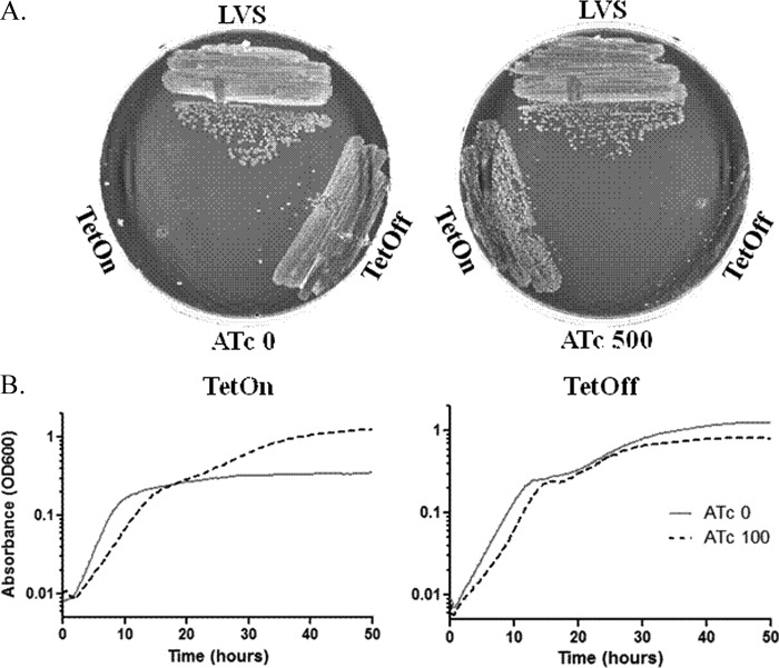
To determine if the TetR and revTetR systems were suitable for silencing gene expression, an essentiality test was performed on SecA, the protein translocase ATPase. SecA is a component of the general secretory pathway (Sec). The Sec pathway consists of three integral membrane proteins, SecY, SecE, and SecG, and two cytosolic proteins, SecA and SecB. The SecYEG complex forms a heterotrimeric channel in the cytoplasmic membrane where the unfolded substrates pass through (32). SecB is a cytosolic chaperone that specifically targets preproteins to SecA (39). SecA is central to the pathway, as it is the ATPase that energizes the transport through SecYEG, as well as recognizing and delivering preproteins to the SecYEG channel (8, 11). In a transposon screen in the closely related Francisella novicida there are two hits in the 3′ end of the secA gene (20), and a study by Margolis et al. found that these secA mutants were defective in biofilm formation, suggesting a defect in protein secretion; however, they were unable to delete the full-length F. novicida secA (31), as SecA is likely essential in all prokaryotes (1, 5, 9, 16, 17, 23). To silence expression of SecA in LVS, we deleted the chromosomal copy of secA in strains bearing either plasmid pEDL54 (FTRp-secA; TetR) or pEDL58 (FTRp-secA; revTetR), which are referred to as the TetOn and TetOff strains, respectively. We streaked LVS, TetOn, and TetOff strains on plates containing 0 or 500 ng ml−1 ATc. The LVS control grew normally in both conditions, and as expected, TetOn grew only in the presence of ATc and TetOff in the absence of ATc (Fig. 6A). There were a few colonies in the primary streak of the silenced strains, suggesting the selection of suppressors. To resolve the nature of the suppression, three overnight cultures of the TetOn strain, grown in 50 ng ml−1 ATc, were plated onto chocolate agar lacking ATc, and 15 colonies were chosen for further analysis. Each strain that grew without ATc was found to harbor a mutation in tetR or tetAo, allowing constitutive production of SecA. These results established SecA as essential; however, the growth kinetics of the two strains on depletion of SecA in CDM might explain whether depletion was bacteriostatic or bactericidal. As expected, the TetOn strain grew without ATc until SecA was depleted but grew to WT levels with ATc present (P < 0.01), and the TetOff strain followed the opposite pattern (P < 0.01) (Fig. 6B). Even though it has been suggested that the lack of SecA or inhibition of SecA is bactericidal (35), these results suggest that depletion was bacteriostatic in LVS; however, at later time points, suppression of the secA deletion did occur in both strains, which obfuscates the results. As it was difficult to maintain a clean population of the TetOn strain because of constant selective pressure, the revTetR system would likely be a better system to use when studying a potentially essential gene.
Fig 6.
SecA is essential. (A) The strains LVS, TetOn (FTRp-secA; TetR), and TetOff (FTRp-secA; revTetR) were streaked onto chocolate agar plates that contained either 0 or 500 ng ml−1 ATc. The plates were allowed to incubate for 72 h at 37°C. (B) Growth curves of either TetOn or TetOff were performed in media containing 0 or 100 ng ml−1 ATc. The absorbance at OD600 was measured every 15 min for 50 h. The growth curves were plotted on a log10 scale. There was a significant difference in final growth as measured by absorbance at OD600, with TetOn being unable to reach WT levels of growth without ATc (P < 0.01) and TetOff unable to reach WT levels with ATc (P < 0.01).
In this study, we developed TetR-based gene regulation systems for Francisella tularensis that allow for inducible or repressible gene expression with addition of ATc and demonstrated that the systems are useful in both broth and cell culture. We further established that the systems can be used effectively for gene silencing. We anticipate these systems will be useful for studies investigating gene dosage, gene timing, and essentiality testing, as well as for expression of toxic proteins. Thus, these systems will be invaluable in addressing protein function in Francisella tularensis.
ACKNOWLEDGMENTS
This work was supported by NIH grant AI082870 to T.H.K. and NIH grant AI068013 to M.S.P.
We thank Miriam Braunstein (University of North Carolina at Chapel Hill), Sabine Ehrt (Weill Cornell Medical College), and Dirk Schnappinger (Weill Cornell Medical College) for TetR r1.7, James Bina (University of Pittsburgh) for the lux plasmid pXB173, and Audrey Chong (Rocky Mountain Laboratories, NIH/NIAID) for thoughtful discussion.
Footnotes
Published ahead of print 20 July 2012
REFERENCES
- 1. Akerley BJ, et al. 1998. Systematic identification of essential genes by in vitro mariner mutagenesis. Proc. Natl. Acad. Sci. U. S. A. 95:8927–8932 [DOI] [PMC free article] [PubMed] [Google Scholar]
- 2. Ausubel FM, et al. 1987. Current protocols in molecular biology. John Wiley and Sons, Inc, New York, NY [Google Scholar]
- 3. Bakshi CS, et al. 2006. Superoxide dismutase B gene (sodB)-deficient mutants of Francisella tularensis demonstrate hypersensitivity to oxidative stress and attenuated virulence. J. Bacteriol. 188:6443–6448 [DOI] [PMC free article] [PubMed] [Google Scholar]
- 4. Beare PA, et al. 2011. Dot/Icm type IVB secretion system requirements for Coxiella burnetii growth in human macrophages. mBio 2:e00175–e00111 doi:10.1128/mBio.00175-11. [DOI] [PMC free article] [PubMed] [Google Scholar]
- 5. Benson RE, Gottlin EB, Christensen DJ, Hamilton PT. 2003. Intracellular expression of peptide fusions for demonstration of protein essentiality in bacteria. Antimicrob. Agents Chemother. 47:2875–2881 [DOI] [PMC free article] [PubMed] [Google Scholar]
- 6. Bertram R, Hillen W. 2008. The application of Tet repressor in prokaryotic gene regulation and expression. Microb. Biotechnol. 1:2–16 [DOI] [PMC free article] [PubMed] [Google Scholar]
- 7. Bina XR, Miller MA, Bina JE. 2010. Construction of a bioluminescence reporter plasmid for Francisella tularensis. Plasmid 64:156–161 [DOI] [PMC free article] [PubMed] [Google Scholar]
- 8. Cabelli RJ, Chen L, Tai PC, Oliver DB. 1988. SecA protein is required for secretory protein translocation into E. coli membrane vesicles. Cell 55:683–692 [DOI] [PubMed] [Google Scholar]
- 9. Caspers M, Freudl R. 2008. Corynebacterium glutamicum possesses two secA homologous genes that are essential for viability. Arch. Microbiol. 189:605–610 [DOI] [PubMed] [Google Scholar]
- 10. Chamberlain RE. 1965. Evaluation of live tularemia vaccine prepared in a chemically defined medium. Appl. Microbiol. 13:232–235 [DOI] [PMC free article] [PubMed] [Google Scholar]
- 11. Cunningham K, Wickner W. 1989. Specific recognition of the leader region of precursor proteins is required for the activation of translocation ATPase of Escherichia coli. Proc. Natl. Acad. Sci. U. S. A. 86:8630–8634 [DOI] [PMC free article] [PubMed] [Google Scholar]
- 12. de Lorenzo V, Timmis KN. 1994. Analysis and construction of stable phenotypes in gram-negative bacteria with Tn5- and Tn10-derived minitransposons. Methods Enzymol. 235:386–405 [DOI] [PubMed] [Google Scholar]
- 13. Dennis DT, et al. 2001. Tularemia as a biological weapon: medical and public health management. JAMA 285:2763–2773 [DOI] [PubMed] [Google Scholar]
- 14. Ehrt S, et al. 2005. Controlling gene expression in mycobacteria with anhydrotetracycline and Tet repressor. Nucleic Acids Res. 33:e21 doi:10.1093/nar/gni013. [DOI] [PMC free article] [PubMed] [Google Scholar]
- 15. Ericsson M, Golovliov I, Sandstrom G, Tarnvik A, Sjostedt A. 1997. Characterization of the nucleotide sequence of the groE operon encoding heat shock proteins chaperone-60 and -10 of Francisella tularensis and determination of the T-cell response to the proteins in individuals vaccinated with F. tularensis. Infect. Immun. 65:1824–1829 [DOI] [PMC free article] [PubMed] [Google Scholar]
- 16. Fagan RP, Fairweather NF. 2011. Clostridium difficile has two parallel and essential Sec secretion systems. J. Biol. Chem. 286:27483–27493 [DOI] [PMC free article] [PubMed] [Google Scholar]
- 17. Forsyth RA, et al. 2002. A genome-wide strategy for the identification of essential genes in Staphylococcus aureus. Mol. Microbiol. 43:1387–1400 [DOI] [PubMed] [Google Scholar]
- 18. Fuller JR, et al. 2008. RipA, a cytoplasmic membrane protein conserved among Francisella species, is required for intracellular survival. Infect. Immun. 76:4934–4943 [DOI] [PMC free article] [PubMed] [Google Scholar]
- 19. Fuller JR, Kijek TM, Taft-Benz S, Kawula TH. 2009. Environmental and intracellular regulation of Francisella tularensis ripA. BMC Microbiol. 9:216 doi:10.1186/1471-2180-9-216. [DOI] [PMC free article] [PubMed] [Google Scholar]
- 20. Gallagher LA, et al. 2007. A comprehensive transposon mutant library of Francisella novicida, a bioweapon surrogate. Proc. Natl. Acad. Sci. U. S. A. 104:1009–1014 [DOI] [PMC free article] [PubMed] [Google Scholar]
- 21. Gossen M, Bujard H. 1993. Anhydrotetracycline, a novel effector for tetracycline controlled gene expression systems in eukaryotic cells. Nucleic Acids Res. 21:4411–4412 [DOI] [PMC free article] [PubMed] [Google Scholar]
- 22. Grall N, et al. 2009. Pivotal role of the Francisella tularensis heat-shock sigma factor RpoH. Microbiology 155:2560–2572 [DOI] [PMC free article] [PubMed] [Google Scholar]
- 23. Guo XV, et al. 2007. Silencing Mycobacterium smegmatis by using tetracycline repressors. J. Bacteriol. 189:4614–4623 [DOI] [PMC free article] [PubMed] [Google Scholar]
- 24. Horzempa J, Carlson PE, Jr, O'Dee DM, Shanks RM, Nau GJ. 2008. Global transcriptional response to mammalian temperature provides new insight into Francisella tularensis pathogenesis. BMC Microbiol. 8:172 doi:10.1186/1471-2180-8-172. [DOI] [PMC free article] [PubMed] [Google Scholar]
- 25. Horzempa J, et al. 2010. Utilization of an unstable plasmid and the I-SceI endonuclease to generate routine markerless deletion mutants in Francisella tularensis. J. Microbiol. Methods 80:106–108 [DOI] [PMC free article] [PubMed] [Google Scholar]
- 26. Horzempa J, Tarwacki DM, Carlson PE, Jr, Robinson CM, Nau GJ. 2008. Characterization and application of a glucose-repressible promoter in Francisella tularensis. Appl. Environ. Microbiol. 74:2161–2170 [DOI] [PMC free article] [PubMed] [Google Scholar]
- 27. Lathem WW, Price PA, Miller VL, Goldman WE. 2007. A plasminogen-activating protease specifically controls the development of primary pneumonic plague. Science 315:509–513 [DOI] [PubMed] [Google Scholar]
- 28. LoVullo ED, Molins-Schneekloth CR, Schweizer HP, Pavelka MS., Jr 2009. Single-copy chromosomal integration systems for Francisella tularensis. Microbiology 155:1152–1163 [DOI] [PMC free article] [PubMed] [Google Scholar]
- 29. LoVullo ED, Sherrill LA, Pavelka MS., Jr 2009. Improved shuttle vectors for Francisella tularensis genetics. FEMS Microbiol. Lett. 291:95–102 [DOI] [PMC free article] [PubMed] [Google Scholar]
- 30. LoVullo ED, Sherrill LA, Perez LL, Pavelka MS., Jr 2006. Genetic tools for highly pathogenic Francisella tularensis subsp. tularensis. Microbiology 152:3425–3435 [DOI] [PubMed] [Google Scholar]
- 31. Margolis JJ, et al. 2010. Contributions of Francisella tularensis subsp. novicida chitinases and Sec secretion system to biofilm formation on chitin. Appl. Environ. Microbiol. 76:596–608 [DOI] [PMC free article] [PubMed] [Google Scholar]
- 32. Meyer TH, et al. 1999. The bacterial SecY/E translocation complex forms channel-like structures similar to those of the eukaryotic Sec61p complex. J. Mol. Biol. 285:1789–1800 [DOI] [PubMed] [Google Scholar]
- 33. Rigel NW, et al. 2009. The accessory SecA2 system of mycobacteria requires ATP binding and the canonical SecA1. J. Biol. Chem. 284:9927–9936 [DOI] [PMC free article] [PubMed] [Google Scholar]
- 34. Scholz O, et al. 2004. Activity reversal of Tet repressor caused by single amino acid exchanges. Mol. Microbiol. 53:777–789 [DOI] [PubMed] [Google Scholar]
- 35. Segers K, Anne J. 2011. Traffic jam at the bacterial sec translocase: targeting the SecA nanomotor by small-molecule inhibitors. Chem. Biol. 18:685–698 [DOI] [PubMed] [Google Scholar]
- 36. Shaner NC, et al. 2008. Improving the photostability of bright monomeric orange and red fluorescent proteins. Nat. Methods 5:545–551 [DOI] [PMC free article] [PubMed] [Google Scholar]
- 37. Starr T, et al. 2012. Selective subversion of autophagy complexes facilitates completion of the Brucella intracellular cycle. Cell Host Microbe 11:33–45 [DOI] [PMC free article] [PubMed] [Google Scholar]
- 38. Tarnok A, Dorger M, Berg I, Gercken G, Schluter T. 2001. Rapid screening of possible cytotoxic effects of particulate air pollutants by measurement of changes in cytoplasmic free calcium, cytosolic pH, and plasma membrane potential in alveolar macrophages by flow cytometry. Cytometry 43:204–210 [DOI] [PubMed] [Google Scholar]
- 39. Valent QA, et al. 1998. The Escherichia coli SRP and SecB targeting pathways converge at the translocon. EMBO J. 17:2504–2512 [DOI] [PMC free article] [PubMed] [Google Scholar]


