Abstract
Exposing mice to a social stressor called social disruption (SDR) that involves repeated social defeat during intermale aggression results in increased circulating cytokines, such as interleukin-1α (IL-1α) and IL-1β, and increased reactivity of splenic CD11b+ macrophages to inflammatory stimuli. For example, upon lipopolysaccharide stimulation, macrophages from stressor-exposed mice produce higher levels of cytokines than do cells from nonstressed controls. Moreover, the SDR stressor enhances the ability of these macrophages to kill Escherichia coli both in vitro and in vivo, through a Toll-like receptor 4-dependent mechanism. The present study tested the hypothesis that stressor-enhanced bacterial killing is due to increases in the production of peroxynitrite. Male mice were exposed to the SDR stressor or were left undisturbed. Upon stimulation with E. coli, splenic macrophages from SDR-exposed mice expressed significantly increased levels of inducible nitric oxide synthase mRNA and produced higher levels of peroxynitrite. Blocking the production of peroxynitrite abrogated the SDR-induced increase in microbicidal activity. Studies in IL-1 receptor type 1 knockout mice indicated that the increased microbicidal activity and peroxynitrite production was dependent upon IL-1 signaling. These data confirm and extend the importance of IL-1 signaling for stressor-induced immunopotentiation; the finding that inhibiting superoxide or nitric oxide production inhibits both peroxynitrite production and killing of E. coli demonstrates that peroxynitrite mediates the stressor-induced increase in bacterial killing.
INTRODUCTION
Macrophages play a fundamental role in innate immunity and are highly efficient phagocytes important in the first wave of immune defense. Multiple studies now indicate that macrophage activity can be strongly modified by environmental factors, such as the physiological response to social and psychological stressors (10, 21, 52). The mammalian response to stressors involves activation of many physiological systems, including the sympathetic nervous system (SNS) and the hypothalamic-pituitary-adrenal (HPA) axis (18). These stressor-induced physiological responses have been shown to impact immune system functioning and the outcome of pathogenic challenge. Most studies have focused on the ability of stressors to suppress immunity, and it is now known that factors such as HPA axis-derived glucocorticoid (GC) hormones can suppress important transcription factors, such as NF-κB (61). As a result, prolonged elevations of glucocorticoids have been shown to suppress macrophage activity (2, 10, 21, 53).
Exposing mice to a widely used and well characterized social stressor involving intermale aggression, called social disruption (SDR), reduces splenic macrophage sensitivity to the suppressive effects of stressor-induced GC hormones. This reduced sensitivity is associated with the development of splenomegaly due to an accumulation of myeloid-derived cells (29). These splenic cells are also more reactive to microbial antigens; they produce higher levels of cytokines upon lipopolysaccharide (LPS) challenge (3, 8, 56) and have enhanced microbicidal activity, in part through Toll-like receptor (TLR) signaling (7). While the physiological parameters by which stressor exposure enhances immune activity are becoming more well defined and involve stressor-induced SNS activity (18, 23, 28, 36) and alterations to the intestinal microbiota (1, 6), the cellular events responsible for the increased ability of splenic macrophages to kill bacteria have not been widely studied. Therefore, the purpose of the present study was to determine key cellular events necessary for stressor exposure to enhance the ability of splenic macrophages to kill microbes.
Macrophages use a variety of mechanisms and effector molecules to kill microbes (54). Their primary method, however, involves the rapid production of reactive molecules associated with an “oxidative burst.” These reactive molecules are then able to damage proteins and DNA of microbial cells (13, 40). A major enzyme responsible for reactive molecule production is inducible nitric oxide synthase (iNOS). Shortly after phagocytosis, iNOS is expressed de novo in the cytosol of macrophages where it converts the amino acid arginine into citrulline and the reactive molecule, nitric oxide (49). Nitric oxide then diffuses across the phagosome membrane, where it can damage the engulfed microbe. In addition, the NADPH oxidase complex located on the phagosome membrane produces superoxide anion, which rapidly combines with nitric oxide to produce a highly destructive intermediate, peroxynitrite. Peroxynitrite is a powerful oxidant that can damage and inactivate many crucial bacterial proteins (47). Since these molecules are vital to macrophage killing, and because previous studies have suggested that SDR exposure enhances iNOS gene expression and the assembly of the NADPH oxidase complex (7), we hypothesized that SDR-induced increases in bacterial killing involve heightened expression of iNOS and subsequent peroxynitrite production.
Previous studies have shown that interleukin-1 (IL-1) is increased in the circulation and secondary lymphoid organs of stressor-exposed mice (30). This increased IL-1 is thought to be responsible for the insensitivity of splenic macrophages to the suppressive effects of glucocorticoid hormones; IL-1R1 knockout mice did not develop glucocorticoid insensitivity after exposure to the SDR stressor. Whether IL-1R1 signaling also impacts the microbicidal activity of splenic macrophages, however, has not been tested. This is a logical hypothesis, because it has already been shown that Toll-like receptor signaling is necessary for the enhanced microbicidal activity to occur (7), and IL-1R1 signaling shares significant downstream pathways with Toll-like receptors (11, 45, 50). Moreover, IL-1R1 signaling is known to induce iNOS gene expression in part through signaling through the mitogen-activated protein kinases (MAPKs) (19, 35, 39, 59). Thus, we tested the hypothesis that SDR-induced increases in microbicidal activity and peroxynitrite production were IL-1R1 dependent.
MATERIALS AND METHODS
Animal handling.
Male C57BL/6 and CD-1 mice between the ages of 4 and 6 weeks were purchased from Charles River Laboratories (Wilmington, MA). IL-1R1−/− mice were bred at Ohio State University and were descendants of a breeding pair originally obtained from Jackson Laboratory (Bar Harbor, ME). They were shown to exhibit a normal phenotype without overt abnormalities in the immune and hematopoietic systems and their response to LPS challenge was equivalent to wild-type (WT) mice (30, 33). Prior to experimentation, the mice were housed three to five per cage and allowed to adjust to the animal vivarium for 1 week. They were kept on a 12:12-h light-dark schedule with lights on at 6:00 a.m. and with food and water available ad libitum. The Ohio State University's Animal Care and Use Committee approved all experimental procedures.
Social disruption.
The social disruption (SDR) stressor is a widely used and extensively characterized murine stressor (4, 5, 55) that entails repeated social defeat from agonistic interactions between an aggressive intruder male mouse and the resident male mice. The SDR stressor occurred between 5:00 p.m. and 7:00 p.m., which spanned the beginning of the dark (i.e., active) cycle. The aggressive intruders used to defeat the home cage residents were mice of the same strain as the residents that had been previously identified as aggressive toward their cagemates and isolated from the rest of the colony. SDR began by placing an aggressive mouse into the home cage of the resident mice. The mice were observed for the first 10 min to ensure that the aggressor fought with and defeated all of the residents. If after 5 min the aggressor had not fought and defeated all of the residents, a different aggressor was added until this occurred. After each 2-h SDR session, the aggressor was removed, and the resident mice were left undisturbed until the next day. After the sixth repeated day of SDR, the defeated home cage residents were sacrificed. Although fighting in mice can result in wounding, significant wounding typically occurs in <5% of mice. In this experiment, wounds were limited to superficial scratches on the hind quarters and tail. Control mice were left undisturbed in their home cages throughout the experiment and are referred to as home cage control (HCC) mice.
Splenocyte processing.
Mice were euthanized via CO2 asphyxiation and cardiac blood immediately taken via cardiac puncture. The spleens were removed and macerated with a Stomacher 80 Biomaster Lab System (Seward, Bohemia, NY) in 5 ml of ice-cold Hanks balanced salt solution (HBSS) for 1 min. The resultant cell suspensions were washed in 10 ml of ice-cold HBSS, and the cells were washed and then pelleted by centrifuging at 650 × g for 10 min at 4°C. After centrifugation, the supernatants were removed, and 1 ml of red blood cell lysis buffer (4.4 g of NH4Cl, 0.5 g of KHCO3, 0.019 g of EDTA, 500 ml of distilled H2O [pH 7.3]) was added to the cell pellet for 2 min. To stop the lysis reaction, 10 ml of HBSS containing 10% heat-inactivated fetal bovine serum (FBS) was added to the cells. After washing in HBSS plus 10% FBS, the cells were filtered using 70-μm-pore-size filters, washed again, and resuspended in 10 ml of RPMI 1640 supplemented with 0.075% sodium bicarbonate, 10 mM HEPES buffer, 100 U of penicillin G/ml, 100 μg of streptomycin sulfate/ml, 1.5 mM l-glutamine, 0.00035% 2-mercaptoethanol, and 10% heat-inactivated FBS. The cells were counted using a Z2 Coulter particle counter and size analyzer (Beckman Coulter, Brea, CA), and each sample was adjusted to contain 5 × 10⁶ cells/ml. A total of 2.5 × 10⁵ cells were then stained with 1 μl of fluorescein isothiocyanate-labeled anti-Gr1 and APC-labeled anti-CD11b to determine the number of monocytes/macrophages, neutrophils, and lymphocytes in each suspension using a FACSCalibur flow cytometer (BD Biosciences, San Jose, CA). The cell suspension was then adjusted so that the final concentration of monocytes/macrophages was 5 × 10⁴ monocytes/macrophages per ml. The cells were cultured in 1 ml per well in a 24-well tissue culture plate for 2 h at 37°C and 5% CO2 to allow the macrophages to adhere.
Bacteria.
Escherichia coli strain K-12 (ATCC 10798), originally obtained from the American Type Culture Collection (Manassas, VA), was grown from frozen stocks in Trypticase soy broth overnight at 37°C.
Microbicidal assay.
A total of 5 × 10⁴ CD11b+ monocytes/macrophages from individual animals were plated in duplicate in a volume of 1 ml per well on 24-well tissue culture plates to determine phagocytosis and bacterial killing by the adherent splenocytes after 20 and 90 min, respectively. The cells were incubated for 2 h to allow the macrophages to adhere. After the 2-h incubation, the nonadherent cells were washed from the plates by three rinses with 1 ml of RPMI 1640. After the final wash, 1 ml of a mixture containing 5 × 10⁷ CFU of E. coli in RPMI 1640 was added to each well, and the plates pulse-centrifuged to help pull the E. coli onto the adherent cells. The E. coli were opsonized by adding 5% fresh serum (pooled from nonstressed HCC mice and stressed SDR mice) to the bacterial suspension. After the bacteria were incubated for 20 min, the extracellular bacteria were washed away by pipetting 1 ml of RPMI 1640 into the well for three washes. After the final wash, 1 ml of 1% Triton X-100 was added to one of the wells to lyse the splenocytes. The lysate was collected into a sterile tube for pour plate enumeration of phagocytized bacteria. The duplicate well was washed three times with RPMI 1640 supplemented with 10% FBS to remove extracellular bacteria. After the final wash, 1 ml of RPMI 1640 supplemented with 10% FBS was added to the duplicate wells, and the plates were incubated for an additional 70 min. After 70 min, the cells were again washed and then lysed with 1% Triton X-100 to quantify bacteria remaining alive within the macrophages via standard pour plate analysis.
Pharmacological inhibitors of macrophage function.
To inhibit the production of nitric oxide by iNOS during the microbicidal assay, 500 μM NG-methyl-l-arginine acetate salt (L-NMMA; Sigma-Aldrich, St. Louis, MO) was added to the RPMI 1640 supplemented with 10% FBS during both the 2 h adherence and during coincubation with E. coli. To scavenge peroxynitrite, 25 μM 5,10,15,20-tetrakis(4-sulfonatophenyl)prophyrinato-iron(III) chloride (FeTPPS; Sigma-Aldrich) was added during the 2-h adherence and during coincubation with E. coli. To inhibit NADPH oxidase production of superoxide, 300 μM apocynin was added during the 2-h adherence and during coincubation with E. coli.
RNA isolation and cDNA synthesis.
Total splenocytes from individual SDR and HCC control mice were cultured in duplicate sets at 5 × 10⁴ adherent macrophages per ml in 24-well tissue culture plates at 37°C and 5% CO2 for 2 h. After the 2-h incubation, the nonadherent cells were washed off by three rinses with RPMI 1640. After the final wash, 1 ml of RPMI 1640 supplemented with 10% FBS was added to half of the wells, and the other half of the cells were stimulated with 1 μg of LPS/ml in 1 ml of RPMI 1640 supplemented with 10% FBS. After incubation for 90 min, the cells were washed with RPMI 1640, and 1 ml of TRIzol reagent (Gibco, Rockville, MD) was added to each well to isolate RNA. RNA was isolated according to the TRIzol protocol provided by the manufacturer (Gibco). Total RNA was reverse transcribed using a commercially available kit (Promega, Madison, WI) according to the manufacturer's instructions. Briefly, 1 μg of total RNA was combined with 5 mM MgCl2, a 1 mM concentration of each deoxynucleoside triphosphate, 1× reverse transcriptase buffer, 1 U of RNasin/μl, and 15 U of avian myeloblastosis virus reverse transcriptase/μg and primed with 1 μg of random hexamers and diethyl pyrocarbonate-H2O to a volume of 20 μl. The reaction was first incubated for 10 min at room temperature and then at 42°C for 1 h. This was followed by 5 min of incubation in boiling water and then a cooling period of 5 min on ice. The volume was adjusted to 50 μl by adding 30 μl of nuclease-free water.
RT-PCR.
Primers and probes were synthesized by Applied Biosystems (Foster City, CA). The 5′-3′ sequences were as follows: iNOS (forward, CAG CTG GGC TGT ACA AAC CTT; reverse, TGA ATG TGA TGT TTG CTT CGG, probe: CGG GCA GCC TGT GAG ACC TTT GA) and 18S (forward, CGG CTA CCA CAT CCA AGG AA; reverse, GCT GGA ATT ACC GCG GCT; probe, TGC TGG CAC CAG ACT TGC CCT C). The PCR mixture consisted of 2.5 μl of cDNA, 2.5 μl of primer mix (900 nM), 2.5 μl of probe, 5 μl of sterile distilled H2O, and 12.5 μl of TaqMan universal master mix (Applied Biosystems) for a final volume of 25 μl. After an initial 2 min cycle at 50°C, followed by 10 min at 95°C, the reaction ran for 40 total cycles consisting of a 15-s denaturing phase (90°C) and a 1-min annealing/extension phase (60°C). The change in fluorescence was measured using an Applied Biosystems 7000 Prism sequence detector (Applied Biosystems) and analyzed using Sequence Detector v1.0. The relative amount of transcript was determined using the comparative threshold cycle (CT) method as described by the manufacturer. In these experiments, gene expression in the untreated cells from the spleens of the nonstressed HCC mice was used as the calibrator. Gene expression, therefore, is expressed as the fold increase in expression over nonstressed HCC control mice.
Peroxynitrite and superoxide measurement.
After spleen cells from individual animals were processed to a single cell suspension, CD11b+ cells were magnetically separated from the total cells using anti-CD11b+ microbeads and MACS MS separation columns according to the manufacturer's directions (Miltenyi Biotec, Auburn, CA). The separated CD11b+ cells were suspended in 1 ml of RPMI 1640 supplemented with 10% FBS and counted using a Coulter counter. After counting, the cells were adjusted to 3 × 10⁶ cells/ml and were plated in two sets of duplicate wells on opaque 96-well tissue culture plates in a volume of 200 μl/well. The cells were incubated for 2 h at 37°C and 5% CO2 to allow the cells to adhere. After the 2-h incubation, the adherent cells were rinsed three times with HBSS without phenol red to remove nonadherent cells. To all wells, 100 μl of HBSS without phenol red containing 25 μM 123-dihydrorhodamine (catalog no. D23806; Invitrogen, Eugene, OR), a peroxynitrite detecting dye, or 5 μM dihydroethidium (DHE; Sigma-Aldrich), a superoxide detecting dye, were added. Half of the wells were stimulated with 5 ng of phorbol 12-myristate 13-acetate (PMA; Sigma-Aldrich)/ml, 1 μg of E. coli LPS (Sigma)/ml, and 5 ng of recombinant mouse gamma interferon (IFN-γ; Thermo Scientific, Rockford, IL)/ml. Immediately after the media were added, the peroxynitrite or superoxide production was measured using a fluorescence microplate reader (Synergy HT Biotek, Winooski, VT) every 15 min for 90 min using excitation and emission wavelengths of 544 nm and 618 nm, respectively, for peroxynitrite detection using 123 DHR, or 544 nm and 618 nm for superoxide detection using DHE.
Nitrite assay.
Splenic CD11b+ cells from individual animals were enriched via magnetic bead separation and placed in 24-well tissue cultures plates for 2 h to allow the macrophages to adhere. The adherent macrophages were then stimulated with E. coli-derived LPS (1 μg/ml) with or without IFN-γ (5 ng/ml) for 24 h. To determine whether nitrite production was dependent upon IFN-γ dose, cells were also stimulated with a higher dose of IFN-γ (i.e., 10 ng/ml). Nitrite levels in the culture supernatants were determined via the Griess reaction using a colorimetric assay (Cayman Chemical Co., Ann Arbor, MI).
Data and statistical analysis.
All data are from cells from individual animals. Each experiment was replicated two to five times. In experiments involving superoxide and peroxynitrite assessments, duplicate or triplicate wells containing cells from the same mouse were averaged prior to calculating the group average. All data are presented as means ± the standard errors. Differences in iNOS mRNA and nitrite levels were determined by using the Student t test. A mixed-factor analysis of variance (ANOVA) was used to determine statistical significance in the number of bacteria remaining alive in coculture with splenic cells, with time (i.e., 20 min versus 90 min) as the repeated variable and group (i.e., HCC control versus SDR stressor) as the between-subjects factor. A mixed-factor ANOVA was also used to determine statistical significance for the peroxynitrite and superoxide anion measurements with time (i.e., 0, 15, 30, 45, 60, 75, and 90 min) as the within-subjects factor and group (i.e., HCC control versus SDR stressor) as the between-subjects factor. Protected t tests were used as post hoc tests. In all cases, the level of significance was set at P < 0.05. All statistics were calculated using SPSS for Windows version 17 (SPSS, Chicago, IL).
RESULTS
Reactive nitrogen species are partly responsible for the enhanced bactericidal activity in CD11b+ cells from SDR-exposed mice.
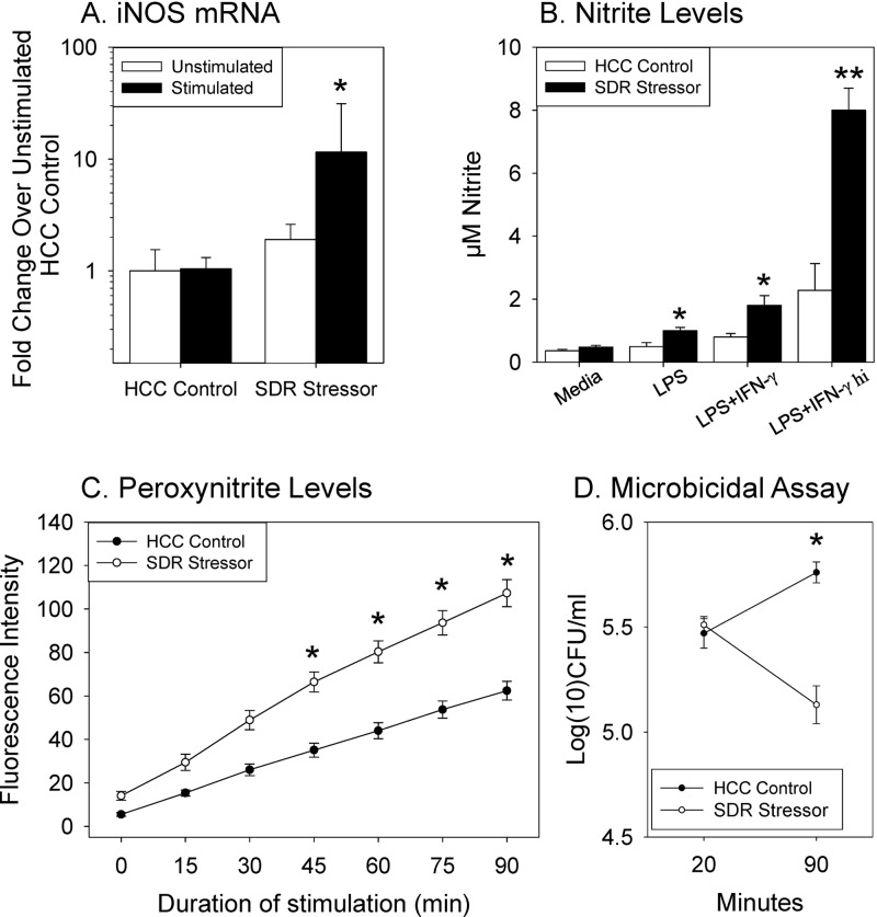
Splenocytes from mice exposed to SDR had significantly increased iNOS mRNA expression in comparison to nonstressed HCC controls [F(1,28) = 4.96, P < 0.05; Fig. 1A]. Although the mean level of iNOS gene expression was increased in unstimulated cells from mice exposed to SDR, the addition of E. coli to the cells for 90 min increased iNOS mRNA ∼10-fold over levels found in cells from HCC mice (P < 0.05; Fig. 1A). The increased iNOS mRNA was associated with a significant stressor-induced increase in the production of nitric oxide-derived nitrite after stimulation [F(3,44) = 36.50, P < 0.001; Fig. 1B]. Nitrite production was in part dependent upon the dose of IFN-γ, with cells from SDR stressor-exposed mice stimulated with LPS and the highest dose of IFN-γ producing more nitrite than all other groups (P < 0.05; Fig. 1B). Moreover, the increase in nitrite production was associated with a significant increase in the production of peroxynitrite (Fig. 1C). Cells from mice exposed to SDR produced significantly higher amounts of peroxynitrite upon stimulation with PMA/LPS/IFN-γ than did cells from nonstressed HCC control mice [F(6,186) = 20.77, P < 0.001]. This was evident at every time point tested (P < 0.05). Importantly, when cells from mice exposed to the SDR stressor were cocultured with E. coli, they killed significantly more E. coli over the 90 min time course than did cells from nonstressed control mice (Fig. 1D). This effect was evident when the CFU/ml were determined [F(1,24) = 44.39, P < 0.001].
Fig 1.
Exposure to the SDR stressor significantly enhances splenic macrophage activity. (A) Significant increase in iNOS mRNA expression in SDR mice stimulated with E. coli compared to mRNA in nonstressed HCC control mice. Values are means ± the strandard errors (SE) (n = 9 SDR and 8 HCC from three different experiments; *, P < 0.05 versus HCC E. coli). (B) Significant increase in the levels of nitric oxide-derived nitrite by LPS-, LPS + IFN-γ (5 ng/ml)-, and LPS + IFN-γ hi (10 ng/ml)-stimulated splenic macrophages from mice exposed to SDR compared to nonstressed HCC control mice. Values are means ± the SE (n = 8 SDR and n = 8 HCC from two different experiments; *, P < 0.05 versus cells from HCC controls; **, P < 0.05 versus all other groups). (C) Significant increase in production of peroxynitrite in PMA/LPS/IFN-γ-stimulated splenic macrophages from mice exposed to the SDR stressor compared to nonstressed HCC mice. Values are fluorescence intensity means ± the SE (n = 15 SDR and n = 18 HCC from five different experiments; *, P < 0.001). (D) Significant decrease in E. coli bacterial counts in SDR splenocytes at 90 min. Bacterial counts determined via plate counts obtained 20 min after addition of bacteria (to calculate number of bacteria phagocytosed) and 90 min after addition of bacteria (to calculate number of remaining phagocytosed bacteria). Values are means ± the SE (n = 13 SDR and n = 13 HCC from three different experiments; *, P < 0.05 versus SDR 20 min).
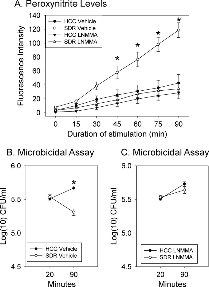
To determine whether increased nitric oxide production was responsible for the stressor-induced increase in peroxynitrite production and the enhanced macrophage bactericidal activity, cells from SDR stressor-exposed and nonstressed HCC control mice were treated with L-NMMA, which blocks nitric oxide production (31, 51). Upon stimulation, the vehicle-treated cells from mice exposed to the SDR stressor produced higher levels of peroxynitrite than did vehicle-treated cells from HCC control mice [F(6,66) = 21.90, P < 0.001] (Fig. 2A). However, after L-NMMA was added to the culture, there was no difference in peroxynitrite production in stimulated cells from stressor-exposed and nonstressed control mice [F(6,66) = 0.14, not significant; Fig. 2A]. This finding is consistent with data obtained in the microbicidal activity assay. Although splenic macrophages from stressor-exposed mice significantly reduced the number of E. coli within the cells during the 90-min period in the presence of vehicle [F(1,10) = 5.20, P < 0.05], splenic macrophages from the nonstressed mice did not kill the phagocytosed E. coli (Fig. 2B). Blocking the production of nitric oxide with L-NMMA significantly affected the ability of cells to kill E. coli (Fig. 2C). In this case, neither the splenic macrophages from the SDR stressor-exposed or the nonstressed HCC control mice were able to kill the E. coli, and there was no significant effect of the stressor on the number of bacteria killed when L-NMMA was in the cultures [F(1,10) = 1.13, not significant].
Fig 2.
Inhibiting iNOS activity abrogates the stressor-induced increase in microbicidal activity. (A) Peroxynitrite production was significantly higher in PMA/LPS/IFN-γ-stimulated splenic macrophages derived from mice exposed to the SDR stressor (*, P < 0.05 versus all other groups at designated time points). However, the addition of L-NMMA to the cultures prevented the stressor-induced increase in peroxynitrite production. Values are fluorescence intensity means ± the SE (n = 6 HCC and n = 7 SDR from two different experiments). (B) Exposure to the SDR stressor significantly increased the number of bacteria killed at the 90 min time point (*, P < 0.05 versus HCC control). However, the addition of the iNOS inhibitor L-NMMA prevented this stressor-induced increase in bacterial killing (n = 6 HCC and n = 6 SDR from two different experiments). Overall, the bacterial levels were lower in the cultures containing cells from SDR-exposed mice (main effect: *, P < 0.05). (C) Cells from SDR-exposed mice killed a significantly greater percentage of the E. coli than did cells from the nonstressed HCC control mice (*, P < 0.05). Addition of L-NMMA prevented the stressor-induced increase in the number of bacteria killed (n = 6 HCC and n = 6 SDR from two different experiments).
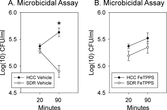
Because the iNOS enzyme, and thus the production of nitric oxide, can lead to the production of reactive molecules other than peroxynitrite (12, 27), we next confirmed whether the enhanced microbicidal activity was indeed abolished upon treatment with FeTPPS, which is a peroxynitrite scavenger (38, 43, 44). Vehicle-treated splenic macrophages from mice exposed to the SDR stressor significantly reduced E. coli levels during the 90 min culture in comparison to levels in splenic macrophages from nonstressed control mice [F(1,14) = 23.88, P < 0.001; Fig. 3A]. However, blocking the effects of peroxynitrite abolished the stressor-induced increase in microbicidal activity as indicated by a lack of a reduction in the number of E. coli by cells in mice from either the HCC control or SDR stressor-exposed mice after 90 min in culture [F(1,16) = 0.01, not significant; Fig. 3B]. These data indicate that the capacity of spleen cells to kill E. coli is dependent upon peroxynitrite production.
Fig 3.
Use of a peroxynitrite scavenger prevented the stressor-induced increase in microbicidal activity. (A) The number of E. coli remaining alive in the cultures at the 90-min time point was significantly lower in the presence of splenic macrophages from SDR-exposed mice compared to nonstressed controls. (B) When the peroxynitrite scavenger FeTPPS was added to the cultures, there was no difference in E. coli levels in cultures containing cells from SDR exposed or from HCC control mice (*, P < 0.05; n = 7 HCC and n = 10 SDR from two different experiments).
Reactive oxygen species are partly responsible for the enhanced bactericidal activity in CD11b+ cells from SDR-exposed mice.
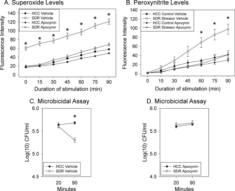
Since peroxynitrite is the reaction product of nitric oxide and superoxide anion, we assessed the role of superoxide anion production in contributing to the increase in peroxynitrite that is needed for the stressor-induced increase in bacterial killing. Superoxide anion production was induced by in vitro stimulation of CD11b+ splenocytes from stressed and nonstressed animals with PMA/LPS/IFN-γ. Vehicle-treated cells from SDR stressor-exposed animals produced significantly more superoxide anion than did cells from nonstressed HCC control mice or from cells treated with apocynin to block NADPH oxidase activity [F(6,60) = 10.17, P < 0.001; Fig. 4A]. In addition, vehicle-treated cells from SDR stressor-exposed animals produced significantly more peroxynitrite than did cells from nonstressed HCC control mice or from cells treated with apocynin to block NADPH oxidase activity [F(6,60) = 57.10, P < 0.05; Fig. 4B]. Importantly, stressor-exposure increased killing of E. coli in vehicle-treated mice [F(1,27) = 27.20, P < 0.001; Fig. 4C], whereas inhibition of NADPH oxidase activity eliminated the stressor-enhanced bacterial killing during in vitro culturing [F(1,28) = 3.21, not significant; Fig. 4D].
Fig 4.
Stressor exposure increases both superoxide anion and nitric oxide production; these intermediates react to form peroxynitrite.(A) Stressor exposure increases the production of superoxide anion, and in vitro inhibition of NADPH oxidase activity eliminates this increase (n = 6 HCC and n = 6 SDR from two different experiments; *, P < 0.05). (B) Inhibiting the activity of NADPH oxidase significantly reduces stressor-induced peroxynitrite production (*, P < 0.05 at 45, 60, 75, and 90 min; n = 6 HCC and n = 6 SDR from two different experiments). (C) Vehicle-treated stressor-exposed mice killed more E. coli in vitro than nonstressed controls (n = 15 HCC and n = 15 SDR from four different experiments] (P < 0.05 versus HCC control). (D) Reduction of peroxynitrite production through blocking the formation of superoxide anion significantly reduces the stress-induced increase in bacterial killing (*, P < 0.05 versus HCC; n = 15 HCC and n = 15 SDR from four different experiments).
IL-1 signaling contributes to enhanced bactericidal activity in CD11b+ cells from SDR-exposed mice.
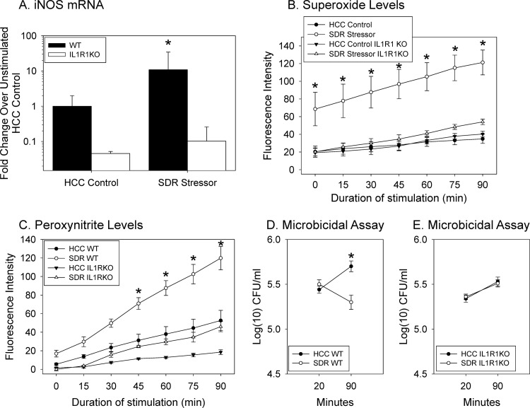
Previous studies have shown that IL-1α and IL-1β mRNA expression and protein production are increased in secondary lymphoid organs following SDR (30). Also, elimination of IL-1R1 signaling through the use of an IL-1R1 knockout (IL-1R1−/−) mouse strain demonstrated that some of the effects of SDR on the immune system, namely, trafficking of CD11b+ cells to the spleen and the development of glucocorticoid insensitivity in this cell population, fail to appear (30). Therefore, we further examined whether IL-1R1 signaling was necessary for the SDR stressor-enhanced iNOS gene expression, superoxide anion and peroxynitrite production, and microbicidal activity. Splenic macrophages from wild-type C57BL/6 mice had a significant increase in iNOS mRNA after E. coli stimulation in comparison to iNOS mRNA in splenic macrophages from nonstressed control mice [t(15) = 2.19, P < 0.05; Fig. 5A]. This stressor-induced increase in iNOS mRNA was not evident in the IL-1R1−/− mice [t(4) = 1.16, not significant] (Fig. 5A). Likewise, cells from stressor-exposed wild-type C57BL/6 mice produced higher levels of both superoxide anion [F(6,120) = 9.64, P < 0.001; Fig. 5B] and peroxynitrite [F(6,95) = 11.01, P < 0.001; Fig. 5C]. Importantly, both superoxide and peroxynitrite production was significantly lower in the SDR-exposed IL-1R1−/− mice compared to wild-type mice exposed to SDR (P < 0.05). Stressor-exposed WT mice killed more E. coli than nonstressed controls [F(1,14) = 36.40, P < 0.001; Fig. 5D]. However, stressor-exposure failed to increase microbicidal activity in the splenic macrophages from IL-1R1−/− mice (Fig. 5E). The number of E. coli within the cells from stressor-exposed mice was similar to the number found within the nonstressed controls after 90 min in culture [F(1,27) = 0.21, not significant] (Fig. 5E), and neither the stressor-exposed nor the nonstressed IL-1R1−/− mice were able to kill E. coli (Fig. 5E).
Fig 5.
Stressor-induced increases in splenic macrophage activity do not manifest in IL-1R1−/− mice. (A) Exposure to the SDR stressor did not enhance iNOS mRNA in the splenic macrophages from IL-1R1−/− mice stimulated with E. coli for 90 min. The stressor did increase iNOS mRNA in wild-type mice (*, P < 0.05; n = 8 WT HCC, n = 7 WT SDR, n = 3 IL-1R1−/− HCC, and n = 3 IL-1R1−/− SDR from two different experiments). (B) Superoxide production did not differ with stress in IL-1R1−/− mice (*, P < 0.05, WT SDR versus all other groups; n = 7 WT HCC, n = 5 IL-1R1−/− HCC, n = 7 WT SDR, and n = 5 IL-1R1−/− SDR from two different experiments). (C) The production of peroxynitrite by PMA/LPS/IFN-γ-stimulated splenic macrophages from WT mice exposed to the SDR stressor was significantly higher than peroxynitrite production by all other groups (*, P < 0.05 versus all other groups at designated time points). Peroxynitrite production by splenic macrophages from IL-1R1−/− mice exposed to the SDR stressor was significantly lower than peroxynitrite production by splenic macrophages from the WT mice exposed to the SDR stressor (*, P < 0.05 at 45, 60, 75, and 90 min; n = 6 WT HCC, n = 6 WT SDR, n = 5 IL-1R1−/−, and n = 5 IL-1R1−/− from two different experiments). (D) The number of E. coli killed in cultures containing splenic macrophages from WT mice exposed to the SDR stressor was significantly different at the 90-min time point (*, P < 0.05 versus HCC control). (E) However, there was no difference in the number of bacteria killed by the splenic macrophages from IL-1R1−/− mice exposed to the SDR stressor compared to the nonstressed HCC IL-1R1−/− mice (n = 8 WT HCC, n = 8 WT SDR, n = 14 IL-1R1−/− HCC, and n = 15 IL-1R1−/− SDR from three different experiments).
DISCUSSION
This study confirms and extends previous reports showing that stressor exposure can enhance the bactericidal ability of macrophages by identifying key cellular events that are responsible for the enhanced killing. The SDR-enhanced killing was observed early after phagocytosis (<90 min), which is consistent with the amount of time needed to develop reactive intermediates (22, 24). Thus, we hypothesized that a possible target for stress-induced modification is the production of reactive molecules which begins immediately following phagocytosis. In support of this hypothesis, macrophages from stressed animals stimulated with whole E. coli expressed an ∼10-fold increase in iNOS gene expression over gene expression in E. coli-stimulated cells from nonstressed control mice. Moreover, when splenic macrophages from mice exposed to SDR were incubated with the iNOS inhibitor L-NMMA (a synthetic form of arginine that cannot be converted to produce nitric oxide), this increase in stress-induced killing was abolished. This finding is consistent with reports from others that have shown that stressor exposure can enhance macrophage phagocytosis (46) and microbicidal activity through the enhanced production of nitric oxide (23, 25, 32).
It is not likely that nitric oxide was directly responsible for the enhanced microbicidal activity since nitric oxide has only moderate microbicidal activity in comparison to other reactive species (13, 16, 47). Moreover, in the presence of superoxide anion, nitric oxide rapidly reacts with superoxide to produce peroxynitrite, which is a powerful oxidant that is able to severely damage bacteria through oxidation and nitration of proteins (47, 50, 54). Studies in vitro have shown that ligating neurotransmitter receptors, namely, the α-adrenergic receptor, on macrophages increases peroxynitrite production (65), which has been suggested to be important for E. coli killing (14, 34, 63, 67). Because previous studies have indicated that NADPH oxidase, and thus superoxide, is enhanced in the spleens of mice exposed to the SDR stressor prior to intravenous E. coli challenge (7), and because of the observed stressor-induced increase in iNOS gene expression, we assessed whether splenic macrophages from mice exposed to the SDR stressor would produce higher levels of peroxynitrite. As predicted, peroxynitrite production was significantly increased. Importantly, blocking the effects of the iNOS enzyme in turn blocked the increase in peroxynitrite as well as bacterial killing. In addition, blocking superoxide anion production also blocked peroxynitrite production and bacterial killing. These data, along with the finding that scavenging peroxynitrite abolished the stressor-induced increase in the microbicidal activity of splenic macrophages, support the hypothesis that stressor-induced increases in microbicidal activity is ultimately due to the increased production of peroxynitrite from reactive nitrogen and oxygen intermediates.
The use of IL-1R1−/− mice indicates that IL-1 signaling is necessary for the stressor-induced increase in microbicidal activity to occur. This finding is important because studies in humans demonstrate that both prolonged natural stressors and acute laboratory stressors often result in increased circulating cytokines, such as IL-6, TNF-α, and IL-1 (57). This is consistent with findings from laboratory animals that have shown that a variety of different stressors, such as tail shock, acute restraint, and social interactions, also elevate circulating cytokines (25, 26, 48, 66). Exposure to the SDR stressor, in particular, has been shown to elevate circulating levels of IL-6 and TNF-α (3, 56), as well circulating and lymphoid tissue levels of IL-1α and IL-1β (30). The mechanisms by which stressor exposure enhances these cytokines are becoming more well defined and are thought to involve stressor-induced activation of the sympathetic nervous system (35), as well as stressor-induced alterations of the microbiota (1, 6). The biological importance of stressor-induced increases in cytokine levels has not been systematically studied, but our results suggest that increased macrophage activity is one biological outcome of stressor-induced increases in cytokine levels.
Signaling through the IL-1R1 is not the only way in which stressor exposure can enhance the activity of splenic macrophages. The expression of TLR2 and TLR4 is enhanced on splenic macrophages from mice exposed to SDR (7), and our previous results demonstrated that exposing mice to the SDR stressor prior to intravenous challenge with E. coli significantly increases the rate at which bacteria can be cleared from the blood and from the spleen (7). Importantly, the SDR-induced increase in clearance was not evident in C3H/HeJ mice that lack functional TLR4, suggesting that signaling through TLR4 is also necessary for the stressor-enhanced killing to manifest. This may not be surprising, since the IL-1R1 and TLR4 utilize many overlapping intermediates in their signaling cascades, and the proteins needed for producing reactive nitrogen and oxygen intermediates have close signaling links to TLR4 and IL-1R1 (17, 19, 39, 59). Interestingly, a recent microarray study quantifying the expression of over 45,000 assayed transcripts found overexpression of key transcription factors, such as NF-κB, PU.1, and NRF2 and evidence of MAPK signaling (N. D. Powell et al., unpublished data). A key component of TLR4 and IL-1R1-induced iNOS and NADPH oxidase activation are the MAPKs (17, 19, 35). Thus, it is possible that the physiological response to the SDR stressor primes cells for enhanced MAPK signaling through an upregulation of TLR4 and IL-1R1 (7, 30). Such priming would in turn increase peroxynitrite-dependent microbicidal activity. This hypothesis will be further tested in future studies.
The physiological stress response has long been considered to be largely immunosuppressive, but accumulating evidence indicates that in some circumstances the stress response instead enhances immunity. This enhanced immune activity can be adaptive, particularly in laboratory animals challenged with bacteria (7, 15, 23, 32) or during squamous cell carcinoma (26). It must be recognized, however, that the reactive molecules modified upon stressor exposure have important roles in numerous physiologic responses that can tip the balance between health and disease (9, 20, 42, 64). Although important for their bactericidal effects, high levels of nitric oxide and superoxide anion can be destructive to the host, causing tissue damage and exacerbating inflammatory conditions such as sepsis, asthma, arthritis, ulcerative colitis, and periodontitis (37, 41, 58, 60, 62). Thus, beneficial outcomes, e.g., increased bacterial clearance, and negative outcomes, e.g., tissue damage, must be balanced when the stress-response modifies innate immunity. Understanding how specific stressors and their associated physiological stress response impact infection and the inflammatory response could lead to more effective treatments, particularly in susceptible patient populations.
ACKNOWLEDGMENTS
We gratefully acknowledge the assistance of Jeffrey D. Galley and Amy Hufnagle.
This study was supported by NIH RO3 AI069097-01A1, Ohio State University start-up funds, and an Ohio State University, College of Dentistry seed grant to M.T.B. R.G.A. was supported via T32 training grant DE014320.
Footnotes
Published ahead of print 23 July 2012
REFERENCES
- 1. Allen RG, et al. 2011. The intestinal microbiota are necessary for stressor-induced enhancement of splenic macrophage microbicidal activity. Brain Behav. Immun. 26:371–382 [DOI] [PMC free article] [PubMed] [Google Scholar]
- 2. Almawi WY, Melemedjian OK. 2002. Negative regulation of nuclear factor-κB activation and function by glucocorticoids. J. Mol. Endocrinol. 28:69–78 [DOI] [PubMed] [Google Scholar]
- 3. Avitsur R, Kavelaars A, Heijnen C, Sheridan JF. 2005. Social stress and the regulation of tumor necrosis factor-alpha secretion. Brain Behav. Immun. 19:311–317 [DOI] [PubMed] [Google Scholar]
- 4. Avitsur R, Stark JL, Dhabhar FS, Sheridan JF. 2002. Social stress alters splenocyte phenotype and function. J. Neuroimmunol. 132:66–71 [DOI] [PubMed] [Google Scholar]
- 5. Avitsur R, Stark JL, Sheridan JF. 2001. Social stress induces glucocorticoid resistance in subordinate animals. Horm. Behav. 39:247–257 [DOI] [PubMed] [Google Scholar]
- 6. Bailey MT, et al. 2011. Exposure to a social stressor alters the structure of the intestinal microbiota: implications for stressor-induced immunomodulation. Brain Behav. Immun. 25:397–407 [DOI] [PMC free article] [PubMed] [Google Scholar]
- 7. Bailey MT, Engler H, Powell ND, Padgett DA, Sheridan JF. 2007. Repeated social defeat increases the bactericidal activity of splenic macrophages through a Toll-like receptor-dependent pathway. Am. J. Physiol. Regul. Integr. Comp. Physiol. 293:R1180–R1190 [DOI] [PubMed] [Google Scholar]
- 8. Bailey MT, Kinsey SG, Padgett DA, Sheridan JF, Leblebicioglu B. 2009. Social stress enhances IL-1β and TNF-α production by Porphyromonas gingivalis lipopolysaccharide-stimulated CD11b+ cells. Physiol. Behav. 98:351–358 [DOI] [PMC free article] [PubMed] [Google Scholar]
- 9. Blos M, et al. 2003. Organ-specific and stage-dependent control of Leishmania major infection by inducible nitric oxide synthase and phagocyte NADPH oxidase. Eur. J. Immunol. 33:1224–1234 [DOI] [PubMed] [Google Scholar]
- 10. Bosch JA, Engeland CG, Cacioppo JT, Marucha PT. 2007. Depressive symptoms predict mucosal wound healing. Psychosom. Med. 69:597–605 [DOI] [PubMed] [Google Scholar]
- 11. Brikos C, O'Neill LA. 2008. Signalling of Toll-like receptors. Handb. Exp. Pharmacol. 183:21–50 [DOI] [PubMed] [Google Scholar]
- 12. Brown GC, Borutaite V. 2006. Interactions between nitric oxide, oxygen, reactive oxygen species and reactive nitrogen species. Biochem. Soc. Trans. 34:953–956 [DOI] [PubMed] [Google Scholar]
- 13. Brunelli L, Crow JP, Beckman JS. 1995. The comparative toxicity of nitric oxide and peroxynitrite to Escherichia coli. Arch. Biochem. Biophys. 316:327–334 [DOI] [PubMed] [Google Scholar]
- 14. Campisi J, Fleshner M. 2003. Role of extracellular HSP72 in acute stress-induced potentiation of innate immunity in active rats. J. Appl. Physiol. 94:43–52 [DOI] [PubMed] [Google Scholar]
- 15. Campisi J, Leem TH, Fleshner M. 2002. Acute stress decreases inflammation at the site of infection: a role for nitric oxide. Physiol. Behav. 77:291–299 [DOI] [PubMed] [Google Scholar]
- 16. Campisi J, Leem TH, Fleshner M. 2003. Stress-induced extracellular Hsp72 is a functionally significant danger signal to the immune system. Cell Stress Chaperones 8:272–286 [DOI] [PMC free article] [PubMed] [Google Scholar]
- 17. Chan ED, Riches DW. 2001. IFN-gamma + LPS induction of iNOS is modulated by ERK, JNK/SAPK, and p38(mapk) in a mouse macrophage cell line. Am. J. Physiol. Cell Physiol. 280:C441–C450 [DOI] [PubMed] [Google Scholar]
- 18. Charmandari E, Tsigos C, Chrousos G. 2005. Endocrinology of the stress response. Annu. Rev. Physiol. 67:259–284 [DOI] [PubMed] [Google Scholar]
- 19. Chen CC, Wang JK. 1999. p38 but not p44/42 mitogen-activated protein kinase is required for nitric oxide synthase induction mediated by lipopolysaccharide in RAW 264.7 macrophages. Mol. Pharmacol. 55:481–488 [PubMed] [Google Scholar]
- 20. Choi HS, Rai PR, Chu HW, Cool C, Chan ED. 2002. Analysis of nitric oxide synthase and nitrotyrosine expression in human pulmonary tuberculosis. Am. J. Respir. Crit. Care Med. 166:178–186 [DOI] [PubMed] [Google Scholar]
- 21. Christian LM, Graham JE, Padgett DA, Glaser R, Kiecolt-Glaser JK. 2006. Stress and wound healing. Neuroimmunomodulation 13:337–346 [DOI] [PMC free article] [PubMed] [Google Scholar]
- 22. Coombes JL, Robey EA. 2010. Dynamic imaging of host-pathogen interactions in vivo. Nat. Rev. Immunol. 10:353–364 [DOI] [PubMed] [Google Scholar]
- 23. Deak T, Nguyen KT, Fleshner M, Watkins LR, Maier SF. 1999. Acute stress may facilitate recovery from a subcutaneous bacterial challenge. Neuroimmunomodulation 6:344–354 [DOI] [PubMed] [Google Scholar]
- 24. DeLeo FR, Allen LA, Apicella M, Nauseef WM. 1999. NADPH oxidase activation and assembly during phagocytosis. J. Immunol. 163:6732–6740 [PubMed] [Google Scholar]
- 25. Dhabhar FS, McEwen BS. 1997. Acute stress enhances while chronic stress suppresses cell-mediated immunity in vivo: a potential role for leukocyte trafficking. Brain Behav. Immun. 11:286–306 [DOI] [PubMed] [Google Scholar]
- 26. Dhabhar FS, et al. 2010. Short-term stress enhances cellular immunity and increases early resistance to squamous cell carcinoma. Brain Behav. Immun. 24:127–137 [DOI] [PMC free article] [PubMed] [Google Scholar]
- 27. Ding AH, Nathan CF, Stuehr DJ. 1988. Release of reactive nitrogen intermediates and reactive oxygen intermediates from mouse peritoneal macrophages: comparison of activating cytokines and evidence for independent production. J. Immunol. 141:2407–2412 [PubMed] [Google Scholar]
- 28. Elenkov IJ, Chrousos GP. 2006. Stress system–organization, physiology and immunoregulation. Neuroimmunomodulation 13:257–267 [DOI] [PubMed] [Google Scholar]
- 29. Engler H, Bailey MT, Engler A, Sheridan JF. 2004. Effects of repeated social stress on leukocyte distribution in bone marrow, peripheral blood and spleen. J. Neuroimmunol. 148:106–115 [DOI] [PubMed] [Google Scholar]
- 30. Engler H, et al. 2008. Interleukin-1 receptor type 1-deficient mice fail to develop social stress-associated glucocorticoid resistance in the spleen. Psychoneuroendocrinology 33:108–117 [DOI] [PMC free article] [PubMed] [Google Scholar]
- 31. Fazal N. 1996. The effect of NG-monomethyl-l-arginine (L-NMMA), an NO-synthase blocker on the survival of intracellular BCG within human monocyte-derived macrophages. Biochem. Mol. Biol. Int. 40:1033–1046 [DOI] [PubMed] [Google Scholar]
- 32. Fleshner M, Nguyen KT, Cotter CS, Watkins LR, Maier SF. 1998. Acute stressor exposure both suppresses acquired immunity and potentiates innate immunity. Am. J. Physiol. 275:R870–R878 [DOI] [PubMed] [Google Scholar]
- 33. Glaccum MB, et al. 1997. Phenotypic and functional characterization of mice that lack the type I receptor for IL-1. J. Immunol. 159:3364–3371 [PubMed] [Google Scholar]
- 34. Hickman-Davis JM, et al. 2002. Killing of Klebsiella pneumoniae by human alveolar macrophages. Am. J. Physiol. Lung Cell. Mol. Physiol. 282:L944–L9456 [DOI] [PubMed] [Google Scholar]
- 35. Huang H, Rose JL, Hoyt DG. 2004. p38 Mitogen-activated protein kinase mediates synergistic induction of inducible nitric-oxide synthase by lipopolysaccharide and interferon-gamma through signal transducer and activator of transcription 1 Ser727 phosphorylation in murine aortic endothelial cells. Mol. Pharmacol. 66:302–311 [DOI] [PubMed] [Google Scholar]
- 36. Johnson JD, et al. 2005. Catecholamines mediate stress-induced increases in peripheral and central inflammatory cytokines. Neuroscience 135:1295–1307 [DOI] [PubMed] [Google Scholar]
- 37. Kanwar JR, Kanwar RK, Burrow H, Baratchi S. 2009. Recent advances on the roles of NO in cancer and chronic inflammatory disorders. Curr. Med. Chem. 16:2373–2394 [DOI] [PubMed] [Google Scholar]
- 38. Kawano T, et al. 2007. iNOS-derived NO and nox2-derived superoxide confer tolerance to excitotoxic brain injury through peroxynitrite. J. Cereb. Blood Flow Metab. 27:1453–1462 [DOI] [PubMed] [Google Scholar]
- 39. Kim YJ, Hwang SY, Oh ES, Oh S, Han IO. 2006. IL-1beta, an immediate-early protein secreted by activated microglia, induces iNOS/NO in C6 astrocytoma cells through p38 MAPK and NF-κB pathways. J. Neurosci. Res. 84:1037–1046 [DOI] [PubMed] [Google Scholar]
- 40. Knight JA. 2000. Review: free radicals, antioxidants, and the immune system. Ann. Clin. Lab. Sci. 30:145–158 [PubMed] [Google Scholar]
- 41. Kubes P, McCafferty DM. 2000. Nitric oxide and intestinal inflammation. Am. J. Med. 109:150–158 [DOI] [PubMed] [Google Scholar]
- 42. Kun JF, et al. 2001. Nitric oxide synthase 2 (Lambarene) (G-954C), increased nitric oxide production, and protection against malaria. J. Infect. Dis. 184:330–336 [DOI] [PubMed] [Google Scholar]
- 43. Lancel S, et al. 2004. Peroxynitrite decomposition catalysts prevent myocardial dysfunction and inflammation in endotoxemic rats. J. Am. Coll. Cardiol. 43:2348–2358 [DOI] [PubMed] [Google Scholar]
- 44. Lauzier B, et al. 2007. A peroxynitrite decomposition catalyst: FeTPPS confers cardioprotection during reperfusion after cardioplegic arrest in a working isolated rat heart model. Fundam. Clin. Pharmacol. 21:173–180 [DOI] [PubMed] [Google Scholar]
- 45. Loiarro M, Ruggiero V, Sette C. 2010. Targeting TLR/IL-1R signalling in human diseases. Mediators Inflamm. 2010:674363. [DOI] [PMC free article] [PubMed] [Google Scholar]
- 46. Lyte M, Nelson SG, Thompson ML. 1990. Innate and adaptive immune responses in a social conflict paradigm. Clin. Immunol. Immunopathol. 57:137–147 [DOI] [PubMed] [Google Scholar]
- 47. McLean S, Bowman LA, Sanguinetti G, Read RC, Poole RK. 2010. Peroxynitrite toxicity in Escherichia coli K-12 elicits expression of oxidative stress responses and protein nitration and nitrosylation. J. Biol. Chem. 285:20724–20731 [DOI] [PMC free article] [PubMed] [Google Scholar]
- 48. Moraska A, et al. 2002. Elevated IL-1beta contributes to antibody suppression produced by stress. J. Appl. Physiol. 93:207–215 [DOI] [PubMed] [Google Scholar]
- 49. Mori M, Gotoh T. 2000. Regulation of nitric oxide production by arginine metabolic enzymes. Biochem. Biophys. Res. Commun. 275:715–719 [DOI] [PubMed] [Google Scholar]
- 50. O'Neill LA. 2008. The interleukin-1 receptor/Toll-like receptor superfamily: 10 years of progress. Immunol. Rev. 226:10–18 [DOI] [PubMed] [Google Scholar]
- 51. Poteser M, Wakabayashi I. 2004. Serum albumin induces iNOS expression and NO production in RAW 267.4 macrophages. Br. J. Pharmacol. 143:143–151 [DOI] [PMC free article] [PubMed] [Google Scholar]
- 52. Saint-Mezard P, et al. 2003. Psychological stress exerts an adjuvant effect on skin dendritic cell functions in vivo. J. Immunol. 171:4073–4080 [DOI] [PubMed] [Google Scholar]
- 53. Schaffner A, Schaffner T. 1987. Glucocorticoid-induced impairment of macrophage antimicrobial activity: mechanisms and dependence on the state of activation. Rev. Infect. Dis. 9(Suppl 5):S620–S629 [DOI] [PubMed] [Google Scholar]
- 54. Serbina NV, Jia T, Hohl TM, Pamer EG. 2008. Monocyte-mediated defense against microbial pathogens. Annu. Rev. Immunol. 26:421–452 [DOI] [PMC free article] [PubMed] [Google Scholar]
- 55. Sheridan JF, Padgett DA, Avitsur R, Marucha PT. 2004. Experimental models of stress and wound healing. World J. Surg. 28:327–330 [DOI] [PubMed] [Google Scholar]
- 56. Stark JL, Avitsur R, Hunzeker J, Padgett DA, Sheridan JF. 2002. Interleukin-6 and the development of social disruption-induced glucocorticoid resistance. J. Neuroimmunol. 124:9–15 [DOI] [PubMed] [Google Scholar]
- 57. Steptoe A, Hamer M, Chida Y. 2007. The effects of acute psychological stress on circulating inflammatory factors in humans: a review and meta-analysis. Brain Behav. Immun. 21:901–912 [DOI] [PubMed] [Google Scholar]
- 58. Su F, et al. 2010. Effects of a selective iNOS inhibitor versus norepinephrine in the treatment of septic shock. Shock 34:243–249 [DOI] [PubMed] [Google Scholar]
- 59. Sung CS, et al. 2005. Inhibition of p38 mitogen-activated protein kinase attenuates interleukin-1β-induced thermal hyperalgesia and inducible nitric oxide synthase expression in the spinal cord. J. Neurochem. 94:742–752 [DOI] [PubMed] [Google Scholar]
- 60. Ugar-Cankal D, Ozmeric N. 2006. A multifaceted molecule, nitric oxide in oral and periodontal diseases. Clin. Chim. Acta 366:90–100 [DOI] [PubMed] [Google Scholar]
- 61. Unlap T, Jope RS. 1995. Inhibition of NFkB DNA binding activity by glucocorticoids in rat brain. Neurosci. Lett. 198:41–44 [DOI] [PubMed] [Google Scholar]
- 62. Vasanthi P, Nalini G, Rajasekhar G. 2009. Status of oxidative stress in rheumatoid arthritis. Int. J. Rheum. Dis. 12:29–33 [DOI] [PubMed] [Google Scholar]
- 63. Vazquez-Torres A, Jones J, Balish E. 1996. Peroxynitrite contributes to the candidacidal activity of nitric oxide-producing macrophages. Infect. Immun. 64:3127–3133 [DOI] [PMC free article] [PubMed] [Google Scholar]
- 64. Voskuil MI, et al. 2003. Inhibition of respiration by nitric oxide induces a Mycobacterium tuberculosis dormancy program. J. Exp. Med. 198:705–713 [DOI] [PMC free article] [PubMed] [Google Scholar]
- 65. Weatherby KE, Zwilling BS, Lafuse WP. 2003. Resistance of macrophages to Mycobacterium avium is induced by α2-adrenergic stimulation. Infect. Immun. 71:22–29 [DOI] [PMC free article] [PubMed] [Google Scholar]
- 66. Yamasu K, Shimada Y, Sakaizumi M, Soma G, Mizuno D. 1992. Activation of the systemic production of tumor necrosis factor after exposure to acute stress. Eur. Cytokine Netw. 3:391–398 [PubMed] [Google Scholar]
- 67. Zhu L, Gunn C, Beckman JS. 1992. Bactericidal activity of peroxynitrite. Arch. Biochem. Biophys. 298:452–457 [DOI] [PubMed] [Google Scholar]


