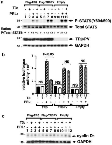
Figure 5.
Liganded TRβ but not TRβPV represses STAT5 signaling and target gene expression in T47D cells. (a) Western blot on whole-cell lysates from T47D cells for P-STAT5 (Y694/ 699), total STAT5, Flag, and GAPDH (glyceraldehyde-3-phosphate dehydrogenase) as a loading control. T47D cells expressing Flag-TRβ (lanes 1–4), Flag-TRβPV (lanes 5–8) or with the empty vector (lanes 9–12) were incubated with or without T3 and PRL for 5 h, as described in Materials and methods. (b) STAT5 activity in T47D cells determined by reporter assay. T47D cells were co-transfected with pcDNA3.1-TRβ (bars 1–4), pcDNA3.1-TRβPV (bars 5–8) or with pcDNA3.1 (bars 9–12) and the pGL4-CISH reporter gene containing four STAT5 binding sites. Cells were incubated with or without T3 and PRL for 5 h, as described in Materials and methods. (c) Western blot on whole-cell lysates from T47D cells for cyclin D1, and GAPDH as a loading control. T47D cells infected with Flag-TRβ (lanes 1–4), Flag-TRβPV (lanes 5–8)adenovirus constructs or with the vector only adenovirus (lanes 9–12) were incubated with or without T3 and PRL for 24 h, as described in Materials and methods.

