Abstract
Objective
Brachymesophalangia – V (BMP-V), the general term for a short and broad middle phalanx of the 5th digit, presents both alone and in a large number of complex brachydactylies and developmental disorders. Past anthropological and epidemiological studies of growth and development have examined the prevalence of BMP-V because small developmental disorders may signal more complex disruptions of skeletal growth and development. Historically, however, consensus on qualitative phenotype methodology has not been established. In large-scale, non-clinical studies such as the Fels Longitudinal Study and the Jiri Growth Study, quantitative assessment of the hand is not always the most efficient manner of screening for skeletal dysmorphologies. The current study evaluates qualitative phenotyping techniques for BMP-V used in past anthropological studies of growth and development in order to establish a useful and reliable screening method for large study samples.
Methods
A total of 1,360 radiographs from Jiri Growth Study participants aged 3 – 18 years were evaluated. BMP-V was assessed using three methods: 1) subjective evaluation of length and width of the bone; 2) comparison with skeletal age-matched radiographs; and 3) subjective evaluation of the length of the middle 4th and 5th phalanges.
Results
We found that the method that uses skeletal age-matched reference radiographs is the better tool for assessing BMP-V because it considers the shape, rather than solely the length and width of the bone, which can be difficult to judge accurately without measurement. This study highlights the complexity of phenotypic assessment of BMP-V and by extension other brachydactylies.
Keywords: brachydactyly, BMP-V, phenotype, skeletal dysplasia, hand
Introduction
Brachymesophalangia – V (BMP-V), a short and broad middle phalanx of the 5th digit, is the most common of all skeletal anomalies of the hand (Poznanski, 1984). Despite this fact, it is not common in all populations, but rather varies across populations from 0-20.7%, with Asian and Native American populations consistently displaying the highest prevalence (see Williams et al. 2007). It is commonly observed alone, as part of a suite of traits that characterize other forms of brachydactyly, as well as in many developmental syndromes (e.g., Down Syndrome). When this feature appears alone, it is clinically known as brachydactyly type A3 (BDA3). From a clinical perspective, when assessing a single patient or a small number of patients general metacarpophalangeal pattern profiling is used (Poznanski et al. 1972) which involved measuring phalanges and metacarpals and applying cut-offs to determine abnormal development.
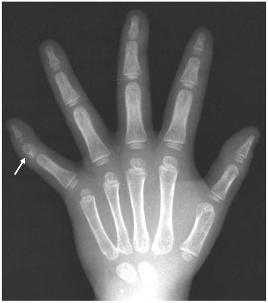
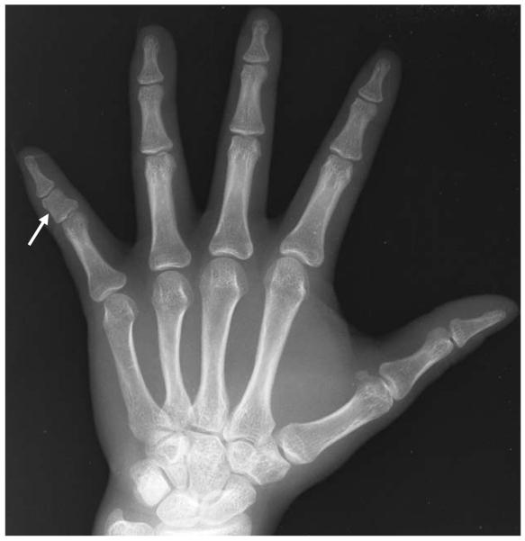
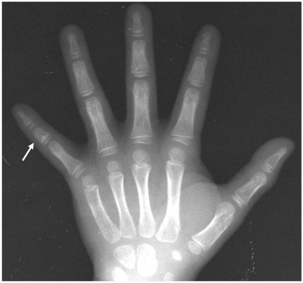
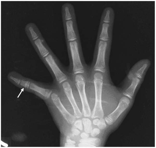
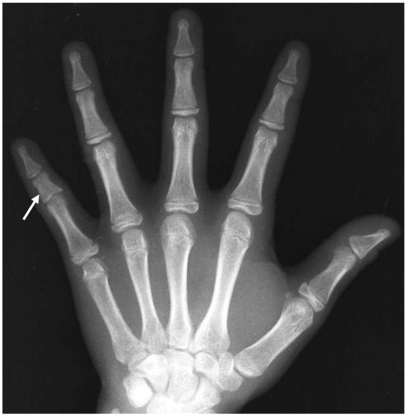
Over the years, researchers have defined BMP-V in several ways. Unfortunately, inconsistent definitions have made population comparisons difficult. Some definitions incorrectly call as “affected” phalanges that do not have the hallmark shape and size features of BMP-V. This is typically due to definitions that rely too much on the relative dimensions of phalanges and metacarpals and that do not consider the characteristic shape of an affected bone. Additionally, many studies have been conducted on adults, whose growth and development has been completed. Phenotyping BMP-V in children and adults differs because of the ongoing changes in bone shape and size that occur in growing children versus the end result of a phalanx with fused epiphyses and completed linear growth (examples at skeletal age 4, 6, 10, 15, and 18 are illustrated in Figures 1 - 5).
Figure 1.
Skeletal Age: 4.0 – 4.99 years
Figure 5.
Skeletal Age: 18.0 – 18.99 years
At least nine different methods, both quantitative and qualitative, have been used in past anthropological and epidemiological studies of BMP-V (see Table 1). Qualitative assessment of the trait has been a more popular choice, most likely because of the relative ease of making a “yes/no” assessment based on a visual inspection. Thus, an affected bone has been defined by many as simply a “shortened” middle phalanx of the 5th digit (Abbie, 1970; Greulich, 1970; Miura et al., 1986; Poznanski, 1984; Roche, 1961). Bell (1951) defined BDA3 (BMP-V alone, in isolation from other forms of brachydactyly or developmental disorders) as shortening of radial side of the middle phalanx of the 5th digit, which may or may not result in clinodactyly. Other textbooks on hand dysplasia reiterate these earlier definitions of the trait. More recent descriptions, however, acknowledge the altered shape of the bone in addition to its shortness. For example, Schmitt and Lanz (2008) describe a short middle phalanx, and further describe the trait as having a trapezoidal configuration. And, Temtamy and Aglan (2008) describe BDA3 as a short middle phalanx with slanting of the distal articular surface leading to radial deflection of the distal phalanx.
Table 1.
Definitions of affected middle phalanx of the 5th digit (BMP-V or BDA3).
| Description | Reference |
|---|---|
| short middle phalanx, 5th digit |
Abbie, 1970; Greulich, 1970; Miura et al., 1986; Poznanski, 1984; Roche, 1961 |
| shortened radial side of middle phalanx, 5th digit (with or without curvature - clinodactyly) |
Bell, 1951 |
| short middle phalanx, 5th digit with trapezoidal shape | Schmidt and Lanz, 2008 |
| short middle phalanx 5th digit with slanting of the distal articular surface leading to radial deflection of the distal phalanx. |
Temtamy and Aglan, 2008 |
| ratio of lengths of 5th digit middle phalanx: proximal phalanx | Garn, 1976; Garn, 1972 |
| ratio of lengths - 5th digit middle phalanx: 4thdigit middle phalanx |
Blanco et al., 1973; Garn, 1976; Hertzog, 1967; Singer, 1980 Bauer, 1907 |
| ratio of lengths - 5th digit middle phalanx: 2nd metacarpal | Garn, 1976 |
| Ratio - length 5th digit middle phalanx: width 5th digit middle phalanx |
Garn, 1976; Garn, 1967 |
| discriminant function analysis using lengths of the 4th and 5th digit middle phalanges and 2nd metacarpal | Garn, 1967 |
Despite it being clear that BMP-V prevalence data that is collected using different methods may not be directly comparable, no work has compared different BMP-V assessment techniques. Lack of a standard definition of BMP-V especially complicates identification of the trait in studies that include children. Although the study of these traits has not received the amount of attention in recent anthropological studies as in the past when documenting population differences in prevalence rates was the primary focus, re-examination of such minor skeletal dysplasia, especially using modern genetic methods, can provide insight into fundamental aspects of skeletal development..
It is well known that tempo of skeletal maturation (as often, for example, assessed from a hand-wrist radiograph) can differ between children due to normal population variation (Greulich and Pyle, 1959; Roche et al., 1988; Todd, 1937). It is possible that using any of the above definitions of BMP-V could over– or under– estimate the prevalence of the trait if children of all ages are included and evaluated with the same strict criteria. This is because the specific growth disruption of the phalanx of the 5th digit that leads to the hallmark morphologic features of BMP-V varies in its appearance during the process of skeletal maturation. This is particularly important when investigating BMP-V given that the epiphyses of the 5th digit are among the last to begin ossifying in the hand (Greulich and Pyle, 1959). Therefore, they may be more susceptible to biological insult given their extended period of vulnerability (Greulich, 1970; Scheur and Black, 2004). In this regard it is important to note that far greater variation has been observed in the length of the middle phalanx of the 5th digit than in that of the other bones of the hand (Hewitt, 1963). On a practical note, in large study samples qualitative assessment of radiographs is often the preferred approach because it is comparatively quick and can be readily employed after some basic training. Furthermore, oftentimes an initial assessment of radiographs is necessary to determine if traits of interest are present at a sufficient level to warrant further study. For these reasons it is worthwhile to have a better standardized method for qualitatively phenotyping BMP-V.
In the present study, we compared three methods for qualitatively phenotyping BMP-V in a large sample of pediatric radiographs. As discussed above, identification of BMP-V in pediatric populations requires special consideration because of the on-going changes in bone form that occur in children that culminate in a phalanx with fused epiphyses and completed linear growth in late childhood. A high prevalence (10.5%) of BMP-V in isolation (i.e., BDA3, sensu stricto) has been observed by us among children in the Jiri Growth Study, a genetic epidemiologic study of normal growth and development conducted in the Jirel ethnic group of eastern Nepal (Williams et al., 2007a; Williams et al., 2007b). The high prevalence of BMP-V in this study population has created an ideal circumstance to examine this aspect of skeletal development of the hand. Understanding the nature of this specific skeletal anomaly in otherwise normal and healthy children will ultimately contribute to our general understanding of genetic influences on limb and digit development. Better defining the BMP-V phenotype is an important task in that endeavor.
Materials and Methods
Study Sample
To date, some 1,600 children aged 3 –18 years have taken part in the Jiri Growth Study. As part of the study, a hand-wrist radiograph is taken annually of each child to monitor their skeletal maturation. For the present study, radiographs of 1,360 Jirel children and adolescents aged 3 –18 years (676 males; 684 females) were assessed for presence or absence of BMP-V by examination of left hand-wrist radiographs viewed on a light box. All radiographs were taken following a standard protocol, including maintenance of a film-to-tube distance of 36 inches. Data used in this present study were obtained from the most recent radiograph available at the time of analysis. The Wright State University Institutional Review Board for Human Subjects Research, and the Nepal Health Research Council, approved all informed consent procedures. Informed consent was obtained from the parents of all children studied, and the children themselves assented to examination after all procedures were explained to them.
Phenotyping
Three qualitative methods were used to determine BMP-V affected/unaffected status. All three methods were based solely on visual inspection. Two trained skeletal research specialists assessed the set of 1,360 radiographs independently of one another. Both raters have several years experience in radiograph assessment, but Rater 1 has been assessing radiographs for various studies for much longer. The first method (Qualitative Method 1) asked raters to answer two questions (yes/no) for each radiograph: 1) “Is the middle phalanx of the 5th digit too short?” and 2) “Is the middle phalanx of the 5th digit too broad?” BMP-V-affected status was later assigned by one of us (KDW) when the answer to both questions was “yes.” BMP-V-unaffected status was assigned when the answer to either or both questions was “no.”
The second method (Qualitative Method 2) asked raters to compare each radiograph in question to a skeletal age (“bone age”)-matched radiograph demonstrating classic BMP-V expression that had been selected as a reference radiograph by one of the authors (KDW). These skeletal age-based reference radiographs were selected to represent single skeletal age years (i.e., skeletal age 3.0 – 3.99 years; 4.0 – 4.99 years, etc.). Thus, there were a total of 16 BMP-V reference radiographs that were used (radiographs are included as electronic Figures 1-16). To facilitate the use of these reference radiographs by other researchers who may not have skeletal age assessments of subjects in their study, in each reference radiograph the skeletal age of the child and his or her chronologic age at the time the radiograph was taken are not different by more than one year. In other words, the skeletal age and chronologic age in each of the 16 reference radiographs are roughly congruent. Separate male and female reference radiographs are not necessary because the trait these radiographs will be used to assess, the middle phalanx of the 5th digit, does not differ dramatically between males and females like other skeletal features of the hand.
Raters were asked if BMP-V was present/absent (affected/unaffected) using skeletal age-matched reference radiographs as a guide. Skeletal age had been previously assessed for all 1,360 study radiographs using the Fels method (Roche et al., 1988).
The third method (Qualitative Method 3) was based on the definition of BMP-V used by Stanley Garn (Garn et al. 1967) in the Fels Longitudinal Study (formerly conducted at the Fels Research Institute affiliated with Antioch College, Yellow Springs, OH; conducted since 1974 at Wright State University, Dayton, OH). This method assigns affected status when the middle phalanx of the 5th digit is less than 75% of the length of the middle phalanx of the 4th digit. In practice, it is a qualitative method because the bones are not actually measured, but rather relative phalanx lengths are estimated by visual inspection. Qualitative Method 3 was used by both raters, but each rated a non-overlapping subset of radiographs (1,282 for Rater 1 and 78 for Rater 2). Although it is therefore not possible to determine inter-rater agreement for Qualitative Method 3, it is still considered here for the purpose of comparing the three different methods.
Data Analysis
Sample prevalences of BMP-V-affected status were calculated using each method. Inter-rater agreement for qualitative methods 1 and 2, and inter-method agreement for each rater, were measured by the tetrachoric correlation coefficient (Drasgow, 1986; Olsson, 1979) and computed using the polychor (v. 0.7-3) function in R (v. 2.5.1, www.r-project.org). Use of this correlation coefficient assumes that the trait being assessed is a binary summary of a continuous normally distributed latent variable, and that rater errors on the latent variable are independent and normally distributed with a homogenous variance relative to the latent value (Uebersax, 2006). Given the design of the experiment, and that the raters were qualitatively judging an inherently continuous trait, these assumptions are reasonable.
Results
Prevalence
For each method we calculated the prevalence of BMP-V as determined by each rater (see Tables 2 and 3). The prevalence determined by Rater 1 was estimated to be 12.1% using both qualitative methods 1 and 2, and 10.0% using the qualitative method 3. Rater 2 identified BMP-V in 18.9% of the radiographs using qualitative method 1, 8.5% of the radiographs using qualitative method 2, and 12.8% of the radiographs using qualitative method 3.
Table 2.
Inter-rater prevalence agreement statistics.
| Method | r | Rater 1 | Rater 2 | Proportion of Overall Agreement |
|---|---|---|---|---|
| 1 | 0.96 | 12.1% | 18.9% | 92% |
| 2 | 0.99 | 12.1% | 8.5% | 96% |
| 3 | NA | 10.0% | 12.8% | NA |
Table 3.
Inter-rater agreement, by method
| Method 1 | ||||
| Rater 2 | ||||
| Rater 1 | Unaffected | Affected | Total Assessed | |
| Unaffected | 1,096 | 99 | 1,195 | |
| Affected | 7 | 158 | 165 | |
|
Total
Assessed |
1,103 | 257 | 1,360 | |
| Method 2 | ||||
| Rater 2 | ||||
| Rater 1 | Unaffected | Affected | Total Assessed | |
| Unaffected | 1,193 | 2 | 1,195 | |
| Affected | 52 | 113 | 165 | |
|
Total
Assessed |
1,245 | 115 | 1,360 | |
Inter-rater Agreement
Using qualitative method 1, the two raters agreed on BMP-V affected/unaffected status for 1,254 of the total of 1,360 radiographs (92%; tetrachoric correlation (SE.) = 0.96 (0.009); Table 2). The mismatches, however, tended to all be in one direction; when Rater 1 concluded “affected” Rater 2 almost always agreed, but when Rater 2 concluded “affected” Rater 1 disagreed 39% of the time (Table 3).
Agreement between the two raters was higher using qualitative method 2 (Table 2). Using this method the raters agreed on BMP-V affected/unaffected status for 1,306 of the total of 1,360 radiographs (96%; tetrachoric correlation (SE) = 0.99 (0.005); Table 2). As with the first method, the mismatches tended to be in one direction. In this case, however, Rater 2 slightly underestimated the prevalence of BMP-V compared to Rater 1 (Table 3).
Inter-Method Agreement
Rater 2 is comparatively inconsistent across methods (Table 4). While this might be due to a difference between the methods, the greater consistency seen for Rater 1, combined with Rater 1’s greater experience, make it more likely that the inconsistency is due to the rater.
Table 4.
Inter-method statistics by rater
| Rater 1 | ||
| Method | r |
Proportion of Overall
Agreement |
| 1 & 2 | 0.97 | 0.96 |
| 1 & 3 | 0.92 | 0.94 |
| 2 & 3 | 0.95 | 0.95 |
| Rater 2 | ||
| Method | r |
Proportion of Overall
Agreement |
| 1 & 2 | 0.96 | 0.89 |
| 1 & 3 | 0.996 | 0.76 |
| 2 & 3 | 0.999 | 0.95 |
There is a high amount of agreement between the three methods as applied by Rater 1, with the strongest agreement being between qualitative methods 1 and 2 (tetrachoric correlation (SE) = 0.97 (0.007); Table 5). The assessments made by Rater 1 using either of these two methods also had high levels of agreement with Rater 1’s application of qualitative method 3 (tetrachoric correlation (SE) = 0.92 (0.018) and 0.95 (0.012), respectively (rater 1 assessed 1,282 radiographs using method 3 while Rater 2 assessed only 78 of the radiographs, thus this comparison between the three methods for this one rater Table 6). The two comparisons involving qualitative method 3, however, led to a skewed pattern of discordant classifications. While the first and second qualitative methods were equally likely to result in a positive BMP-V classification, qualitative method 3 was less likely to lead to a positive classification.
Table 5.
Inter-method agreement, Rater 1
| Method 1 | ||||
| Unaffected | Affected | Total | ||
|
Method
2 |
Unaffected | 1,170 | 25 | 1,195 |
| Affected | 25 | 140 | 165 | |
| Total | 1,195 | 165 | 1,360 | |
| Method 1 | ||||
| Unaffected | Affected | Total | ||
|
Method
3 |
Unaffected | 1,101 | 53 | 1,154 |
| Affected | 28 | 100 | 128 | |
| Total | 1,129 | 153 | 1,282 | |
| Method 2 | ||||
| Unaffected | Affected | Total | ||
|
Method
3 |
Unaffected | 1,114 | 40 | 1,154 |
| Affected | 21 | 107 | 128 | |
| Total | 1,135 | 147 | 1,282 | |
Table 6.
Inter-method agreement, Rater 2
| Method 1 | ||||
| Unaffected | Affected | Total | ||
|
Method
2 |
Unaffected | 1,102 | 1 | 1,103 |
| Affected | 143 | 114 | 257 | |
| Total | 1,245 | 115 | 1,360 | |
| Method 1 | ||||
| Unaffected | Affected | Total | ||
|
Method
3 |
Unaffected | 49 | 19 | 68 |
| Affected | 0 | 10 | 10 | |
| Total | 49 | 29 | 78 | |
| Method 2 | ||||
| Unaffected | Affected | Total | ||
|
Method
3 |
Unaffected | 68 | 0 | 68 |
| Affected | 4 | 6 | 10 | |
| Total | 72 | 6 | 78 | |
Discussion
BMP-V phenotype assessment is a more complex problem than has been previously appreciated. The goal of this study was to evaluate different relatively easy to implement qualitative methods for phenotyping BMP-V. This is important because large scale anthropological and epidemiological studies of growth and development often use such qualitative screening methods to ascertain the presence/absence of skeletal dysplasia.
This study compared three qualitative methods of BMP-V assessment and found that each method yielded good agreement between two independent raters. Method 1, which asked raters to evaluate the length and width of the bone separately, yielded more discrepant findings between the two raters, and overall appeared to overestimate the prevalence of the BMP-V. Method 2, which asked raters to evaluate the shape of the bone based on an example of the classic presentation of BMP-V at whole year skeletal age stages of the hand, yielded better agreement between raters. Method 2 is a better tool for phenotyping affected or unaffected status of BMP-V because it considers the shape of the bone rather than solely the length and width of the bone. In the analyses presented here, we found evidence that considering shape, instead of length and width alone, is a better method of BMP-V assessment in children. Rater 1’s assessment using Method 3, the standard in longitudinal growth and development studies such as the Fels Longitudinal Study, was compared with Rater 1’s Method 1 and Method 2 assessments. We found that Method 3 underestimated the prevalence of BMP-V because of the subjective nature of the assessment in which the rater is asked to estimate relative length of the 4th and 5th middle phalanges.
Analyses of the three methods of qualitative assessment of BMP-V demonstrate that they have a high degree of overall inter-rater and inter-method agreement. But, while overall agreement was very high, we observed systematic differences between raters in the results of the application of these three methods (Table 2). That the prevalences as determined by Rater 1 are more consistent across methods may be due, in part, to Rater 1’s greater experience in evaluating skeletal maturation. Based on the results of all three methods from the more experienced Rater 1, qualitative Methods 1 and 2 were the most consistent.
The development of the bones of the hand has been well studied, and patterns of skeletal maturation have been well documented (e.g., see Greulich and Pyle, 1959; Roche et al., 1988; Todd, 1937). It is well known that the tempo of skeletal maturation varies even within populations of healthy children. Variability in shape and width versus length resulting from variation in the tempo of maturation may lead to presence/absence misidentification of BMP-V (and other anomalies). Using skeletal age-matched radiographs removes some potential error in BMP-V phenotyping since the size and shape of the bones in children who are close in skeletal age are more likely to be similar in size and shape than children simply of the same chronological age.
Conclusion
While all three methods produced similar findings, there were systematic differences in ratings between the more experienced rater and the less experienced one, as well as between all three methods for the less experienced rater. These systematic differences suggest that there is an underlying quantitative distribution to the presentation of BMP-V upon which the raters based their assessments either consciously or unconsciously. In this case, the added years of experience possessed by Rater 1 benefited the assessment as compared to the less experienced rater who was less consistent.
Assessment of skeletal maturation is an important aspect of the study of growth and development, especially when evaluating congruency of skeletal maturation with chronological age in the cases of serious growth disruption or disease. Variation in tempo of growth may give the appearance of dysmorphology, but in fact represent only a transient growth disruption. It is therefore advisable to consider the current skeletal maturational stage of the individual. We recommend using a qualitative method of phenotyping BMP-V that considers the skeletal maturational stage of the child. This is readily achieved by comparing the subject’s radiograph with a skeletal age-matched reference radiograph, or a delimited set of such radiographs bracketing the subject’s chronological age if skeletal age is not available.
Figure 2.
Skeletal Age: 6.0 – 6.99 years
Figure 3.
Skeletal Age: 10.0 – 10.99 years
Figure 4.
Skeletal Age: 15.0 – 15.99 years
Acknowledgments
We thank Mr. Robin Singh, Mr. Suman Jirel, and all current and former staff members of the Jiri Growth Study and the Jiri Helminth Project, for their hard work and dedication. And, we respectfully acknowledge and thank the members of the Jirel community for their generous participation in this study.
This work was supported by NIH grants F32HD053206 (Williams), R01HD40377 (Towne), R01AI370919 (Williams-Blangero), R01AI44406 (Williams-Blangero), and R37MH59490 (Blangero).
Literature Cited
- Abbie AA. Brachymesophalangy V in Australian Aborigines. Med J Australia. 1970;2:736–737. doi: 10.5694/j.1326-5377.1970.tb63148.x. [DOI] [PubMed] [Google Scholar]
- Bell J. On brachydactyly and symphalangism. In: Penrose LS, editor. The Treasury of Human Inheritance. University Press; Cambridge: 1951. pp. 1–31. [Google Scholar]
- de Iturriza JR, Tanner JM. Cone-shaped epiphyses and other minor anomalies in the hands of normal British children. J Pediatr. 1969;75(4):265–272. doi: 10.1016/s0022-3476(69)80397-5. [DOI] [PubMed] [Google Scholar]
- Drasgow F. Polychoric and polyserial correlations. In: Kotz S, Johnson N, editors. The Encyclopedia of Statistics. Wiley; 1986. [Google Scholar]
- Garn S, Fels S, Israel H. Brachymesophalangia of digit five in ten populations. Am J Phys Anthropol. 1967;27:205–210. [Google Scholar]
- Giedion A. Cone-shaped epiphyses (C.S.E.) Ann Rad. 1965;8:135–145. [Google Scholar]
- Greulich W. The incidence of dysplasia of the middle phalanx of the fifth finger in normal Japanese, in some American Indian groups, and in caucasians with Down’s Syndrome. Environmental Influences on Genetic Expression. 1970:91–105. [Google Scholar]
- Greulich WW, Pyle SI. Radiographic Atlas of Skeletal Development of the Hand and Wrist. 2nd edition Stanford University Press; Stanford: 1959. [Google Scholar]
- Hewitt D. Pattern of correlations in the skeleton of the growing hand. Ann Human Genet. 1963;27:157–169. doi: 10.1111/j.1469-1809.1963.tb00208.x. [DOI] [PubMed] [Google Scholar]
- Miura T, Torii S, Nakamura R. Brachymetacarpia and brachyphalangia. The J Hand Surg. 1986;11A(6):829–836. doi: 10.1016/s0363-5023(86)80231-3. [DOI] [PubMed] [Google Scholar]
- Olsson U. Maximum likelihood estimation of the polychoric correlation coefficient. Psychometrika. 1979;44:443–460. [Google Scholar]
- Poznanski A. The Hand in Radiologic Diagnosis. 2nd Edition W.B. Saunders; Philadelphia: 1984. [Google Scholar]
- Poznanski AK, Garn SM, Gall JC, Stern AM. Metacarpophalangeal pattern profiles in the evaluation of skeletal malformations. Radiol. 1972;104:1–11. doi: 10.1148/104.1.1. [DOI] [PubMed] [Google Scholar]
- Roche A. Clinodactyly and brachymesophalangia of the fifth finger. Acta Paediatrica. 1961;50:387–391. doi: 10.1111/j.1651-2227.1961.tb08190.x. [DOI] [PubMed] [Google Scholar]
- Roche AF, Chumlea WC, Thissen D. Assessing the Maturity of the Hand Wrist: Fels Method. Charles C. Thomas; Springfield, IL: 1988. [Google Scholar]
- Scheur L, Black S. The Juvenile Skeleton. Elsevier Academic Press; London: 2004. [Google Scholar]
- Schmitt R, Lanz U. Diagnostic Imaging of the Hand. Thieme; New York: 2008. [Google Scholar]
- Temtamy SA, Aglan MS. Brachydactyly. Orphanet J Rare Dis. 2008;13:3–15. doi: 10.1186/1750-1172-3-15. [DOI] [PMC free article] [PubMed] [Google Scholar]
- The Ten State Nutrition Survey, 1968-1970, 1972. C.D.C.; Atlanta: USDep. Health, Education Welfare Publ. (HSM) 72-8134. [Google Scholar]
- Todd TW. Atlas of Skeletal Maturation. Mosby; Part I: The Hand St. Louis: 1937. [Google Scholar]
- Uebersax JS. The tetrachoric and polychoric correlation coefficients. Statistical Methods for Rater Agreement web site. 2006 http://ourworld.compuserve.com/homepages/jsuebersax/tetra.htm.
- Williams KD, Blangero J, Cottom CR, Lawrence S, Choh AC, Czerwinski SA, Lee M, Duren DL, Sherwood RJ, Dyer TD, et al. Heritability of brachydactyly type A3 in children, adolescents, and young adults from an endogamous population in eastern Nepal. Hum Biol. 2007a;79(6):609–622. doi: 10.1353/hub.2008.0016. [DOI] [PubMed] [Google Scholar]
- Williams KD, Blangero J, Cottom CR, Lawrence S, Jha B, Subedi J, Williams-Blangero S, Towne B. Methods for phenotyping brachydactyly type A3 and related anomalies in an endogamous population from eastern Nepal (Abstract 97) Am J Hum Biol. 2007b;19(2):288. [Google Scholar]
- Williams KD, Blangero J, Duren DL, Cottom CR, Lawrence S, Dyer T, Jha B, Subedi J, Williams-Blangero S, Towne B. High prevalence of brachymesophalangia-V in an endogamous population from eastern Nepal. Am J Hum Genet.Mtg. 2005 Abstracts:143. [Google Scholar]
- Williams KD, Blangero J, Duren DL, Cottom CR, Lawrence S, Dyer T, Jha B, Subedi J, Williams-Blangero S, Towne B. Heritability of brachymesophalangia-V and related phenotypes in an endogamous population from eastern Nepal. 11th Int Congr Hum Genet Mtg; 2006. Abstracts:233. [Google Scholar]


