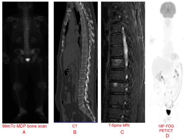
A 54-year-old male with biopsy proven Gleason 9 prostate cancer presented with rapidly increasing serum PSA level of 12.4 ng/ml and PSA doubling time <1 month after radical prostatectomy. Conventional imaging scans (chest, abdomen and pelvis CT and 99mTc-MDP bone scintigraphy) were obtained which identified a single equivocal mildly sclerotic and active lesion in T8 vertebral body (Fig. 1). The patient was then enrolled in an ongoing prospective clinical imaging trial designed to assess the diagnostic utility of 18F-FDG PET/CT and 18F-NaF PET/CT in men with biochemical recurrence of prostate cancer and negative or equivocal standard imaging. While 18F-FDG PET/CT was negative, 18F-NaF PET/CT demonstrated numerous randomly distributed fluoride avid lesions in the skeleton (cervical/thoracic/lumbar spine, bilateral acetabuli pelvis, right clavicle) compatible with skeletal metastases (Fig. 2). Follow-up MRI of the spine confirmed the bony lesions (see Fig. 1). Patient was then started on androgen deprivation therapy (bicalutamide/leuprolide acetate) and radiotherapy to a painful area in lower-thoracic spine (3250 cGy to T7–T11) after which his PSA declined to 0.6 ng/ml. Follow-up 18F-NaF PET/CT showed increase in sclerosis on CT and decline in NaF uptake at sites of skeletal metastases. Conventional bone scintigraphy and CT are standard in the imaging evaluation of men with suspected metastatic disease based on rising serum PSA level. However, when available, 18F-NaF PET/CT should be considered since it may be useful in early detection of occult skeletal metastases as clearly demonstrated here in our case.1–3
Fig. 1.
Initial imaging evaluation of a man with biochemical relapse of prostate cancer: (A) equivocal 99mTc MDP bone scan, (B) equivocal sagittal view of thoracic spine CT, (C) sagittal view of follow-up thoracic spine MRI (performed after 18F-NaF PET/CT) demonstrating osseous metastases, and (D) negative maximum intensity projection image of 18F-FDG PET/CT.
Fig. 2.
Demonstration of widespread osseous metastatic disease with 18F-NaF PET/CT: maximum intensity projection images of 18F-NaF PET/CT (A) at baseline, (B) at 6 months after start of therapy, (C) transaxial CT and fused 18F-NaF PET/CT appearance of a lesion in L4 at baseline and at 6 months with demonstration of therapy-indfguced decline in lesion 18F-NaF uptake and increase in sclerosis.
Acknowledgments
Supported by the National Institutes of Health, National Cancer Institute Grant no. R01-CA111613.
References
- 1.Even-Sapir Mester U, Mishani E, Lievshitz G, Lerman H, Leibovitch I. The detection of bone metastases in patients with high-risk prostate cancer: 99mTc-MDP planar bone scintigraphy, single- and multi-filed-of-view SPECT, 18F-fluoride PET, and 18F-fluoride PET/CT. J Nucl Med. 2006;47:287–97. [PubMed] [Google Scholar]
- 2.Even-Sapir E, Mishani E, Flusser G, Metser U. 18F-Fluoride positron emission tomography and positron emission tomography/computed tomography. Semin Nucl Med. 2007;37:462–9. doi: 10.1053/j.semnuclmed.2007.07.002. [DOI] [PubMed] [Google Scholar]
- 3.Grant FD, Fahey FH, Packard AB, Davis RT, Alavi A, Treves ST. Skeletal PET with 18F-fluoride: applying new technology to an old tracer. J Nucl Med. 2008;49:68–78. doi: 10.2967/jnumed.106.037200. [DOI] [PubMed] [Google Scholar]


