Abstract

Aim
Reconstructing or augmenting the lumbo-pelvic junction after resection of L5 and part of the sacrum is challenging. Numerous lumbo-pelvic reconstruction methods based on posterior construct and anterior cages have been proposed for cases involving total sacrectomy and lumbar vertebrectomy. These constructs create long lever arms and generate high cantilever forces across the lumbo-sacral junction, resulting in implant failure or breakage. Biomechanical studies have shown that placing implants anterior to lumbo-sacral pivot point provides a more effective moment arm to resist flexion force and improves the ultimate strength of the construct. We present here a novel method to augment a lumbo-pelvic construction using a pelvic ring construct.
Methods
A 69-year-old lady presented with implant failure of her two previous posterior lumbo-pelvic reconstructions performed by the authors. She initially presented, two and a half years previously with 6 months history of back pain with normal neurological function. MRI scans of her whole spine showed isolated secondaries in the lumbar spine (L4, L5) and sacrum (S1). An abdominal CT scan revealed a primary tumour in her right kidney. Briefly, the first surgery involved a single-stage removal of posterior elements of L4 and L5 and posterior stabilisation from L2 to pelvis, anterior resection of L4 and L5 and partially S1 with implantation of an expandable Synex II cage. The cage was replaced with an anterior rod construct from L2 and L3 to a trans-sacral screw a week later as it had dislodged. The second revision, 9 months later, involved removal of two posterior broken rods which were replaced and converted into a modified four-rod construct. While monitoring her progress, it was subsequently noted that the trans-sacral rod had broken. Therefore, it was decided to augment her lumbo-pelvic construct to prevent eventual catastrophic posterior construct failure. From a posterior approach, contoured rods were passed bilaterally along the inner table of the pelvis under the iliacus muscle up to the anterior border of the pelvis. Using T-connectors, the rods were connected to the posterior lumbo-pelvic construct. Thereafter, two anterior supra-acetabular pelvic screws were connected to a subcutaneously placed rod matched to the shape of the anterior abdominal wall. The pelvic ring construct was completed on connecting this rod with T-connectors to the free ends of the contoured iliac rods.
Results and conclusion
There were no intra-operative complications. At the end of 12 months, she was mobilising with a frame, with no radiological evidence of failure of the construct. However, she died due to disease progression at the end of 15 months. Experience from one clinical case shows that such a construct is feasible and adds a technical option to the difficult reconstruction of lumbo-pelvic junction after tumour surgery.
Keywords: Lumbo-pelvic instrumentation, Pelvic ring construct, Spinal metastasis, Revision construct
Case presentation
This 69-year-old lady presented with implant failure of her two previous posterior lumbo-pelvic reconstructions performed by the authors. She initially presented two and a half years previously with 6 months history of back pain with normal neurological function. MRI scans of her whole spine showed isolated secondaries in the lumbar spine (L4, L5) and sacrum (S1). An abdominal CT scan demonstrated a primary tumour in her right kidney.
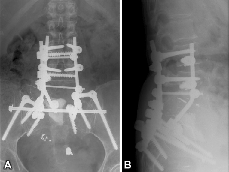
The first surgery involved a single stage removal of posterior elements of L4 and L5 and posterior stabilisation from L2 to pelvis including screws for S1/2 followed by right nephrectomy, anterior resection of L4 and L5 and partially S1 with implantation of an expandable Synex II cage (Fig. 1). The cage dislodged from the sacrum within the first postoperative week and subsequent revision was performed with an anterior rod construct from L2 and L3 to a trans-sacral screw (Fig. 2). Postoperatively she suffered temporary tetra-paresis. All investigations, however, were unremarkable and she recovered completely.
Fig. 1.
Postoperative anteroposterior (a) and lateral (b) radiographs of the pelvis following the index surgery (anterior and posterior resection of tumour and reconstruction with SynexII cage anteriorly and posterior stabilisation from L2 to pelvis). The cage had dislodged inferiorly from its base in the sacrum
Fig. 2.
Postoperative anteroposterior (a) and lateral (b) radiographs following removal of SynexII cage and anterior reconstruction with contoured titanium rod from L2 to a trans-sacral screw
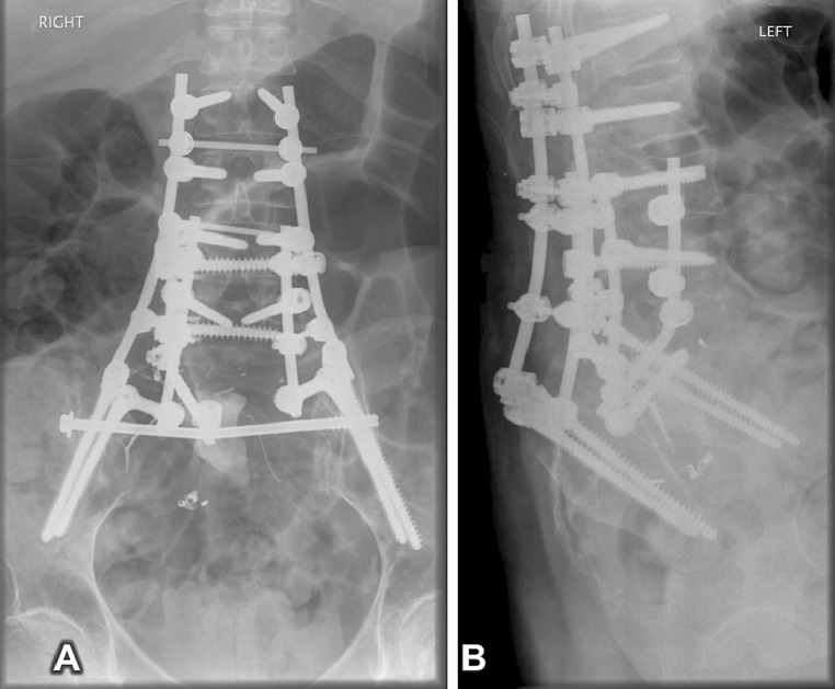
The second revision procedure was performed 9 months later. This involved removal of two posterior broken rods which were replaced and converted into a modified four-rod construct by adding two more rods and extending from T12 to an additional set of pelvic screws (Fig. 3).
Fig. 3.
Postoperative anteroposterior (a) and lateral (b) radiographs of the lumbo-sacral spine showing posterior modified four-rod reconstruction from T12 to pelvis
While monitoring her progress, it was subsequently noted that the trans-sacral rod had broken. Therefore, it was decided to augment her lumbo-pelvic construct to prevent eventual catastrophic posterior construct failure. At this stage, she was maintaining her mobility using a Zimmer frame with normal motor power and intact bladder and bowel function.
Historical review
Lumbo-pelvic reconstruction is a challenging problem despite the advances in tumour reconstructive surgery [1–8]. The anatomy of this region, poor quality bone and non-availability of stable anchoring sites frequently place enormous loads at this junction [9]. They include axial, translational and rotational loads, and bending moments [6, 10]. The potential for survival of patients over several years demands long-term stability from the reconstruction [14]. Added to this is the problem of revision surgery for stabilisation and the restrictions posed by the confines of extraspinal soft tissues and visceral structures [10]. As a result, implant failures are high [9]. A number of augmentation techniques have been described [1–8] to address these issues. Broadly, they can be divided as proximal anchor to lumbar vertebrae and distally into the sacrum and pelvis. Proximal anchoring techniques include use of cement, hydroxyapatite screws and expandable screws. Distally expandable cages, Galveston rods, mesh cages and cage supported on trans-iliac rods have been described.
Rationale of treatment and evidence-based literature
As a result of these challenges, lumbo-pelvic reconstruction has been evolving over the years. Various techniques include Galveston intra-sacral post [1], Jackson intra-sacral bar [2], spino-pelvic trans-iliac fixation [3], sacral bar [4], ilio-sacral screws [5], four-rod technique [6], custom-made prosthesis [7] and double trans-iliac rods to support anterior and posterior constructs [8]. The key areas in the lumbo-pelvic reconstructions are proximal and distal fixation points of the construct. The distal part of the construct has to have a stable purchase, provide a stable platform for implants like cages to stand on and at the same time have biomechanical distal fixation points anterior to the lumbo-sacral pivot point. The four-rod technique after biomechanical studies [13] has shown significantly reduced motion in the L5-pelvic level in flexion and extension. In our case, a modified four-rod technique was used in the second surgery to revise the posterior broken rods. However, this did not prevent failure of the trans-sacral screw in our case. The double trans-iliac rods technique [8] has provided the base for distractible anterior cage and augmentation of the posterior construct after total sacrectomy.
Our pelvic ring rods technique provides a greater base of stability around the transverse flexion extension axis of the spinal column by placing implants anterior to the lumbo-sacral pivot point [11, 12, 15, 16]. This provides a more effective moment arm to resist flexion force and improve the ultimate strength of the construct [10, 13]. The technique is relatively atraumatic and is mostly minimally invasive. The anterior abdominal rod used in our construct has been used successfully to treat unstable pelvic fractures in a case series of 17 cases, with only two cases of reported transient femoral cutaneous nerve palsy [17]. The blood loss in our case was negligible.
Procedure
Principle of surgical technique
The procedure involved building a pelvic ring construct skirting along the ‘inner table’ of the pelvis under the iliacus muscle. Using a minimally invasive technique, two 6-mm contoured titanium hard rods were inserted through a posterior approach skirting along the ‘inner table’ of the pelvis (both iliac bones) to reach the proximity of the anterior superior iliac spine (ASIS) anteriorly. During this manoeuvre, constant contact of the leading tip of the curved rod with the iliac bone was maintained to minimise any damage to the soft tissues. The posterior and anterior free ends of these rods were connected to the outer rods of the posterior construct and an anterior construct, respectively. The anterior construct consisted of a subcutaneously passed 6-mm contoured titanium hard rod across the anterior abdominal wall supported by two 8-mm pedicle screws in the supra-acetabular portion of the ilium, all performed through an minimally invasive surgery (MIS) technique. These anterior pedicle screws had good purchase in the sciatic buttress.
Technical details
The patient was placed in a prone position over bolsters. Using image intensifier guidance, two incisions were made over the distal part of the outer rods in the posterior construct.
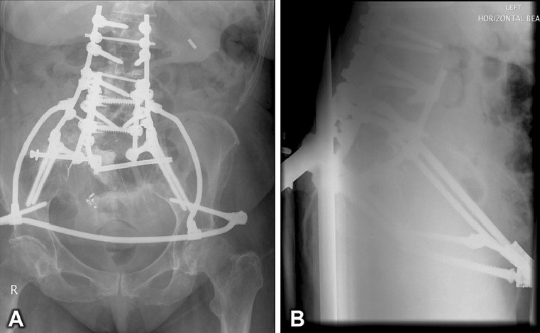
The posterior superior iliac spine (PSIS) on either side was then exposed and minor subperiosteal dissection performed along the inside curvature of the posterior iliac crest. A 6 mm × 250 mm-long contoured hard titanium rod, shaped like a ‘C’, was then passed from the dissected entry point of the pelvis at the PSIS along the inner table of the pelvis under fluoroscopic control to reach the subcutaneous skin near the ASIS (Fig. 4a–d). Care was taken to keep the tip of the rod in constant contact with the inner table of the pelvis during the passage. Very little force was required for the passage of the rods. The same procedure was performed on both sides. “T” clamps were then used to connect these ‘C’-shaped rods to the lateral rods of the posterior construct. The anterior free ends of the rods were left to be palpable under the skin near the ASIS. The posterior wounds were closed without tension to the skin and the patient turned over to a supine position. Two small transverse incisions were made over the palpable rod near the ASIS on both sides. Using imaging, an 8-mm × 90-mm polyaxial pedicle screw was inserted through both the ASIS between the two tables of the pelvis to engage into the sciatic buttress (Fig. 4e, f). A third 6-mm hard titanium rod contoured like a ‘bow’ to match the shape of the anterior abdominal wall was passed subcutaneously from left ASIS to reach the right ASIS. This was passed just below the lower abdominal crease to avoid pressure on the bowel or vascular structures below the inguinal ligament. At each end, this third rod was connected to the polyaxial screw in the ASIS and the anterior end of the ‘C’-shaped rod using a “T” clamp (Figs. 4e, f, 5, 6, 7). Stable construct was achieved. Wounds were then closed in the usual manner and the procedure completed without complication. The estimated blood loss for the procedure was <250 ml.
Fig. 4.
a–d Intraoperative photographs and fluoroscopic ‘spot’ images showing sequential insertion of contoured rod from posterior to anterior along the inner table of the pelvis to reach the skin subcutaneously near the ASIS. e, f ‘T’ Clamp connecting the anterior contoured rod and the anterior end of the posterior construct (red line posterior rod; green line anterior contoured rod; yellow line contour of the pelvis)
Fig. 5.
Final anteroposterior (a) and lateral (b) radiographs of the pelvis showing the pelvic ring construct in situ
Fig. 6.
Surface-shaded 3D reconstruction of a computed tomography (CT) scan of the pelvis showing the pelvic reconstruction in situ from anterior (a) and anterior-oblique perspectives (b)
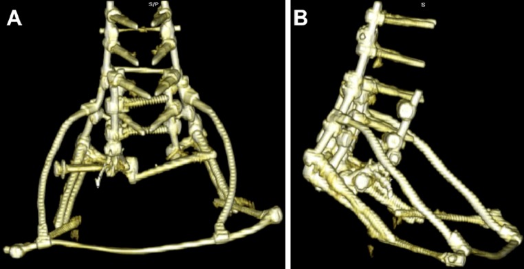
Fig. 7.
Surface-shaded 3D reconstruction of a computed tomography (CT) scan of the pelvis with windowing adjusted to show only the metalwork used in the pelvic ring construct. Anterior (a) and lateral (b) perspectives
Procedure imaging section
Outcome and follow-up
Postoperatively, the patient’s neurological examination revealed grade 5/5 power with intact bladder and bowel function. She mobilised with a frame without the need for a brace. She was discharged to further rehabilitation from the ward. At 12 months, she was ambulating independently with a frame. X-rays showed no evidence of implant loosening or failure despite local progression of the remnant of the sacral metastasis. Fifteen months after this surgery, the patient died as a result of disease progression.
Conflict of interest
None.
References
- 1.Gokaslan ZL, Romsdahl MM, Kroll SS, et al. Total sacrectomy and Galveston L-rod reconstruction for malignant neoplasms. J Neurosurg. 1997;87:781–787. doi: 10.3171/jns.1997.87.5.0781. [DOI] [PubMed] [Google Scholar]
- 2.Jackson RP, McManus AC. The iliac buttress: a computed tomographic study of sacral anatomy. Spine. 1993;18:1318–1328. doi: 10.1097/00007632-199308000-00011. [DOI] [PubMed] [Google Scholar]
- 3.Schildhauer TA, McCulloch P, Chapman JR, et al. Anatomic and radiographic considerations for placement of transiliac screws in lumbopelvic fixation. J Spinal Disord Tech. 2002;15:199–205. doi: 10.1097/00024720-200206000-00005. [DOI] [PubMed] [Google Scholar]
- 4.Widmann RF, Hresko MT, Hall JE. Lumbosacral fusion in children and adolescents using the modified sacral bar technique. Clin Orthop Relat Res. 1999;1:85–91. doi: 10.1097/00003086-199907000-00012. [DOI] [PubMed] [Google Scholar]
- 5.Farcy JP, Rawlings BA, Glassman SD. Technique and results of fixation to the sacrum with iliosacral screws. Spine. 1992;17:S190–S195. doi: 10.1097/00007632-199206001-00022. [DOI] [PubMed] [Google Scholar]
- 6.Shen FH, Harper M, Foster WC, et al. A novel “four-rod technique” for lumbo-pelvic reconstruction: theory and technical considerations. Spine. 2006;31:1395–1401. doi: 10.1097/01.brs.0000219527.64180.95. [DOI] [PubMed] [Google Scholar]
- 7.Wuisman P, Lieshout O, Dijk MV, et al. Reconstruction after total en bloc sacrectomy for osteosarcoma using a custom-made prosthesis: a technical note. Spine. 2001;26:431–439. doi: 10.1097/00007632-200102150-00021. [DOI] [PubMed] [Google Scholar]
- 8.Gallia GL, Suk I, Witham TF, et al. Lumbopelvic reconstruction after combined L5 spondylectomy and total sacrectomy for en bloc resection of a malignant fibrous histiocytoma. Neurosurgery. 2010;67(2):E498–E502. doi: 10.1227/01.NEU.0000382972.15422.10. [DOI] [PubMed] [Google Scholar]
- 9.An HS, Jenis LG. Complications of surgery: treatment and prevention. USA: Lippincott William & Wilkins; 2006. pp. 136–137. [Google Scholar]
- 10.Benzel E (2001) Biomechanics of spine stabilisation. Thieme, pp 302–307
- 11.Dickman CA, Fehlings MG, Gokaslan ZL (2006) Spinal cord and spinal column tumour: principles and practice. Thieme, pp 600–606
- 12.McCord DH, Cunningham BW, Shono Y. Biomechanical analysis of lumbosacral fixation. Spine. 1992;17(Suppl 8S):235–243. doi: 10.1097/00007632-199208001-00004. [DOI] [PubMed] [Google Scholar]
- 13.Kelly BP, Shen FH, Schwab JS, et al. Biomechanical testing of a novel four-rod technique for lumbo-pelvic reconstruction. Spine. 2008;33(13):E400–E406. doi: 10.1097/BRS.0b013e31817615c5. [DOI] [PubMed] [Google Scholar]
- 14.Tomita K, Kawahara N, Baba H, et al. Total en bloc spondylectomy for solitary spinal metastases. Int Orthop. 1994;18:291–298. doi: 10.1007/BF00180229. [DOI] [PubMed] [Google Scholar]
- 15.Gunterberg B, Romanus B, Stener B. Pelvic strength after major amputation of the sacrum. An experimental study. Acta Orthop Scand. 1976;47:635–642. doi: 10.3109/17453677608988751. [DOI] [PubMed] [Google Scholar]
- 16.Kawahara N, Murakami, Yoshida A. Reconstruction after total sacrectomy using a new instrumentation technique: a biomechanical comparison. Spine. 2003;28(14):1567–1572. [PubMed] [Google Scholar]
- 17.Vaidya R, Tonnos F, Colen R, Sethi A. Minimally invasive treatment of unstable pelvic ring injuries with an internal anterior fixator (INFIX) and posterior iliosacralscrews. J Bone Jt Surg Br. 2011;93-B(Suppl III):263. [Google Scholar]


