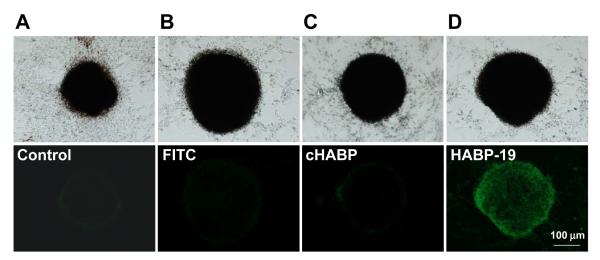
Figure 3.
Images of HA in VSMCs. HA calcification of VSMCs was induced by osteogenic factors and a 3D environment. For corroborative visualization of the extent of HA calcification, cells cultured in SM for 4 days were stained with FITC, cHABP, or HABP-19 and observed by fluorescent microscopy. Bright field (top) and fluorescence images (bottom) from mineral layers of VSMCs. HA deposits of VSMCs induced by AA, β-GP, LPC, and 3D-Ns were stained with control (A), FITC (B), cHABP (C) and HABP-19 (D) probes.

