Abstract
Objective
The formation of thrombi at the site of atherosclerotic lesions plays a central role in atherothrombosis. Impaired fibrinolysis may exacerbate pre-existing coronary artery disease and potentiate its evolution. While the fibrinogen plasma level has been strongly associated with the severity of coronary artery disease, its relevance in the evaluation of plasminogen in coronary artery disease patients remains unclear. This study evaluated fibrinogen and plasminogen levels in subjects with coronary artery disease as diagnosed by angiography.
Methods
This is a cross-sectional study. Blood samples obtained from 17 subjects with angiographically normal coronary arteries (controls), 12 with mild/moderate atheromatosis and 28 with severe atheromatosis were evaluated. Plasma plasminogen and fibrinogen levels were measured by chromogenic and coagulometric methods, respectively.
Results
Fibrinogen levels were significantly higher in the severe atheromatosis group compared to the other groups(p-value < 0.0001). A significant positive correlation was observed between the severity of coronary artery diseaseand increasing fibrinogen levels (r = 0.50; p-value < 0.0001) and between fibrinogen and plasminogen levels (r =0.46; p-value < 0.0001). There were no significant differences in the plasminogen levels between groups.
Conclusion
Plasma fibrinogen, but not plasminogen levels were higher in patients with coronary artery disease compared to angiographically normal subjects. The plasma fibrinogen levels also appear to be associated with the severity of the disease. The results of this study provide no evidence of a significant correlation between plasma plasminogen levels and the progress of coronary stenosis in the study population.
Keywords: Plasminogen, Fibrinogen, Coronary artery disease
Introduction
The formation of thrombi at the site of atherosclerotic lesions plays a central role in the hypothesis of atherothrombosis(1). A decreased endogenous fibrinolytic system and prothrombotic factors are supposed to influence coronary thrombosis. Impaired fibrinolysis may exacerbate already existing coronary artery disease (CAD) and potentiate its evolution(2).
Several studies have shown that fibrinogen is an important and independent risk factor for CAD(3,4) and this is currently used as a marker of inflammation(4,5). The increase in the plasma fibrinogen concentration is related to the development of CAD through changes in the mechanisms of platelet aggregation due to the influence of plasma fibrinogen on the quantity of fibrin formed and accumulated and its connection with the evolution of atherosclerotic plaque(6,7) and with the increase in blood viscosity related to the risk of thrombosis(8).
Plasminogen, a zymogen that is usually present in plasma(9,10), is a single-chain glycoprotein synthesized in the liver; it is considered an inactive proenzyme that is converted to plasmin(11). This serine protease degrades fibrin through the action of physiological activators, including tissue-type plasminogen activator (t-PA) and urokinase-type plasminogen activator (u-PA), secreted by endothelial cells(12). The endothelium itself controls the production of these activators through the synthesis of specific inhibitors of the fibrinolytic system, such as type 1-plasminogen activator inhibitor (PAI-1)(12,13). Some researchers have shown an independent and unexpected association between high plasminogen levels and the risk of CAD(14,15). However, the relevance of evaluating plasminogen levels in CAD patients remains unclear.
This study aimed to investigate the association between both fibrinogen and plasminogen plasma levels and the presence of increasing degrees of coronary atheromatosis in subjects submitted to coronary angiography.
Methods
Patients
Fifty-seven subjects, with ages ranging from 40 to 65 years, who had been submitted to coronary angiography in the Department of Hemodynamics of the Hospital Socor, Belo Horizonte, Brazil were enrolled in the study. Blood samples were collected before coronary angiography. Based on results of the coronary angiography, the participants were classified into three groups: angiographically normal arteries (n = 17), mild/moderate atheromatosis (n = 12) and severe atheromatosis (n = 28).
All patients were referred for cardiac catheterization due to chest pain and/or the presence of demographic profiles and risk factors for CAD. All patients had a history of stable angina. Even so, no patients had had recent myocardial infarction or unstable angina (within the three months preceding blood collection),congestive heart failure, coagulation disorders, renal problems, hepatic or auto-immune diseases or cancer or were on warfarin treatment or undergoing lipid-modifying therapy.
The protocol of this study was approved by the ResearchEthics Committees of both of the Hospital Socor (#05/04) and the Universidade Federal de Minas Gerais (UFMG - #137/04), Belo Horizonte, Brazil. The subjects enrolled in this study were informed about the aims of the investigation and those who agreed signed informed consent forms.
Angiography
Coronary angiography was performed by the percutaneoustransfemoral approach. The images were recorded digitally andall angiograms were analyzed by three experienced cardiologists. The extent of angiographically documented CAD was quantified asfollows: angiographically normal coronary arteries (no stenosis),mild disease (stenosis of up to 30% of the luminal diameter in oneor more coronary arteries), moderate disease (stenosis of 31% to70%) or severe disease (stenosis of more than 70%).
Blood collection and laboratory tests
Venous blood samples were drawn after 12 hours of fasting in vacuum tubes containing 3.2 w/v sodium citrate as anticoagulant. Blood samples were centrifuged at 2500 rpm for 10 minutes to obtain plasma.
Plasma fibrinogen levels were measured using a commerciallyavailable kit (Fibrinogen "O") according to the Clauss method. Theassay was performed as instructed by the manufacturer (DiaMed(r)AG, Cressier Sur Morat, Switzerland) in a ST4BIO Coagulometer device (Stago(r)Diagnostica, Asnieres-sur-Seine, France). The lowerlimit of detection by the assay is 45 ng/mL. The intra and inter assay coefficients of variation are 2.9% and 6.8%, respectively.
Plasma plasminogen levels were determined usingthe electrochromeTM plasminogen assay according to the manufacturer's instructions (Hemoliance(r)Instrumentation Laboratory, Lexington, USA). The lower limit of detection for the assay is 5%. The intra and inter assay coefficients of variation are 1.9% and 4.1%, respectively. For both fibrinogen and plasminogen, no significant cross-reactivity or interference from other coagulation factors have been observed for the assays and commercial control-plasma was used to validate the assay.
Statistical analysis
The sample size was defined using published coefficients ofvariation for the studied parameters, considering a 10% variationfrom the average and two degrees of freedom, using a minimum ofeleven subjects in each group so that possible statistical differencescould be demonstrated with a significance level of 5%.
Data presented normal distribution and were analyzed using the Sigma Stat version 1.0 software system with one-way analysis of variance followed by Tukey's test. Pearson's correlation coefficient was used to measure the linear association between plasma fibrinogen and plasminogen levels, and Spearman's correlation coefficient was used to measure the linear association between CAD and continuous variables.
Results
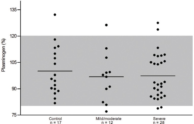
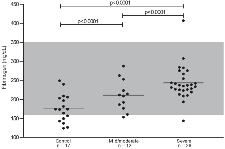
The three study groups showed homogeneity in relation to age, gender and body mass index (BMI). Results are shown as means ± standard deviation (SD) in Table 1 and graphically in Figures 1 and 2. Plasma fibrinogen levels were significantly higher in patients with severe atheromatosis compared to those with mild/moderate atheromatosis (p-value < 0.0001) or controls (p-value <0.0001). The difference between controls and the mild/moderate atheromatosis group was also significant (p-value < 0.0001). Forplasminogen, significant differences were not found for mean values between the three groups.
Table 1.
Characterization of the studied groups and the biomarker assays results
| Parameter | Control | Mild/moderate atheromatosis | Severe atheromatosis | p-value |
| Gender m/f | 8/9 | 8/4 | 16/12 | |
| Age (years) | 58.9 ± 7.7 | 61.0 ± 11.6 | 60.9 ± 9.6 | 0.515 |
| Body mass index (Kg/m2) | 24.9 ± 4.6 | 26.1 ± 4.9 | 24.3 ± 3.2 | 0.231 |
| Smokers - n (%) | 3 (17.6) | 3 (25.0) | 9 (32.1) | 0.432 |
| Arterial hypertension – n (%) | 14 (82.4) | 9 (75.0) | 24 (85.7) | 0.842 |
| Sedentary lifestyle – n (%) | 16 (94.1) | 9 (75.0) | 21 (75.0) | 0.501 |
| Family history – n (%) | 7 (41.2) | 5 (41.7) | 12 (42.9) | 0.347 |
| Fibrinogen (mg/dL) | 177.1 ± 36.7 | 210.9 ± 41.0a | 243.4 ± 45.1a,b | < 0.0001a,b |
| Plasminogen - % | 100.1 ± 13.9 | 96.8 ± 14.1 | 97.3 ± 12.9 | 0.622 |
Data are shown as means ± standard deviation or percentages. Signifcant differences between the groups are indicated by the letters: a. versus control and b. versus mild/moderate atheromatosis.
Figure 1.
Plasma plasminogen levels in control (angiographically normal arteries), mild/moderate and severe atheromatosis groups. No significant differences were found between the three groups. The shaded area corresponds to the normal range (80 – 120%) for plasminogen levels; horizontal lines represent the mean values of groups.
Figure 2.
Distribution of plasma fibrinogen levels in control (angiographically normal arteries), mild/moderate and severe atheromatosis groups. Statistically significant differences were found between the three groups (p-value < 0.05). The shaded area corresponds to the normal range (160 – 350 mg/dL) for fibrinogen levels; horizontal lines represent the mean values of groups.
The plasma fibrinogen levels showed a significant positive correlation with plasminogen levels (r = 0.46; p-value < 0.0001). The presence of CAD was associated with elevated plasma fibrinogen levels (r = 0.50; p-value < 0.0001), while no association was observed for the other variables.
Discussion
There are several systemic changes in hemostasis and fibrinolysis in atherosclerosis(16). In this study, the association between the degree of coronary atheromatosis and two key components of the coagulation system were evaluated. Once a thrombus resulting from a hypercoagulability condition is formed it is dissolved by the fibrinolytic system. So, researchers have hypothesized that a decrease in fibrinolytic activity could be a risk factor for ischemic events(17).
It is still unclear in the literature whether the quantity ofplasminogen that is transformed into plasmin is linearly correlatedto plasminogen levels. Plasminogen plasma concentration does notvary significantly in normal blood coagulation(18). In the present study, the average values obtained for plasminogen did not show statistically significant differences between the three groups (Table 1) and remained within the normal range indicated for themethod. An independent and unexpected association between high plasminogen levels and CAD risk has been reported previously(4,8), since decreased plasminogen levels suggest the generation of less plasmin and signal impaired fibrinolytic activity, which favors thedeposit of fibrin and contributes to atherothrombosis. However,plasminogen has several non-fibrinolytic roles the most importantof which is related to the inflammatory process(18). Plasminogenis directly linked to monocyte recruitment during inflammatory response(4) and there is evidence that interleukin-6 may increase theplasminogen gene expression(19). So, it is likely that the increase inplasminogen plasma concentration is secondary to the inflammatory process present in subclinical atherosclerotic lesions(19). This may,at least in part, justify the presence of the positive correlation found in this study between plasminogen and fibrinogen levels (r = 0.46; p-value < 0.0001).
Few studies have shown the participation of plasminogen in the local process of atherothrombosis(4,18). Likewise, in the present study, plasminogen levels were not correlated to the presence or severity of CAD and did not bring additional benefits, at least for subjects with the diagnosis of CAD. Similar observations were made by Hoffmeister et al.(20) in a case-control study; theauthors failed to find any association between elevated levels of plasminogen and stable CAD in German patients.
Some systemic inflammation markers may indicate the severity of the inflammatory process and their levels were recently associated to CAD(4,21). Table 1 indicates that for plasma fibrinogen levels a significant difference was found between the three groups (p-value < 0.0001). Figure 1 shows that the plasma level of fibrinogen increases as coronary atherosclerosis establishes and progresses to the mild/moderate and severe atheromatosis degrees, even though the mean values of groupsremained within the normal range for the method. Similar results were reported by Giannitsis et al.(22) who found that the mean values for the plasma levels of fibrinogen increased progressively as the CAD became more severe, although these values remained within the normal range. This current study also showed that, despite being within the normal range, plasma fibrinogen levels are significantly higher in patients with mild/moderate and severe atheromatosis than in angiographically normal subjects. The association between hyperfibrinogenemia and the progression of CAD has been widely reported(8,23-25). It is well-known that hyperfibrinogenemia is associated to acute coronary events and may be partly a cause and not just a consequence of the lesion as fibrinogen is a protein that appears in the acute phase and as the atherosclerotic process presents inflammatory components(8).
As this is a cross-sectional study, we evaluated associations, not predictions nor causation. Another important limitation is the fact that our observations are based on a small group of patients; further studies involving larger numbers of patients may elucidate the association between plasma levels of plasminogen and the severity of coronary stenosis. Our findings relate to a group of patients that, according to cardiologists, require coronary angiography and thus our subjects with no coronary stenosis may not be representative of the general population.
Conclusion
Plasma fibrinogen, but not plasminogen levels were higher in patients with CAD compared to angiographically normal subjects. The plasma fibrinogen levels also appear to be associated with the severity of the disease. These results give no evidence that there is a significant correlation between plasma plasminogen levels and the progress of coronary stenosis.
Acknowledgments
The authors wish to thank Dr. Cirilo Pereira da Fonseca Neto, Dr. José Carlos Faria Garcia and Dr. Jamil Abdala Saad from the Department of Hemodynamics, the Socor Hospital of Belo Horizonte, Minas Gerais, Brazil, for their help with the selection of subjects. We are very grateful to CAPES and CNPq for financial support.
Funding Statement
Neto, Dr. José Carlos Faria Garcia and Dr. Jamil Abdala Saad from the Department of Hemodynamics, the Socor Hospital of Belo Horizonte, Minas Gerais, Brazil, for their help with the selection of subjects. We are very grateful to CAPES and CNPq for financial support.
Footnotes
Conflict-of-interest disclosure: The author declares no competing financial interests
References
- 1.Saigo M, Hsue PY, Waters DD.Role of thrombotic and fibrinolytic factors in acute coronary syndromes Prog Cardiovasc Dis 200446 (6) 524-538 [DOI] [PubMed] [Google Scholar]
- 2.Selwyn AP.Prothrombotic and antithrombotic pathways in acute coronary syndromes Am J Cardiol 200391 (12A) 3H-11H [DOI] [PubMed] [Google Scholar]
- 3.Hoffmeister HM, Heller W, Seipel L.Blood coagulation and fibrinolysis in atherosclerosis Z Kardiol 199988 (5) 315-323German: [DOI] [PubMed] [Google Scholar]
- 4.Salomaa V, Rasi V, Kulathinal S, Vahtera E, Jauhiainen M, Ehnholm C, et al. Hemostatic factors as predictors of coronary events and total mortality: the FINRISK 92 hemostasis study Arterioscler Thromb Vasc Biol 200222 (2) 353-358 [DOI] [PubMed] [Google Scholar]
- 5.Papageorgiou N, Tousoulis D, Siasos G, Stefanadis C.Is fibrinogen a marker of inflammation in coronary artery disease? Hellenic J Cardiol 201051 (1) 1-9 [PubMed] [Google Scholar]
- 6.De Luca G, Verdoia M, Cassetti E, Schaffer A, Cavallino C, Bolzani V, Marino P, Novara Atherosclerosis Study Group (NAS) High fibrinogen level is an independent predictor of presence and extent of coronary artery disease among Italian population J Thromb Thrombolysis 201131 (4) 458-463 [DOI] [PubMed] [Google Scholar]
- 7.Devendra GP, Hart SA, Whitney EJ, Krasuski RA.Impact of fibrinogen levels on angiographic progression and 12-year survival in the armed forces regression study Angiology 201061 (4) 333-337 [DOI] [PubMed] [Google Scholar]
- 8.Folsom AR, Aleksic N, Park E, Salomaa V, Juneja H, Wu KK.Prospective study of fibrinolytic factors and incident coronary artery disease. The Atherosclerosis Risk in Communities (ARIC) Study Arterioscler Thromb Vasc Biol 200121 (4) 611-617 [DOI] [PubMed] [Google Scholar]
- 9.Cantin B, Després JP, Lamarche B, Moorjani S, Lupien PJ, Bogaty P, Bergeron J, Dagenais GR.Association of fibrinogen and lipoprotein(a) as a coronary Heart disease risk factor in men (The Quebec Cardiovascular Study) Am J Cardiol 200289 (6) 662-666 [DOI] [PubMed] [Google Scholar]
- 10.Luc G, Bard JM, Juhan-Vague I, Ferrieres J, Evans A, Amouyel P, Arveiler D, Fruchart JC, Ducimetiere P, PRIME Study Group C-reactive protein, interleukin-6 and fibrinogen as predictors of coronary heart disease Arterioscler Thromb Vasc Biol 200323 (7) 1255-1261 [DOI] [PubMed] [Google Scholar]
- 11.van Der Bom JG, de Maat MP, Bots ML, Haverkate F, de Jong PT, Hofman A, et al. Elevated plasma fibrinogencause or consequence of cardiovascular disease? Arterioscler Thromb Vasc Biol 199818 (4) 621-625 [DOI] [PubMed] [Google Scholar]
- 12.Jovin IS, Muller-Berghaus G.Interrelationships between the fibrinolytic system and lipoproteins in the pathogenesis of coronary atherosclerosis Atherosclerosis 2004174 (2) 225-233 [DOI] [PubMed] [Google Scholar]
- 13.Scazziota A, Altman R.El mecanismo de la hemostasia normal Rev Iberoamer Thromb Hemostasia 199619-26 [Google Scholar]
- 14.Syrovets T, Simmet T.Novel aspects and new roles for the serine protease plasmin Cell Mol Life Sci 200461 (7-8) 873-885 [DOI] [PMC free article] [PubMed] [Google Scholar]
- 15.Lijnen HR, Collen D.Mechanisms of physiological fibrinolysis Baillieres Clin Haematol 19958 (2) 277-290 [DOI] [PubMed] [Google Scholar]
- 16.Selwyn AP.Prothrombotic and antithrombotic pathways in acute coronary syndromes Am J Cardiol 200391 (12A) 3-11H [DOI] [PubMed] [Google Scholar]
- 17.Zorio E, Gilabert-Estellés J, España F, Ramón LA, Cosín R, Estellés A.Fibrinolysis: the key to new pathogenetic mechanisms Curr Med Chem 200815 (9) 923-929 [DOI] [PubMed] [Google Scholar]
- 18.Ploplis VA, Castellino FJ.Nonfibrinolytic functions of plasminogen Methods 200021 (2) 103-110 [DOI] [PubMed] [Google Scholar]
- 19.Jenkins GR, Seiffert D, Parmer RJ, Miles LA.Regulation of plasminogen gene expression by interleukin-6 Blood 199789 (7) 2394-2403 [PubMed] [Google Scholar]
- 20.Hoffmeister A, Rothenbacher D, Khuseyinova N, Brenner H, Koenig W.Plasminogen levels and risk of coronary artery disease Am J Cardiol 200290 (10) 1168-1170 [DOI] [PubMed] [Google Scholar]
- 21.Park CS, Ihm SH, Yoo KD, Kim DB, Lee JM, Kim HY, et al. Relation between C-reactive protein, homocysteine levels, fibrinogen, and lipoprotein levels and leukocyte and platelet counts, and 10-year risk for cardiovascular disease among healthy adults in the USA Am J Cardiol 2010105 (9) 1284-1288 [DOI] [PubMed] [Google Scholar]
- 22.Giannitsis E, Siemens HJ, Mitusch R, Tettenborn I, Wiegand U, Schmücker G, et al. Prothrombin fragments F1+2, thrombin-antithrombin III complexes, fibrin monomers and fibrinogen in patients with coronary atherosclerosis Int J Cardiol 199968 (3) 269-274 [DOI] [PubMed] [Google Scholar]
- 23.Lindahl B, Toss H, Siegbahn A, Venge P, Wallentin L.Markers of myocardial damage and inflammation in relation to long-term mortality in unstable coronary artery disease. FRISC Study Group. Fragmin during Instability in Coronary Artery Disease N Engl J Med 2000343 (16) 1139-1 47Comment in: N Engl J Med. 2000;343(16):1179-82. [DOI] [PubMed] [Google Scholar]
- 24.Athyros VG, Kakafika AI, Karagiannis A, Mikhailidis DP.Do we need to consider inflammatory markers when we treat atherosclerotic disease? Atherosclerosis 2008200 (1) 1-12 [DOI] [PubMed] [Google Scholar]
- 25.Shlipak MG, Ix JH, Bibbins-Domingo K, Lin F, Whooley MA.Biomarkers to predict recurrent cardiovascular disease: the Heart and Soul Study Am J Med 2008121 (1) 50-57 [DOI] [PMC free article] [PubMed] [Google Scholar]


