Abstract
Increasing numbers of pyelonephritis-associated uropathogenic Escherichia coli (UPEC) are exhibiting high resistance to antibiotic therapy. They include a particular clonal group, the CTX-M-15-producing O25b:H4-ST131 clone, which has been shown to have a high dissemination potential. Here we show that a representative isolate of this E. coli clone, referred to as TN03, has enhanced metabolic capacities, acts as a potent intestine- colonizing strain, and displays the typical features of UPEC strains. In a modified streptomycin-treated mouse model of intestinal colonization where streptomycin was stopped 5 days before inoculation, we show that TN03 outcompetes the commensal E. coli strains K-12 MG1655, IAI1, and ED1a at days 1 and 7. Using an experimental model of ascending UTI in C3H/HeN mice, we then show that TN03 colonized the urinary tract. One week after the transurethral inoculation of the TN03 isolates, the bacterial loads in the bladder and kidneys were significantly greater than those of two other UPEC strains (CFT073 and HT7) belonging to the same B2 phylogenetic group. The differences in bacterial loads did not seem to be directly linked to differences in the inflammatory response, since the intrarenal expression of chemokines and cytokines and the number of polymorphonuclear neutrophils attracted to the site of inflammation was the same in kidneys colonized by TN03, CFT073, or HT7. Lastly, we show that in vitro TN03 has a high maximum growth rate in both complex (Luria-Bertani and human urine) and minimum media. In conclusion, our findings indicate that TN03 is a potent UPEC strain that colonizes the intestinal tract and may persist in the kidneys of infected hosts.
Introduction
Urinary tract infections (UTIs) are one of the bacterial infections that most often affect children, young adults, and also renal transplanted patients. UTIs are mainly due to uropathogenic Escherichia coli (UPEC), which are becoming more resistant, thus hampering the therapeutic management of UTI.
E. coli is a ubiquitous human pathogen responsible for both community- and hospital-acquired infections. Over the past five decades, scientists have witnessed increasing antimicrobial resistance in the community setting. Initially, resistance was previously limited to certain specific antibiotics, such as ampicillin or trimethoprim [1], but recently the horizon of resistance has expanded, with the emergence of broad resistance to large families agents. In particular, plasmid-mediated extended-spectrum ß-lactamases (ESBLs), have become prominent in the community [2] [3]. Unfortunately, in addition to being resistant to most-ß-lactam antibiotics, ESBL producers are also often resistant to aminoglycosides and fluoroquinolones. Over the last decade, the CTX-M enzymes, have gradually replaced the classical TEM and SHV-type ESBLs in many countries [4], [5] and have gained worldwide attention. In 2008, two research groups, analyzing the population of E. coli ESBL-producing strains, described a particular CTX-M-15-producing clonal group. This clone occurred in both in-patients and out-patients worldwide, strongly suggesting that it is widely disseminated [6], [7], which constitutes a major health problem [8], [9]. Indeed, such bacterial resistance frequently delays the establishment of appropriate therapy [10], leading to higher costs and increased use of the “last resort” antimicrobials (i.e. carbapenems) [5]. This clone, which exhibits an O25b:H4 serotype, belongs to phylogenetic group B2 and sequence type (ST) 131 [7], [11]. Interestingly, several studies have confirmed the worldwide prevalence of this E. coli with the ST131 sequence type harboring a broad range of resistance genes on a transferable plasmid, mostly from the CTX-M family, and virulence genes [12]. This ST131 sequence type has also been detected in companion animals, non-companion animals, and food [12].
The clinical spectrum of disease induced by ST131 E. coli is similar to that for other E. coli. UTIs predominate, ranging from uncomplicated cystitis to life threatening sepsis. As this clone is mainly isolated from UTI, we determined the ability of a representative isolate of the O25b:H4 ST131 CTX-M-15 E. coli clone, known as the TN03 strain [11], [13], [14], first to colonize the intestine and then to infect the kidney using experimental mouse models in order to gain insights into its evolutionary success.
Results and Discussion
The TN03 strain is a potent colonizer of the intestine
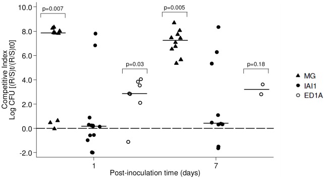
Infection of the urinary tract presumably begins with the colonization of the bowel by a uropathogenic strain [15], as suggested by the fact that the UPEC isolates present in infected urine are almost always detectable in the host's fecal flora at the time of presentation [16]. We therefore determined the ability of E. coli strain TN03 to colonize the gut using a mouse model of competition for intestinal colonization. A classical streptomycin-treated mouse model is usually used [17], because E. coli colonization cannot be studied experimentally in conventional animals due to colonization resistance. Streptomycin is used in this case to eliminate the natural coliform intestinal population and allows E. coli to colonize. Indeed, such experiments require an animal model with open niches where E. coli can colonize in relatively high numbers, but must also have a dense and diverse anaerobic community that matches the native microbiota of the conventional animal as closely as possible. In our case, however, the TN03 strain is resistant to streptomycin in contrast to all its other experimental competitors. Although, in-vitro streptomycin competitor mutants could have been used, these mutations could have been a burden on the fitness of the strains, which would have introduced bias into the experiment. We decided to modify slightly the classical protocol. Six-week-old CD1 female mice were pre-treated with streptomycin (5 g/liter) during five days in order to create the appropriate conditions for colonization. Streptomycin administration was then stopped five days before inoculation, thus avoiding bias linked to antibiotic pressure. Natural coliform removal efficiency was checked just before bacterial inoculation. The absence of residual streptomycin in the stools was assessed by a microbiological assay (data not shown). Mice were given 106 of each E. coli strain per os: the TN03 bacteria, or one of three commensal strains, belonging to different phylogenetic groups: the K-12 MG1655 [18], IAI1 and ED1a strains [19] from phylogenetic groups A, B1 and B2, respectively (Table 1), to check the individual capacities of each strain to colonize the mouse intestinal tract. The sizes of bacterial populations in the intestine were evaluated at days 1 and 7 following administration. All individual strains colonized the mouse intestine at levels ranging from 104 to 109 bacteria/g feces at day 1, and from 103 to 109 bacteria/g feces at day 7, with no difference being found between the strains (data not shown). The observed intra-strain variability can be attributed to the variability of the natural mouse microbiota in the absence of streptomycin treatment. To circumvent this variability, we decided to evaluate the competition capacities of the pathogenic E. coli TN03 isolate. To do this, the bacteria were mixed in a 1∶1 ratio of TN03 with commensal E. coli K-12 MG1655, IAI1, or ED1a bacteria. 106 of this bacterial mix was then administered per os to the mice as described above, The sizes of the bacterial populations in the intestine were evaluated at days 1, 2, 4 and 7 following inoculation. They tended to decrease slowly over time (data not shown). A competitive index (CI) which could be used to compare the colonization success of the different strains was calculated as previously described [20]. Analysis of the CI showed that the TN03 E. coli strain had outcompeted all of the commensal E. coli strains. Significant differences between TN03 with K-12 MG1655 or ED1a as a competitor (CIMG-D1 = 7.87, p = 0.007, CIED-D1 = 2.86, p = 0.03) were already detected day 1 post-bacterial administration, and persisted between TN03 with K-12 MG1655 (CIMG-D7 = 7.25, p = 0.005) at day 7 post-bacterial administration (Fig. 1). It is worth noting that all the competitors did not react equally strongly, and that it was easier for TN03 to compete with K-12 MG1655 or ED1a than with IAI1, although TN03 was always the most effective. These data attested that the TN03 strain has a high capability to colonize the intestine rapidly, for a period of at least one week, despite the presence of competitors.
Table 1. Main characteristics of the E. coli isolates used in this study.
| Strain ID | K-12 MG1655 | IAI1 | ED1a | TN03 | HT7 | CFT073 |
| Origin | Feces | Feces | Feces | Urine | Urine | Blood |
| Pathogenicity | Commensal | Commensal | Commensal | UTI | UTI | Urosepsis |
| Phylogenetic group 1 | A | B1 | B2 | B2 | B2 | B2 |
| ST Achtman 2 | 98 | 1128 | 452 | 131 | 95 | 73 |
| B2 sub-group 3 | / | / | VIII | I | IX | II |
| O-type | O16 | O8w | O81 | O25b | O1 | O6 |
| Virulence factors | ||||||
| Adhesins | ||||||
| fimH | + | + | + | + | + | + |
| papC | − | − | − | − | + | + |
| papG | − | − | − | − | II | II |
| sfa/foc | − | − | − | − | − | + |
| hra | − | − | − | − | − | − |
| iha | − | − | + | + | − | + |
| ibeA | − | − | − | − | − | − |
| Toxins | ||||||
| hly | − | − | − | − | − | + |
| sat | − | + | − | + | − | + |
| cnf1 | − | − | − | − | − | − |
| Siderophores | ||||||
| iroN | − | − | − | − | + | + |
| aer (iucC) | − | − | + | + | + | + |
| fyuA | − | − | + | + | + | + |
| irp2 | − | − | + | + | + | + |
| ireA | − | − | + | − | + | + |
| Capsule | ||||||
| neuC-K1 | − | − | − | − | + | − |
| Miscellaneous | ||||||
| usp | − | − | + | + | + | + |
| ompT | + | − | + | + | + | + |
| traT | − | − | − | + | + | − |
Figure 1. Intestinal colonization.
Intestinal competition tests were performed in streptomycin-pre-treated CD1 mice. Competitive indexes (CIs) are given for day 1 and day 7 post-inoculation. The CI index is a ratio of ratios, in which the ratio between resistant (TN03) and sensitive (K-12 MG1655, IAI1 or ED1a) strains at post-inoculation time points is divided by this same ratio at the initial inoculation time. Horizontal bars represent median log10 CI ratios and compared to a ratio of null effect (0, that is log10 1.0) using a Wilcoxon signed-rank test.
Metabolic capacities of the TN03 strain
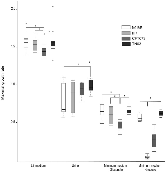
In order to better understand the capacity of this strain to colonize the intestinal tract, we first checked for potential production of bacteriocins by TN03. These antimicrobial peptides are widespread among the Bacteria and Archaea and are active against other bacteria, whether of the same species or across genera [21]. TN03 strain of E. coli was spotted onto a layer of E. coli K-12 MG1655 spread on a mitomycin plate and incubated for 18 h at 37°C. No halo was visible around the spot, indicating that no colicin (or phage) was present. Moreover, TN03 appears to be devoid of the iroN gene [11] (Table 1), which is associated with the main microcin genes (micH47 and micV) [22]. Given these negative results, we then checked whether the TN03 strain had better metabolic capacities than the other strains. This experiment was based on the hypothesis that E. coli is probably a highly successful competitor in the mammalian colon, because it exploits its ability to utilize the available nutrients more efficiently than other resident species [15]. We therefore studied the bacterial metabolic capacities of the TN03 strain by measuring its maximal growth rate (MGR) under three culture conditions using Luria Bertani (LB) broth, a rich medium, and two minimum media: a minimum medium containing gluconate (MMGluconate), as gluconate has been reported to be a major carbon source in the intestine [23], and a minimum medium containing glucose (MMGlc). Indeed, in addition to the capacity of TN03 to colonize the bowel, the question arises as to whether the TN03 strains exhibit better metabolic capacities than other UPEC strains. We therefore compared the metabolic capacity of TN03 to that of the non-pathogenic K-12 MG1655 strain and two B2-UPEC strains, i.e. CFT073 [24] and HT7 [25]. All the strains studied were precultured in the corresponding medium for 24 h with constant stirring. They were each then inoculated into the appropriate medium into plates containing 96 flat-bottomed wells. OD600 was measured every 5 minutes in order to determine the growth over a period of 24 hours. Interestingly, the TN03 strain exhibited high in-vitro growth capacity in all three media tested (Fig. 2). TN03 had a greater MGR than CFT073 in LB (1.58 vs 1.43, p = 0.01), or MMGluconate (0.65 vs 0.47 p<0.01). TN03 also exhibited a higher MGR that the other three strains (TN03-K-12 MG1655 0.64 vs 0.55 p = 0.01; TN03-HT7 0.64 vs 0.06 p<0.01; TN03-CFT073 0.64 vs 0.31 p<0.01) when grown in MMGlc. Taken together, these data suggest that TN03 has high metabolic potential, facilitating its adaptation to different environments, and probably an enhanced ability to establish and maintain intestinal colonization, the first step of uropathogenicity. These data support the hypothesis suggested by Johnson et al [26] that ST131 strains may have enhanced fitness for upstream steps, including colonization.
Figure 2. Maximum growth rate in four media.
Four E. coli strains: K-12 MG1655 (white), HT7 (light grey), CFT073 (grey) and TN03 (dark grey) were grown in four different media: Luria Bertani (LB), urine, minimum medium with gluconate and minimum medium with glucose. Boxplots represent distribution of maximal growth rates (MGRs) calculated during the three repetitions of culture assays with the smooth spline function from R network. Black bars inside each boxplot represent medians. Dots located far from the box represent outliers. Links between boxplots with asterisks represent significant differences between two strains tested by Welch test for observed mean comparison. * p<0.05.
The TN03 strain is able to infect the urinary tract for at least 7 days
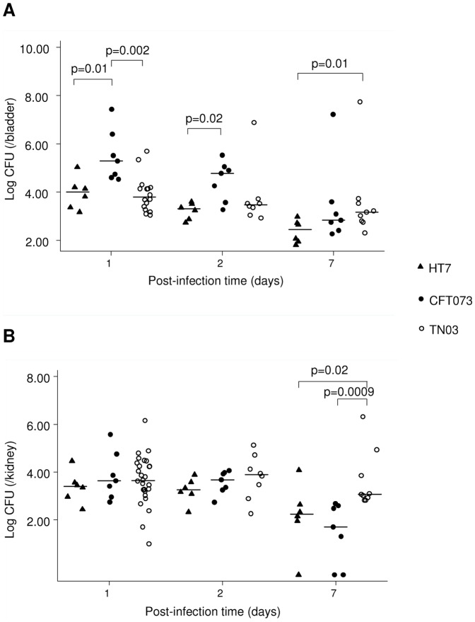
After having colonized the intestinal bowel, UPEC are then able to colonize the periurethral area, and secondly colonize the lower (i.e. the bladder) and the upper (i.e. kidneys) urinary tract. To better understand the uropathogenic abilities of the E. coli TN03 strain, experiments were then performed using an experimental model of ascending UTI as previously described [25], [27]. Renal retrograde urinary tract infection was carried out on C3H/HeN (Lpsn) mice, which were then sacrificed at days 1, 2, and 7 post bacterial inoculation. Median log10 bacterial counts per bladder or per kidney were calculated and compared in the different strains at each of the post-infection time-points (days 1, 2, and 7) using a Kruskal-Wallis equality-of-populations rank test. Murine models of UTI have been established in different strains of mice with different genetic backgrounds, such as C57BL/6, CBA, FVB/NJ or different strains of C3H mice (for review see [28]). However, these strains exhibit large differences in the levels of bacterial loads in the lower and upper urinary tract and in the degree of host inflammatory response. Whereas C57BL/6 mice produce a more robust inflammatory response and resolve acute infection and bacteriuria more rapidly than the other strains, C3H background mouse strains have been reported to develop chronic UTI for up to 2 weeks post-infection [29]–[31]. Studies using CH3/HeN (LPSn) mice exhibiting functional Toll-like receptor 4 (TLR4), and C3H/HeJ (LPSd) mice exhibiting an inactivating mutation in the tlr4 gene have demonstrated the importance of the UPEC-mediated TLR4 signaling pathway (reviewed in [32]). We also showed that different UPEC strains colonize the kidneys of C3H/HeN mice [25]. C3H/HeN mice were therefore used as a model of chronic infection to study the outcome of the early and delayed kinetics of renal colonization and renal inflammatory response caused by TN03, which were compared, as controls, to the same two B2-UPEC strains used above: CFT073 and HT7. These strains belong to B2 subgroups II (ST73) and IX (ST95), respectively, whereas TN03 belongs to subgroup I (ST131) (Table 1) [33]. As CFT073 is cytotoxic for renal tubule epithelial cells, whereas HT7 is not [25], mice were infected either with the TN03 strain or with the CFT073 or the HT7 strains. Like the UPEC CFT073 and HT7 strains [34], [35], the E. coli TN03 strain colonized the bladder and the kidneys 24 h after retrograde inoculation. One and two days after the inoculation of UPEC, the bacterial burdens were significantly greater in the bladders from CFT073-infected mice than those from HT7- or TN03-infected mice, but no significant difference was observed in the bacterial loads at days 1 and 2 in the kidneys (Fig. 3 A and B). However, at day 7, the median value of CFU in the bladder was greater for TN03 than for HT7 (1480 vs 283 CFU/bladder, p = 0.01), and to a lesser extent, for CFT073 (Fig. 3A). In the kidneys, the median value of CFU at day 7 was significantly higher for TN03 than for HT7 (1170 vs 175 CFU/kidney, p = 0.02) or CFT073 (1170 vs 50 CFU/kidney, p = 0.0009) isolates (Fig. 3B). These data, which highlighted the better capacities of this strain to persist in the kidneys, led us to check its metabolic capacities in the urinary tract. We therefore compared the MGR of the TN03 strain, the CFT073 and HT7 UPEC strains and the K-12 MG1655 strain in urine from healthy volunteers as described above. Interestingly, the TN03 strain grew better than K-12 MG1655 (0.98 vs 0.81, p = 0.03), and to a lesser extent, better than the other UPEC strains analyzed (Fig. 2). Collectively, these findings suggest that the TN03 strain is indeed an UPEC strain, in agreement with the study of Totsika et al. [36] on ST131 E. coli strains. We showed, in addition, that the TN03 UPEC strain invades the bladder less rapidly than the CFT073 strain, but persists at higher level in the bladder and kidneys than the UPEC CFT073 and HT7 strains, one week after bacterial inoculation.
Figure 3. Experimental model of UTI.
C3H/HeN mice were infected with the HT7, CFT073, or TN03 strains directly by transurethral inoculation of the bacteria (108 cfu in 50 µl sterile PBS). At day 1, day 2 and day 7 post infection, bladders (A) and kidneys (B) were harvested and their bacterial burdens determined. Horizontal bars represent median log10 bacterial counts. Statistical differences between strains at each post-infection time point were performed using the Kruskal-Wallis equality-of-populations rank test.
TN03 induces a low renal host inflammatory response
We next investigated the host's inflammatory response following urinary tract infection caused by TN03. For this purpose, we analyzed the levels of mRNA expression of the TNF-α cytokine and 6 pro-inflammatory cytokines, such as MIP-2, KC, IL-1ß, RANTES, IL-6, and MCP-1, by real-time PCR as previously described [25] in kidneys from mice infected with TN03 using CFT073 UPEC as a control. Among the proinflammatory mediators tested, only MIP-2/CXCL2, which is a chemoattractant chemokine playing a key role in the migration of polymorphonuclear neutrophils to the site of inflammation [37], [38], showed a significantly higher level in the day-2 TN03 than day-2 CFT073 post-infected kidneys (Fig. 4). The expression level of KC/CXCL1 which also plays a key role in the neutrophils chemoattraction and the expression levels of the other pro-inflammatory mediators tested (Fig. 4), as well as the number of Ly6-G-positive neutrophils attracted to the kidneys, did not differ significantly either in the day 2 post TN03-infection kidneys versus the CFT073-infected kidneys nor in those for day 7 (data not shown). These findings suggest that TN03 causes quite similar inflammatory response compared to that induced by CFT073. Colonization of the urinary tract by UPEC induces the rapid (within the first 6–12 h) activation of proinflammatory mediators and the recruitment of the neutrophils for the efficient phagocytic killing of bacteria. Thereafter, the increase in pro-inflammatory mediators tends to return to normal levels. Given that all measurements were performed 48 h after the inoculation of UPEC, no direct conclusions could be drawn concerning the possibility that TN03 alters the inflammatory response and subsequent recruitment of neutrophils to the site of inflammation. Bacterial attachment to mucosal bladder and renal epithelial cells by fimbrial adhesins constitutes the initial step in UPEC pathogenicity. Pyelonephritis-associated UPEC strains usually express P-fimbriae, such as PapGII, which preferentially binds to the globoseries glycosphingolipids that are abundantly expressed on the surface of renal epithelial cells (see for review [39]). The attachment of fimbriated UPEC to renal epithelial cells triggers the innate host response, mediated in part by TLR4, which recognizes lipopolysaccharide [27], [40]–[42]. The activation of TLR4 signaling stimulates the production of pro-inflammatory mediators leading to the recruitment of neutrophils and macrophages to the site of inflammation for the efficient killing of bacteria. The fact that TN03 does not express PapGII (Table 1), could suggest that the low binding capacity of TN03 to epithelial cells may favor the persistence of long-lived bacteria within the kidneys. The ability of the UPEC TN03 strain to limit the inflammatory response and the subsequent clearance of bacteria, and to persist in the upper urinary tract without activating a significant inflammatory response calls for further studies.
Figure 4. Expression of pro-inflammatory mediators in kidneys infected by UPEC.
Expression of MIP-2, KC, IL-1ß, IL-6, MCP-1, RANTES and TNF-α in the day 2 and day 7 post-infection kidneys following the transurethral inoculation of the CFT073 and TN03 UPEC isolates. mRNA expression of each inflammatory marker was calculated using the −ΔΔCT method. Values are expressed as the relative fold increase in kidneys of each mRNA level of proinflammatory mediator in comparison with that measured in naive mice. Horizontal bars represent the median values. Statistical differences between the strains were determined at each time point using the Kruskal-Wallis equality-of-populations rank test.
Concluding remarks
Special pathogenicity traits and the prevalence of the E. coli strain in the fecal microbiota are both important features required for the occurrence of UTI [16]. Virulence traits and group B2 status were shown to be closely associated with the fecal abundance and dominance of E. coli strains and their pauciclonality – i.e. the fact that no more than 4 different E. coli clones coexist concurrently. Collectively, these various different factors contribute jointly to UTI pathogenesis, in association with other still-unidentified virulence factors and other group B2-associated characteristics. This latter may promote intestinal dominance [33], thereby increasing the probability that the subsequent steps in pathogenesis will occur. Johnson et al [26] also suggest that the epidemiological success of ST131 may reflect the fact that it displays greater fitness during the upstream steps involved in pathogenesis, colonization or in transmission. In this study, we suggest that the TN03 strain has higher metabolic capacities, and that these may explain its enhanced capacities to colonize and subsequently to cause infectious disease. Indeed, epidemiological studies both during UTI in 2003 [43] and on stools of healthy subjects in 2006 [44] have reported a high prevalence of the ST131 clone of E. coli (3% and 7%, respectively). Rogers et al [12] and Johnson et al [45] also confirmed the worldwide prevalence of ST131 harboring a broad range of virulence and resistance genes. Undoubtedly, the increasing prevalence of the ESBL not only at the hospital, but also in the community setting, suggests that this particular clone harboring ESBL is also increasing. However, further investigations are needed to define the current prevalence of this clone more precisely.
The spectrum of clinical infections caused by the ST131 clone appears broadly similar to that of other strains of E. coli. The urinary tract is the most common site of human infection at which E. coli predominates. Pitout et al [46] identified a greater propensity for urinary sepsis above other sites of infection in ST131 than in non-ST131 E. coli bacteriemia. The results from the present work suggest that the specific TN03 ST131 strain expressing CTX-M is a potent uropathogenic strain that can efficiently colonize the intestine, as a result of its greater metabolic capacities or other still-unknown factors, and subsequently infect the urinary tract, conferring on it an intrinsic evolutionary success, regardless of its selection as a result of antibiotic pressure. This TN03 clone possesses a number of virulence factors (Table 1), including the pathogenicity island HPI (fyuA, irp2), siderophores (iutA, iha), the capacity for serum resistance (traT) and a capsule (kpsE) [47], but it does not possess the classical extraintestinal PAIs (PAICFT073, PAI IIJ96 and PAI III536), which may limit the inflammatory host response and the subsequent clearance of bacteria. The group of Frimodt-Møller showed that some B2 E. coli strains, with a higher aggregate virulence factor genes score, are associated with persistence or relapse in recurrent UTI [48]. In this line, TN03 also has the ability to form a biofilm [11], a property which may contribute to its long-term persistence in various environments and to its resistance to antimicrobial agents or disinfectants [49].
However, our data should be balanced by the fact that we have studied only one isolate representing the ST131 clone, i.e. TN03, and that high genetic [45], [50], [51] and intrinsic virulence [26] polymorphism has been reported within isolates of this clone. More isolates clearly need to be studied.
Given the rapid emergence of the ST131 CTX-M-15 E. coli clone, particular attention should be paid to this particular uropathogenic strain, which expresses resistance to classical antibiotic therapy and constitutes a major health problem [8]. Our experimental data on a representative strain of the emerging O25b ST131 clone support the link between the B2 group, intestinal colonization, virulence determinants, and UTI. Further studies will be required to identify the additional factor(s) responsible for intestinal colonization and the ability of the TN03 strain to invade the urinary tract system.
Materials and Methods
Bacterial strains and growth conditions
Experiments were carried out using several different E. coli strains: the TN03 strain, which is a representative isolate of the O25b:H4-ST131 CTX-M-15 E. coli clone [11], two UPEC strains belonging to phylogenetic group B2: the CFT073 strain [24] and the HT7 strain, which was isolated from the infected urine of a young woman hospitalized following an episode of pyelonephritis at the Tenon hospital (Paris, France) [27], [35], two commensal E. coli strains: IAI1 and ED1a, one from phylogenetic group B1 and the other from phylogenetic group B2, and the non-pathogenic laboratory strain E. coli K-12 MG1655 [18] (Table 1). When not otherwise indicated, bacteria were grown in LB broth with shaking for 18 h at 37°C.
For the comparative growth assays, strains were grown at 37°C in four different media: LB (tryptone 10 g, yeast extract 5 g, NaCl 10 g/L), minimum medium with glucose (MMGlc) 20 mM [NaH2PO4,1 H2O 4.69 g, Na2HPO4 11.15 g/L, (NH4)2SO4 2.65 g/L, MgSO4 7 H2O 0.075 g/L, KCl 3 g/L, FeCl3 0.2 ml Molar Solution 0.1%, glucose 20 mM/L], minimum medium with gluconate (MMGluconate) 20 mM (the same as the minimum medium, but with glucose instead of gluconate), and urine. Urine was collected from ten healthy male volunteers who were not taking any medication, pooled, filtered, and stored at −20°C before use. LB and urine are complex media, whereas MMGlc and MMGluconate are minimal media with only one source of carbon. The MMGluconate is used, as this sugar has been reported to be a major source of carbon in the intestine [23]. All the strains studied were grown overnight (O/N) in the three media in flasks at 37°C with constant stirring at 280 rpm. O/N cultures were prediluted at 1/100 in PBS and strains were inoculated in twelve different wells each at 1/100 in a Costar® 96 flat-bottomed well plate. Growth was recorded by an Infinite 200 Tecan®, which measured the OD600 in each well every 5 minutes at 37°C, while stirring for 24 hours. Growth assays were repeated 3 times. The MGR was calculated from growth curves obtained by Tecan®. OD600 in nm were collected and Log-transformed. Curves were calculated from a smoothed spline function. The MGR was defined as the time point at which the maximum value of the derivative of the smoothed function was observed. All MGRs were compared by strain and by medium using a Welch test.
Intestinal colonization
Six-week-old female mice (Charles River CD-1) pretreated with streptomycin were used to monitor the ability of E. coli strains to colonize the intestine of a mammalian host. The mice were isolated and had free access to sterile food and drinking water supplemented with streptomycin sulfate (5 g/liter) for 5 days. The mice did not receive streptomycin for 5 days before bacterial inoculation. This modification of the classical streptomycin-treated mouse colonization assay [17] permits the subsequent colonization of the mouse intestine by streptomycin-sensitive strains. The effectiveness of the antibiotic treatment against the coliform intestinal population was checked by plating a pure suspension of feces on Drigalski's selective agar medium. The absence of streptomycin was also confirmed by assaying the antibiotic in the stools. The day of inoculation, 106 E. coli bacteria were then administered in 200 µl of PBS by oral route to mice free of coliform flora, either alone or mixed at a ratio of 1∶1. On days 1, 2, 4 and 7 after bacterial administration, the sizes of bacterial populations in the intestine of mice were evaluated by plating dilutions of weighed fresh feces on Drigalski's agar with or without 2 µg/ml cefotaxime. At least four mice were used in three independent experiments. As previously described [20], a competitive index (CI) was calculated. The CI index is a ratio of ratios, in which the ratio between resistant and sensitive strains at post-inoculation time points is divided by the same ratio at the initial inoculation time. All experiments were performed in accordance with the recommendations of the French Ministry of Agriculture and approved by the French Veterinary Services (accreditation number A 75-18-05).
Production of colicins and phages
Colicins and phages were detected as previously described [52]. Briefly, they were detected using an overnight (O/N) preculture suspension of E. coli K-12 MG1655 as a sensitive strain. K-12 MG1655 bacteria were plated on a LB agar medium containing mitomycin (25 µm/L). Then, 10 µl of an O/N culture in LB medium of each strain were spotted. After an O/N culture at 37°C, the presence of colicin (or phage) were detected for the strains surrounded by a halo reflecting an inhibition of the culture of the E. coli K-12 strain.
Bacterial determinants
As previously described, the strains were assigned to 1 of the 4 main E. coli phylogenetic groups, i.e., groups A, B1, B2, and D, using triplex PCR developed previously by Clermont et al [53]. MLST was performed by gene amplification and sequencing as in [54] and/or [55]. The B2 subgroup O-type was determined as described by Le Gall et al [33]. The presence of 19 virulence factors representative of identified E. coli extraintestinal virulence determinants were tested by PCR [56].
Murine model of ascending urinary tract infection
Renal retrograde urinary tract infection was carried out on 8-week-old female C3H/HeN (Lpsn) mice (Janvier Breeding Center, Le Genest St Isle, France), which were caged individually. Mice subjected to water restriction for 12 h were anesthetized, and then infected with various E. coli strains introduced (108 bacteria diluted in 50 µ l sterile PBS) via the transurethral route into the bladder as described previously [27], [35]. Once the mice had recovered from anesthesia, they were provided with water for the 7 days following the bacterial inoculation. Mice were then sacrificed at days 1, 2, and 7 after bacterial inoculation, and the kidneys and bladder were aseptically removed. Two halves of one kidney were fixed or quickly frozen in liquid nitrogen, and the contralateral kidney was homogenized, diluted in sterile PBS, and plated on LB agar plates to count the number of colony forming units (CFUs). Kidney sections were stained using an anti-Ly6-G antibody (BD Biosciences France SA) to quantify the number of infiltrating neutrophils. Mice were infected either with the TN03 strain or with either the UPEC CFT073 or HT7 strain belonging to phylogenetic group B2. At least ten mice were used for each condition. The experiments were performed in accordance with the guidelines of the French Agriculture Ministry (see above).
Quantitative real time PCR
Total RNA was extracted from kidneys using the RNABle kit (Eurobio, Courtaboeuf, France), and reverse-transcribed using Moloney Murine Leukemia Virus reverse transcriptase (Invitrogen, Cergy-Pontoise, France). cDNA was subjected to real-time PCR, using a Chromo IV sequence detector (MJ Research, Waltham, MA) using mouse primers and TaqMan probes used for 6 different proinflammatory cytokines: chemokine (C-X-C motif) ligand 2 (CXCL2) also called macrophage inflammatory protein 2 α (MIP-2α), chemokine (C-X-C motif) ligand 1 (CXCL1) also called keratinocyte-derived chemokine (KC), Interleukin-1β (IL-1β), Interleukin-6 (IL-6), chemokine (C-C motif) ligand 2 (CCL2) also known as monocyte chemoattractant protein-1 (MCP-1), chemokine (C-C motif) ligand 5 (CCL5) also known as regulated upon activation normal T cell expressed and secreted (RANTES); the tumor necrosis factor-α (TNF-α) cytokine, and ß-actin as previously described earlier [25]. PCR data were reported as the relative increase, after infection, in mRNA transcripts versus non infected kidneys, and corrected by the levels of ß-actin mRNA, used as the internal standard. Values are the means of 6–8 separate determinations.
Statistical analysis
The values are given as medians (IQR) and, unless specified otherwise, comparisons between strains were performed using either the Wilcoxon signed-rank test or the Kruskal-Wallis equality-of-populations rank test. All statistics were computed using STATA (v10.0, College Station, TX, USA) or R (R Development Core Team, 2009, Vienna, Austria) and statistical significance was determined at a p-value of less than 0.05.
Acknowledgments
We would like to thank M.D. Kitzis for assaying the residual rate of streptomycin in the stools of the mice.
Funding Statement
This work was funded in part by a grant from the French National Research Agency under reference ANR-08-MIE-030 (to GA, and AV). AV was in receipt of a “Contrat Hospitalier de Recherche Translationnelle” (CHRP, INSERM-APHP, 2010). A.B. was supported by a “Bourse Médico-Scientifique” from the “Fondation pour la Recherche Médicale”. The funders had no role in study design, data collection and analysis, decision to publish, or preparation of the manuscript.
References
- 1. Gupta K, Scholes D, Stamm WE (1999) Increasing prevalence of antimicrobial resistance among uropathogens causing acute uncomplicated cystitis in women. Jama 281: 736–738. [DOI] [PubMed] [Google Scholar]
- 2. Pitout JD, Nordmann P, Laupland KB, Poirel L (2005) Emergence of Enterobacteriaceae producing extended-spectrum beta-lactamases (ESBLs) in the community. J Antimicrob Chemother 56: 52–59. [DOI] [PubMed] [Google Scholar]
- 3. Rodriguez-Bano J, Paterson DL (2006) A change in the epidemiology of infections due to extended-spectrum beta-lactamase-producing organisms. Clin Infect Dis 42: 935–937. [DOI] [PubMed] [Google Scholar]
- 4. Paterson DL, Bonomo RA (2005) Extended-spectrum beta-lactamases: a clinical update. Clin Microbiol Rev 18: 657–686. [DOI] [PMC free article] [PubMed] [Google Scholar]
- 5. Pitout JD, Laupland KB (2008) Extended-spectrum beta-lactamase-producing Enterobacteriaceae: an emerging public-health concern. Lancet Infect Dis 8: 159–166. [DOI] [PubMed] [Google Scholar]
- 6. Coque TM, Novais A, Carattoli A, Poirel L, Pitout J, et al. (2008) Dissemination of clonally related Escherichia coli strains expressing extended-spectrum beta-lactamase CTX-M-15. Emerg Infect Dis 14: 195–200. [DOI] [PMC free article] [PubMed] [Google Scholar]
- 7. Nicolas-Chanoine MH, Blanco J, Leflon-Guibout V, Demarty R, Alonso M P, et al. (2008) Intercontinental emergence of Escherichia coli clone O25:H4-ST131 producing CTX-M-15. J Antimicrob Chemother 61: 273–281. [DOI] [PubMed] [Google Scholar]
- 8. Johnson JR, Johnston B, Clabots C, Kuskowski MA, Castanheira M, et al. (2010) Escherichia coli sequence type ST131 as the major cause of serious multidrug-resistant E. coli infections in the United States. Clin Infect Dis 51: 286–294. [DOI] [PubMed] [Google Scholar]
- 9. Peirano G, Pitout JD (2010) Molecular epidemiology of Escherichia coli producing CTX-M beta-lactamases: the worldwide emergence of clone ST131 O25:H4. Int J Antimicrob Agents 35: 316–321. [DOI] [PubMed] [Google Scholar]
- 10. Owens RC Jr, Johnson JR, Stogsdill P, Yarmus L, Lolans K, et al. (2011) Community Transmission in the United States of a CTX-M-15-Producing Sequence Type ST131 Escherichia coli Strain Resulting in Death. J Clin Microbiol 49: 3406–3408. [DOI] [PMC free article] [PubMed] [Google Scholar]
- 11. Clermont O, Lavollay M, Vimont S, Deschamps C, Forestier C, et al. (2008) The CTX-M-15-producing Escherichia coli diffusing clone belongs to a highly virulent B2 phylogenetic subgroup. J Antimicrob Chemother 61: 1024–1028. [DOI] [PubMed] [Google Scholar]
- 12. Rogers BA, Sidjabat HE, Paterson DL (2011) Escherichia coli O25b-ST131: a pandemic, multiresistant, community-associated strain. J Antimicrob Chemother 66: 1–14. [DOI] [PubMed] [Google Scholar]
- 13. Eckert C, Gautier V, Saladin-Allard M, Hidri N, Verdet C, et al. (2004) Dissemination of CTX-M-type beta-lactamases among clinical isolates of Enterobacteriaceae in Paris, France. Antimicrob Agents Chemother 48: 1249–1255. [DOI] [PMC free article] [PubMed] [Google Scholar]
- 14. Lavollay M, Mamlouk K, Frank T, Akpabie A, Burghoffer B, et al. (2006) Clonal dissemination of a CTX-M-15 beta-lactamase-producing Escherichia coli strain in the Paris area, Tunis, and Bangui. Antimicrob Agents Chemother 50: 2433–2438. [DOI] [PMC free article] [PubMed] [Google Scholar]
- 15. Kaper JB, Nataro JP, Mobley HL (2004) Pathogenic Escherichia coli . Nat Rev Microbiol 2: 123–140. [DOI] [PubMed] [Google Scholar]
- 16. Moreno E, Andreu A, Pigrau C, Kuskowski MA, Johnson JR, et al. (2008) Relationship between Escherichia coli strains causing acute cystitis in women and the fecal E. coli population of the host. J Clin Microbiol 46 8:2529–2534. [DOI] [PMC free article] [PubMed] [Google Scholar]
- 17. Myhal ML, Laux DC, Cohen PS (1982) Relative colonizing abilities of human fecal and K 12 strains of Escherichia coli in the large intestines of streptomycin-treated mice. Eur J Clin Microbiol 1: 186–192. [DOI] [PubMed] [Google Scholar]
- 18. Blattner FR, Plunkett G 3rd, Bloch CA, Perna NT, Burland V, et al. (1997) The complete genome sequence of Escherichia coli K-12. Science 277: 1453–1474. [DOI] [PubMed] [Google Scholar]
- 19. Touchon M, Hoede C, Tenaillon O, Barbe V, Baeriswyl S, et al. (2009) Organised genome dynamics in the Escherichia coli species results in highly diverse adaptive paths. PLoS Genet 5: e1000344. [DOI] [PMC free article] [PubMed] [Google Scholar]
- 20. Diard M, Garry L, Selva M, Mosser T, Denamur E, et al. (2010) Pathogenicity-associated islands in extraintestinal pathogenic Escherichia coli are fitness elements involved in intestinal colonization. J Bacteriol 192: 4885–4893. [DOI] [PMC free article] [PubMed] [Google Scholar]
- 21. Riley MA, Wertz JE (2002) Bacteriocins: evolution, ecology, and application. Annu Rev Microbiol 56: 117–137. [DOI] [PubMed] [Google Scholar]
- 22. Waters VL, Crosa JH (1991) Colicin V virulence plasmids. Microbiol Rev 55: 437–450. [DOI] [PMC free article] [PubMed] [Google Scholar]
- 23. Chang DE, Smalley DJ, Tucker DL, Leatham MP, Norris WE, et al. (2004) Carbon nutrition of Escherichia coli in the mouse intestine. Proc Natl Acad Sci U S A 101: 7427–7432. [DOI] [PMC free article] [PubMed] [Google Scholar]
- 24. Welch RA, Burland V, Plunkett G 3rd, Redford P, Roesch P, et al. (2002) Extensive mosaic structure revealed by the complete genome sequence of uropathogenic Escherichia coli . Proc Natl Acad Sci U S A 99: 17020–17024 Epub 2002 Dec 05. [DOI] [PMC free article] [PubMed] [Google Scholar]
- 25. Chassin C, Vimont S, Cluzeaud F, Bens M, Goujon JM, et al. (2008) TLR4 facilitates translocation of bacteria across renal collecting duct cells. J Am Soc Nephrol 19: 2364–2374. [DOI] [PMC free article] [PubMed] [Google Scholar]
- 26. Johnson JR, Porter SB, Zhanel G, Kuskowski MA, Denamur E (2012) Virulence of Escherichia coli clinical isolates in a murine sepsis model in relation to sequence type ST131 status, fluoroquinolone resistance, and virulence genotype. Infect Immun 80: 1554–1562. [DOI] [PMC free article] [PubMed] [Google Scholar]
- 27. Chassin C, Goujon JM, Darche S, du Merle L, Bens M, et al. (2006) Renal collecting duct epithelial cells react to pyelonephritis-associated Escherichia coli by activating distinct TLR4-dependent and -independent inflammatory pathways. J Immunol 177: 4773–4784. [DOI] [PubMed] [Google Scholar]
- 28. Hung CS, Dodson KW, Hultgren SJ (2009) A murine model of urinary tract infection. Nat Protoc 4: 1230–1243. [DOI] [PMC free article] [PubMed] [Google Scholar]
- 29. Hopkins WJ, Gendron-Fitzpatrick A, Balish E, Uehling DT (1998) Time course and host responses to Escherichia coli urinary tract infection in genetically distinct mouse strains. Infect Immun 66: 2798–2802. [DOI] [PMC free article] [PubMed] [Google Scholar]
- 30. Schilling JD, Lorenz RG, Hultgren SJ (2002) Effect of trimethoprim-sulfamethoxazole on recurrent bacteriuria and bacterial persistence in mice infected with uropathogenic Escherichia coli . Infect Immun 70: 7042–7049. [DOI] [PMC free article] [PubMed] [Google Scholar]
- 31. Hannan TJ, Mysorekar IU, Hung CS, Isaacson-Schmid ML, Hultgren SJ (2010) Early severe inflammatory responses to uropathogenic E. coli predispose to chronic and recurrent urinary tract infection. PLoS Pathog 6: e1001042. [DOI] [PMC free article] [PubMed] [Google Scholar]
- 32. Song J, Abraham SN (2008) Innate and adaptive immune responses in the urinary tract. Eur J Clin Invest 38 Suppl 2: 21–28. [DOI] [PubMed] [Google Scholar]
- 33. Le Gall T, Clermont O, Gouriou S, Picard B, Nassif X, et al. (2007) Extraintestinal virulence is a coincidental by-product of commensalism in B2 phylogenetic group Escherichia coli strains. Mol Biol Evol 24: 2373–2384. [DOI] [PubMed] [Google Scholar]
- 34. Lane MC, Alteri CJ, Smith SN, Mobley HL (2007) Expression of flagella is coincident with uropathogenic Escherichia coli ascension to the upper urinary tract. Proc Natl Acad Sci U S A 104: 16669–16674. [DOI] [PMC free article] [PubMed] [Google Scholar]
- 35. Chassin C, Hornef MW, Bens M, Lotz M, Goujon JM, et al. (2007) Hormonal control of the renal immune response and antibacterial host defense by arginine vasopressin. J Exp Med 204: 2837–2852. [DOI] [PMC free article] [PubMed] [Google Scholar]
- 36. Totsika M, Beatson SA, Sarkar S, Phan MD, Petty NK, et al. (2011) Insights into a multidrug resistant Escherichia coli pathogen of the globally disseminated ST131 lineage: genome analysis and virulence mechanisms. PLoS One 6: e26578. [DOI] [PMC free article] [PubMed] [Google Scholar]
- 37. Hang L, Haraoka M, Agace WW, Leffler H, Burdick M, et al. (1999) Macrophage inflammatory protein-2 is required for neutrophil passage across the epithelial barrier of the infected urinary tract. J Immunol 162: 3037–3044. [PubMed] [Google Scholar]
- 38. Frendeus B, Godaly G, Hang L, Karpman D, Lundstedt AC, et al. (2000) Interleukin 8 receptor deficiency confers susceptibility to acute experimental pyelonephritis and may have a human counterpart. J Exp Med 192: 881–890. [DOI] [PMC free article] [PubMed] [Google Scholar]
- 39. Lane MC, Mobley HL (2007) Role of P-fimbrial-mediated adherence in pyelonephritis and persistence of uropathogenic Escherichia coli (UPEC) in the mammalian kidney. Kidney Int 72: 19–25. [DOI] [PubMed] [Google Scholar]
- 40. Schilling JD, Martin SM, Hung CS, Lorenz RG, Hultgren SJ (2003) Toll-like receptor 4 on stromal and hematopoietic cells mediates innate resistance to uropathogenic Escherichia coli . Proc Natl Acad Sci U S A 100: 4203–4208. [DOI] [PMC free article] [PubMed] [Google Scholar]
- 41. Song J, Duncan MJ, Li G, Chan C, Grady R, et al. (2007) A novel TLR4-mediated signaling pathway leading to IL-6 responses in human bladder epithelial cells. PLoS Pathog 3: e60. [DOI] [PMC free article] [PubMed] [Google Scholar]
- 42. Hagberg L, Hull R, Hull S, McGhee JR, Michalek SM, et al. (1984) Difference in susceptibility to gram-negative urinary tract infection between C3H/HeJ and C3H/HeN mice. Infect Immun 46: 839–844. [DOI] [PMC free article] [PubMed] [Google Scholar]
- 43. Clermont O, Dhanji H, Upton M, Gibreel T, Fox A, et al. (2009) Rapid detection of the O25b-ST131 clone of Escherichia coli encompassing the CTX-M-15-producing strains. J Antimicrob Chemother 64: 274–277. [DOI] [PubMed] [Google Scholar]
- 44. Leflon-Guibout V, Blanco J, Amaqdouf K, Mora A, Guize L, et al. (2008) Absence of CTX-M enzymes but high prevalence of clones, including clone ST131, among fecal Escherichia coli isolates from healthy subjects living in the area of Paris, France. J Clin Microbiol 46: 3900–3905. [DOI] [PMC free article] [PubMed] [Google Scholar]
- 45. Johnson JR, Nicolas-Chanoine MH, DebRoy C, Castanheira M, Robicsek A, et al. (2012) Comparison of Escherichia coli ST131 pulsotypes, by epidemiologic traits, 1967–2009. Emerg Infect Dis 18: 598–607. [DOI] [PMC free article] [PubMed] [Google Scholar]
- 46. Pitout JD, Gregson DB, Campbell L, Laupland KB (2009) Molecular characteristics of extended-spectrum-beta-lactamase-producing Escherichia coli isolates causing bacteremia in the Calgary Health Region from 2000 to 2007: emergence of clone ST131 as a cause of community-acquired infections. Antimicrob Agents Chemother 53: 2846–2851. [DOI] [PMC free article] [PubMed] [Google Scholar]
- 47. Clermont O, Olier M, Hoede C, Diancourt L, Brisse S, et al. (2011) Animal and human pathogenic Escherichia coli strains share common genetic backgrounds. Infect Genet Evol 11: 654–662. [DOI] [PubMed] [Google Scholar]
- 48. Ejrnaes K, Stegger M, Reisner A, Ferry S, Monsen T, et al. (2011) Characteristics of Escherichia coli causing persistence or relapse of urinary tract infections: phylogenetic groups, virulence factors and biofilm formation. Virulence 2: 528–537. [DOI] [PubMed] [Google Scholar]
- 49. Ghigo JM (2001) Natural conjugative plasmids induce bacterial biofilm development. Nature 412: 442–445. [DOI] [PubMed] [Google Scholar]
- 50. Johnson JR, Urban C, Weissman SJ, Jorgensen JH, Lewis JS 2nd, et al. (2012) Molecular epidemiological analysis of Escherichia coli sequence type ST131 (O25:H4) and blaCTX-M-15 among extended-spectrum-beta-lactamase-producing E. coli from the United States, 2000 to 2009. Antimicrob Agents Chemother 56: 2364–2370. [DOI] [PMC free article] [PubMed] [Google Scholar]
- 51. Coelho A, Mora A, Mamani R, Lopez C, Gonzalez-Lopez JJ, et al. (2011) Spread of Escherichia coli O25b:H4-B2-ST131 producing CTX-M-15 and SHV-12 with high virulence gene content in Barcelona (Spain). J Antimicrob Chemother 66: 517–526. [DOI] [PubMed] [Google Scholar]
- 52. Lescat M, Clermont O, Woerther PL, Glodt J, Dion S, et al. (2012) Commensal Escherichia coli strains in Guiana reveal a high genetic diversity with host-dependant population structure. Submitted [DOI] [PubMed] [Google Scholar]
- 53. Clermont O, Bonacorsi S, Bingen E (2000) Rapid and simple determination of the Escherichia coli phylogenetic group. Appl Environ Microbiol 66: 4555–4558. [DOI] [PMC free article] [PubMed] [Google Scholar]
- 54. Jaureguy F, Landraud L, Passet V, Diancourt L, Frapy E, et al. (2008) Phylogenetic and genomic diversity of human bacteremic Escherichia coli strains. BMC Genomics 9: 560. [DOI] [PMC free article] [PubMed] [Google Scholar]
- 55. Wirth T, Falush D, Lan R, Colles F, Mensa P, et al. (2006) Sex and virulence in Escherichia coli: an evolutionary perspective. Mol Microbiol 60: 1136–1151. [DOI] [PMC free article] [PubMed] [Google Scholar]
- 56. Lefort A, Panhard X, Clermont O, Woerther PL, Branger C, et al. (2011) Host factors and portal of entry outweigh bacterial determinants to predict the severity of Escherichia coli bacteremia. J Clin Microbiol 49: 777–783. [DOI] [PMC free article] [PubMed] [Google Scholar]


