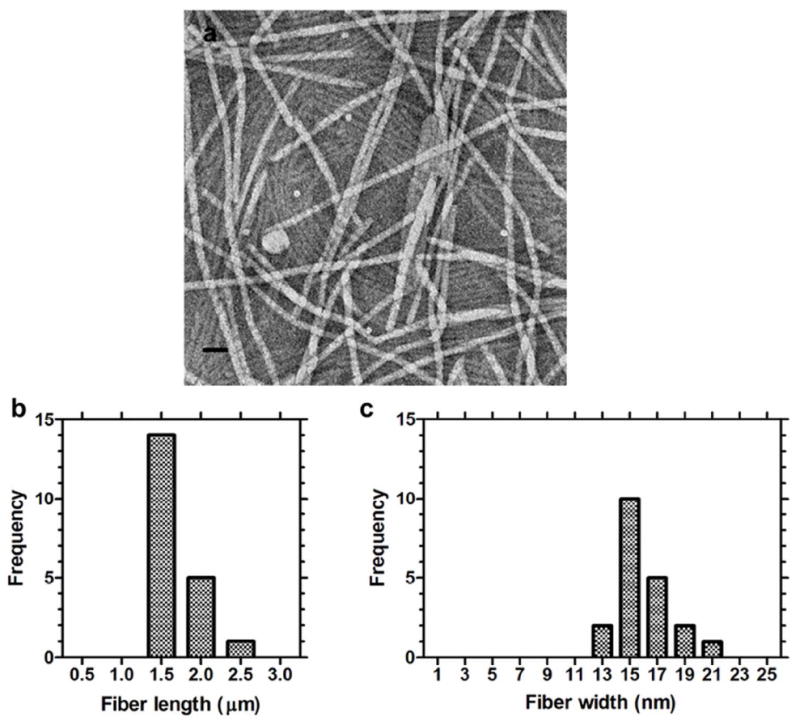
Figure 3. dpA3 self-assembles cylindrical fibers.

The dpA3 (1000 μM) was stained with 1% uranyl acetate and imaged using transmission electron microscopy (TEM). (a) Numerous fiber-like cylindrical micelle structures 1.7 ± 0.2 nm (mean ± SD, n=3) in length were observed. (b) Distribution of fiber lengths. (c) Distribution of fiber widths. Scale bar = 50 nm.
