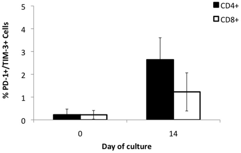
Figure 8. PD-1+/TIM-3+ lymphocytes at the beginning and end of culture.

Cells were stained with antibodies to PD-1 and Tim-3, markers of T-cell exhaustion. At the start of MDLN culture both CD4+ and CD8+ had exhausted phenotype for <1% of cells. At the end of culture exhausted phenotype increased to 2.6 (p=0.003) for CD4+ and 1.2 (p=0.061) for CD8+. Error bars indicate 95% CI. N=4.
