Abstract
fMRI responses to recognition memory test items in two regions of ventral lateral parietal cortex—the angular gyrus and temporo-parietal junction (TPJ)—are enhanced when recognition is accompanied by recollection. According to the ‘episodic buffer’ hypothesis, ventral parietal recollection effects reflect processes involved in maintaining or representing recollected information. According to the ‘attention to memory’ hypothesis, however, the effects reflect attentional re-orienting to the products of recollection. The present experiment addressed the question whether these operations map on to the angular gyrus and TPJ, respectively. Subjects were scanned during a memory test that required a Remember/Know/New and a source memory judgment, allowing recollected items to be segregated by amount of contextual information recollected. Angular gyrus activity tracked amount of recollected information, whereas activity in the TPJ was enhanced for items endorsed as recollected, but was insensitive to amount of information recollected. Thus, the two regions likely support functionally dissociable processes.
Keywords: Human episodic memory, fMRI, Lateral parietal cortex, Source memory, Recollection, Confidence
Introduction
A consistent finding in functional neuroimaging studies of memory retrieval is that neural activity in lateral parietal cortex (LPC) is enhanced when retrieval is successful (for reviews, see Vilberg & Rugg, 2008; Cabeza, Ciaramelli, Olson, & Moscovitch, 2008; Hutchinson, Uncapher, & Wagner, 2009). In recognition memory tests, these ‘retrieval success’ effects are revealed by contrasts between studied items correctly judged old (hits) and studied items incorrectly judged new (misses) or unstudied items correctly judged new (correct rejections). The effects typically involve much of the left LPC, encompassing the intra-parietal sulcus [IPS; Brodmann Area (BA) 7] and extending ventrally into the angular gyrus [BA 39] and, more infrequently, the supramarginal gyrus (BA 40) and its junction with the temporal lobe [the temporo-parietal junction (TPJ), BA40/22]. Analogous effects are sometimes also evident in the right hemisphere (for review, see Cabeza et al., 2008).
There is a consensus that retrieval success effects in the IPS can be dissociated from effects in the supramarginal and angular gyri. Using variants of the ‘Remember/Know’ procedure, studies have investigated retrieval effects according to whether recognition was accompanied by a sense of recollection (a Remember or ‘R’ response) or was based on a sense of familiarity (a Know or ‘K’ response). Whereas items attracting R or K judgments both elicit effects in the vicinity of the IPS, effects in more ventral regions are elicited exclusively by items endorsed R (e.g. Yonelinas, Otten, Shaw, & Rugg, 2005; Montaldi, Spencer, Roberts, & Mayes, 2006; for reviews, see Vilberg & Rugg, 2008; Kim, 2010). Additionally, retrieval success effects in the angular gyrus have been reported to covary with the amount of information recollected about previously studied pictures (Vilberg & Rugg, 2007; Vilberg & Rugg, 2009a), words (Vilberg & Rugg, 2009b) and faces (Guerin & Miller, 2011). The generality of graded recollection effects across different classes of test materials is consistent with the proposal that the representation of recollected information in the angular gyrus is amodal (see below).
The functional significance of retrieval success effects in different regions of the LPC is uncertain. According to one proposal (Vilberg & Rugg, 2008), recollection-selective effects in the angular gyrus reflect the participation of this region in a network that supports something like the episodic buffer proposed by Baddeley (2000). By this argument (see also Shimamura, 2011; Guerin & Miller, 2011) this region contributes to the integration of retrieved information into an amodal episodic representation. Activity in the IPS, by contrast, is held to reflect accumulation of evidence that an item is old, regardless of whether the evidence is based on recollection or familiarity.
An alternative view (the ‘attention to memory’ or A to M hypothesis) links retrieval success effects in the IPS and the supramarginal/angular gyri to different types of attentional processes (Cabeza, Ciaramelli, Olson, & Moscovitch, 2008). This view stems from the proposal these LPC regions are components of two distinct, albeit interactive, attentional networks (Corbetta & Shulman, 2002; Corbetta, Patel, & Shulman, 2008). The dorsal network, to which the IPS belongs, supports the ‘top-down’ direction of attention to task-relevant stimulus events. By contrast, retrieval success effects in the ventral network reflect ‘bottom-up’ re-orienting of attention toward the products of retrieval.
The A to M model has received criticism on the grounds that retrieval success and attentional effects demonstrate little overlap. For example, in a recent review Hutchinson et al. (2009) reported that ‘bottom-up’ attentional effects are most frequently reported in the right TPJ, whereas effects associated with recollection tend to be localized to the left angular gyrus. The idea that attention to perceptual input and attention to memory engage distinct LPC regions also receives support from a study contrasting activity elicited during search for a visual target with activity elicited during a cued memory search (Sesteri, Shulman, & Corbetta, 2010). The two conditions engaged almost completely non-overlapping regions of the LPC. However, in a study that attempted to directly assess the amount of overlap between attentional re-orienting and successful retrieval (Cabeza et al., 2011), the two classes of effect were found to partially overlap. The overlap was confined to the anterior part of the left TPJ, with retrieval but not attentional effects extending into the angular gyrus.
Here, we focus on the functional significance of ventral LPC retrieval success effects, asking whether effects localized to the TPJ and angular gyrus can be functionally dissociated. Several sources of evidence suggest this might be the case. Perhaps most persuasively, findings from resting-state functional connectivity studies indicate that these two regions belong to different brain networks (Vincent, Kahn, Snyder, Raichle, & Buckner, 2008; Andrews-Hanna, Reidler, Sepulcre, Poulin, & Buckner, 2010; Nelson et al., 2010). According to Andrews-Hanna et al. (2010), the TPJ interacts with dorsomedial frontal and inferior lateral temporal cortex to form a network that supports introspection about mental states. By contrast, the angular gyrus is coupled with the medial temporal lobe (MTL), retrosplenial cortex and ventromedial frontal cortex to form a network implicated in episodic memory. From this perspective, the A to M (Cabeza et al, 2008) and episodic buffer (Vilberg & Rugg, 2008) hypotheses might both be relevant to the understanding of retrieval-related ventral parietal activity. Whereas the A to M hypothesis might account for retrieval success effects in the TPJ, effects in the angular gyrus might reflect the engagement of a buffer.
To assess this possibility, fMRI was used to measure the neural activity elicited by test items in a procedure that combined Remember/Know and source memory judgments. For each item, subjects first made an R/K/New judgment. For any item judged R or K, subjects then made a source memory judgment, rating their confidence in the judgment. Following Vilberg & Rugg (2007), we assume that regions supporting attentional re-orienting will demonstrate enhanced activity for items endorsed as recollected, but will be relatively unaffected by the amount or quality of the information recollected. By contrast, retrieval-related activity in regions playing a role in the representation (rather than merely the detection) of retrieved information—such as those that support an episodic buffer—should co-vary with the amount of retrieved information and, therefore, with the confidence and accuracy of the associated source judgment.
Methods
The methods are described below in abbreviated form. See Yu, Johnson, & Rugg (in press) for full details.
Sixteen right-handed, healthy young adults (13 females; age range 18–23 yrs) participated for payment. The experiment involved a study phase outside of the scanner followed by a scanned test phase. The study phase consisted of 150 colored pictures of objects presented sequentially either on the left or right side of a display monitor. Subjects were instructed to indicate by button press whether each object would more likely be found indoors or outdoors. They were not informed that memory for the pictures and their locations would later be tested. A short practice list was administered prior to the study phase proper.
The scanned test phase employed 225 critical items (150 studied pictures and 75 new pictures, pseudo-randomly ordered, with a stimulus onset asynchrony of 5.5s). The items were presented in central vision across three runs, each comprising 75 critical items, with two initial pictures serving as buffer items, and 25 randomly interspersed null trials. For each item the requirement was to first make a ‘Remember/Know/New’ judgment. Instructions were to use the ‘Remember’ (‘R’) response if recognition was accompanied by retrieval of any reportable detail about its study presentation, to use the ‘Know’ (‘K’) response for items judged to be studied in the absence of the retrieval of study details, and to respond ‘New’ (‘N’) to items judged to be unstudied or for which study status was uncertain. A cue appeared for 2.5s after item onset to signal the requirement to make the ‘R/K/N’ response. For any item accorded an ‘R’ or ‘K’ response, a new cue appeared for 2.0s to signal the requirement to judge whether the item had been studied on the left or right side. The source judgment was made on a 3-point confidence scale (High, Moderate, Low). Prior to entering the scanner, subjects practiced on a short list that comprised of pictures presented during the study practice along with new items. To ensure the Remember/Know instructions were comprehended, subjects were required to explain the basis of each Remember judgment that they gave during this practice session. A further short practice session was undertaken inside the scanner prior to administration of the experimental test list.
A 3T Philips Achieva scanner (Philips Medical Systems; Andover, MA) equipped with an 8-channel SENSE head coil was used to acquire T1-weighted anatomical images and T2*-weighted echoplanar images (3 × 3 mm in plane resolution; TR: 2000 ms; slice thickness: 3 mm; 1 mm gap). Three functional runs of 290 volumes were acquired. Each EPI volume consisted of 30 axial slices, oriented parallel to the AC-PC line.
Functional data preprocessing was performed with SPM8 (http://www.fil.ion.ucl.ac.uk/spm). Images were movement corrected, normalized to a standard EPI template [Cocosco, Kollokian, Kwan, & Evans, (1997)], resampled to 3 mm isotropic voxels, and smoothed with an 8 mm FWHM Gaussian kernel. The time series for each voxel was highpass filtered to 1/128 Hz and scaled within session to a constant grand mean.
Statistical analyses were performed using a General Linear Model (GLM). Neural activity elicited by each test item was modeled by a delta function. The BOLD response to each event type was then modeled by convolving these neural functions with a canonical hemodynamic response function (HRF), along with its temporal and dispersion derivatives, yielding the regressors for the GLM. Parameter estimates derived from the canonical HRF were taken to the second level of analysis.
We categorized correctly recognized test items according to whether they were endorsed as ‘R’ or ‘K’ and, for ‘R’ items, according to the accuracy and confidence of the associated source judgment (‘R-High’, ‘R-Mod’, ‘R-Low’). Because of low trial numbers, ‘R’ items attracting low confidence and incorrect source judgments were collapsed to form an ‘R-Weak’ category. For the same reason, we created a ‘K-All’ category that was comprised of ‘Know’ trials associated with both accurate and inaccurate source judgments. R-High, R-Mod, R-Weak, and K-All categories were carried forward to fMRI analyses as events of interest. Mean trial numbers (and ranges) for the four categories of interest were 38 (18–58), 25 (10–50), 36 (10–59), and 36 (9–82), respectively for R-High, R-Mod, R-Weak, and K-All categories. Misses, correct rejections (CRs), false alarms and items where one or both responses were omitted were also modeled. Six regressors modeling movement-related variance and session-specific constant terms modeling the mean over scans were also entered into the design matrix. In a secondary analysis, we formed a ‘K-Weak’ response category, which comprised only those K trials on which the associated source judgment was inaccurate or of low confidence (i.e., directly analogous to the R-Weak response category). It should be noted that these estimates are based on substantially fewer trials than those used to form the K-All category, [means (and range) of 36 (9–82) vs. 22 (5–37), respectively] and hence are less stable across subjects.
To identify voxels that differentiated the four events of interest, their respective voxel-wise parameter estimates were subjected to one-way ANOVA using the statistical methods implemented in SPM8. The ANOVA was thresholded at P < .001 (2-tailed). Control of Type I error was effected with a corrected cluster-wise threshold of P < .05. The threshold was set at 20 contiguous voxels on the basis of a Monte Carlo simulation based on 10,000 iterations of randomized data (http://afni.nimh.gov/afni). As described below, specific contrasts, each employing the common error term of the ANOVA, were employed to identify voxels where recollection-related activity was either sensitive or insensitive to the confidence/accuracy of the associated source judgment.
In a complementary analysis we adopted a region-of interest (ROI) approach to contrast the patterns of activity elicited by test items in the TPJ and the angular gyrus. The ROIs were derived from the review of fMRI studies of the R/K procedure by Cabeza et al. (2008), which listed the co-ordinates of each left parietal cluster identified by the contrast between R and K trials (Cabeza et al., 2008, supplementary table 3). We considered recollection effects for which the y-coordinate of the peak voxel was greater than −55 to fall within the vicinity of the TPJ, whereas effects for which the y-coordinate was less than −60 were considered to fall within the angular gyrus. The mean co-ordinates (after conversion from the Talairach to the MNI coordinate system; Brett, Johnsrude, & Owen, 2002) of the two sets of effects (Ns of 6 and 7 for the TPJ and angular gyru, respectively) were −50 −47 25 (TPJ) and −42 −75 31 (angular gyrus).
Results
Behavioral performance
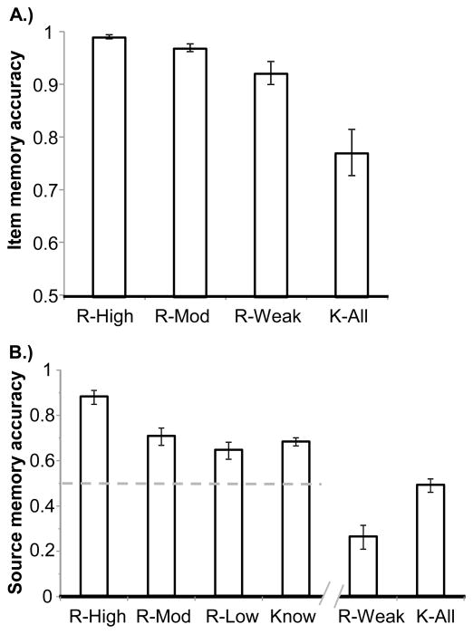
The behavioral data are presented here in summary form (see Yu et al., in press, for full details). Accuracy was calculated for each category of recognition judgment as the ratio of hits to hits plus false alarms (accuracy scores for R-Weak items comprised the trial-weighted mean of the accuracies of R items associated with low confidence and incorrect source judgments). These data are illustrated in Figure 1a. ANOVA contrasting the scores for the four response categories, and follow-up pairwise t-tests on each pair of immediately neighboring response types, confirmed the impression that recognition accuracy was markedly higher for R-Weak than K-All items, and then grew more modestly with source confidence/accuracy (F1.5, 21.8= 19.46, P < .001; all pairwise Ps <0.025). Figure 1b depicts the proportions of trials in each response category that were associated with an accurate source memory judgment (accuracy for R-Weak items was calculated as the trial-weighted mean of the accuracies for R-Low and R-Incorrect categories). ANOVA contrasting the accuracy scores for the R-High, R-Mod, and R-Low response categories revealed a significant main effect (F1.6, 24.5= 30.67, P < .001). Follow-up pairwise t-tests on immediately neighboring response types revealed that source accuracy increased monotonically with response confidence (all Ps <0.05). Consistent with the impression given by Figure 1b, source accuracy for items endorsed K exceeded the chance value of 0.5.
Figure 1.
A: Mean accuracy of item memory judgments as a function of subsequent source memory judgments. B: Proportion of trials associated with accurate source memory judgments. Note: ‘Know’ proportions are collapsed across source confidence. Also shown are source accuracies for ‘R-Weak’ and ‘K-All’ response categories, which were employed in the fMRI analyses. Dotted line indicates chance performance. Error bars signify within-category standard errors of the mean.
fMRI findings
As described in the Methods section we employed two complementary approaches to address the question whether the TPJ and angular gyrus demonstrated dissociable patterns of recollection-related activity. The first approach was based on the ANOVA model described in the Methods, and the second involved analysis of parameter estimates extracted from ROIs determined from a set of independent studies that each employed an R > K contrast (Cabeza et al., 2008).
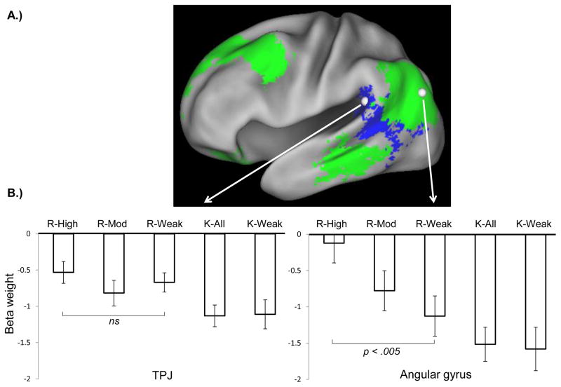
In the first approach, we performed two sets of pre-experimentally determined contrasts. The first set of contrasts identified voxels that were sensitive to the R/K distinction, but not to level of source confidence/accuracy. This was achieved by exclusively masking the weighted contrast between R-High, R-Mod, R-Weak and K-All items (weights of 1 1 1 −3; thresholded at P < .001) with the contrast between R-High and R-Weak items (thresholded at P < .05, one-tailed; note that the more liberal the threshold applied to an exclusive mask, the more conservative is the analysis). The second contrast, between R-High and R-Weak items (thresholded at P < .001), identified voxels sensitive to the accuracy and confidence of the source judgments made on recollected items. The outcomes of the two contrasts are illustrated in Figure 2a. The first contrast revealed a cluster in the vicinity of the TPJ (peak voxel −51 −49 16). The second contrast revealed a large cluster in the angular gyrus (peak voxel −48 −79 28). As is evident from Figure 2a, the two clusters encompass the TPJ and angular gyrus ROIs employed in the analysis described next (see Methods).
Figure 2.
A: Green: outcome of the contrast between the R-High and R-Weak response categories. Blue: voxels where activity was greater for items endorsed R than for items endorsed K, but where activity did not differ between R-High and R-Weak. Effects are mapped onto the standardized brain of the PALS-B12 atlas implemented in Caret5 (Van Essen, 2005). White spheres indicate the loci of the TPJ and angular gyrus ROIs (see methods). B: Across-subjects mean peak parameter estimates for each response category in the TPJ (left) and angular gyrus (right). Error bars signify within-category standard errors of the mean.
Parameter estimates from the TPJ and angular gyrus ROIs are illustrated in Figure 2b for the four principal response categories, and additionally, for the K-Weak category (see Methods). A 2x4 ANOVA of these data (factors of region and response category; dfs Greenhouse-Geisser corrected for non-sphericity) gave rise to a main effect of category (F1.9, 21.0= 21.10, P < .001) and, crucially, to a region by response category interaction (F2.5, 37.8= 5.28, P < .01). Follow-up analyses indicated that the interaction was carried by R-High and R-Weak trials, with an ANOVA restricted to the data for these two response categories giving rise to a significant category x region interaction (F1, 15= 12.21, P < .005). Pairwise t-tests indicated that the difference between the two response categories was significant in the angular gyrus, but not in the TPJ (t15= 3.53, P < .005, and t15= 1.17, P > .1, two-tailed, respectively). Together, the reliable interaction effect and subsequent pair-wise contrasts indicate that the angular gyrus, but not the TPJ, was sensitive to the accuracy/confidence of the source judgments associated with items endorsed R, consistent with our pre-experimental hypothesis. Additional pair-wise contrasts indicated that whereas the R-Weak and K-All categories differed reliably in the TPJ (t15= 4.22, P < .001, two-tailed) they did not quite do so in the angular gyrus (t15= 2.04, P < .06, two-tailed).
The foregoing ANOVA was repeated using the data from the K-Weak rather than the K-All response category, minimizing any confounding effects of source confidence and accuracy that may have biased the R-Weak and K-All contrast (albeit at the expense of less precise K parameter estimates; see Methods). Critically, this second ANOVA also gave rise to a reliable region by response category interaction (F2.6, 38.9= 6.37, P < .005). Again echoing the prior findings, pairwise contrasts between the R-Weak and K-Weak categories revealed a significant difference in the TPJ (t15 = 3.09, P < .001, two-tailed). Unlike in the previous analysis, however, the contrast between these categories was also significant in the angular gyrus (t15= 2.61, P < 0.05, two-tailed). The parameter estimates for the two classes of K items did not significantly differ in either region (both Ps > .6).
In summary, the two sets of analyses of the fMRI data described above converge to indicate that recollection-related activity in LPC dissociates according to whether it is located in the TPJ or the angular gyrus. Whereas activity in the TPJ differed solely according to whether test items were endorsed as recollected or familiar, activity in the angular gyrus co-varied with the confidence/accuracy of the associated source judgment.
General Discussion
Behavioral findings
Recognition accuracy was markedly higher for items endorsed R than K. Thus, as has been noted previously (Wixted & Squire, 2011), R and K judgments confound the presence versus the absence of a phenomenal sense of recollection with memory ‘strength’ (as this is indexed by recognition accuracy). A second noteworthy feature of the behavioral findings is that the accuracy of source judgments associated with items endorsed R co-varied with confidence, replicating prior findings (e.g. Slotnick & Dodson, 2005; Wixted & Mickes, 2010). Thus, response confidence indexed the amount of source-specifying information that was retrieved, consistent with the view that source recollection is supported by a continuous rather than a thresholded signal (Wixted & Mickes, 2010). From this perspective, unless the amount of retrieved information non-diagnostic for source declined with increasing source confidence, the amount of information recollected about items endorsed R can be assumed to have co-varied with confidence.
An alternative interpretation of the positive relationship between the accuracy and confidence of the source judgments made on items endorsed R is that, other than at the highest confidence level, these judgments were supported by a sense of familiarity rather than recollection of qualitative information about the study episode (e.g. Parks & Yonelinas, 2007). This proposal, however, has been strongly challenged on the basis of behavioral evidence (Wixted, 2007; but see Parks & Yonelinas, 2007). In the absence of a plausible mechanism through which the familiarity of a test item presented in central vision could signal the location of the item when it was studied, we assume that the relationship between the confidence and accuracy of source judgments is indicative of a graded recollection signal.
A related issue concerns the finding that items endorsed K were associated with above-chance source accuracy (for similar results see Hickes, Marsh, & Ritschel, 2002 and Wais, Mickes, & Wixted, 2008). According to one view (e.g. Wixted and Mickes, 2010), this finding reflects the influence of a recollection signal too weak to lead to an R endorsement but, strong enough to lift source judgments above chance. According to an alternative view (e.g. Parks & Yonelinas, 2007), accurate source judgments on K trials are familiarity-driven. Since the fMRI responses elicited by K-All and K-Weak items in the TPJ and angular gyrus did not significantly differ (see Figure 2b), for present purposes little hangs on this issue.
fMRI findings
Our findings for the angular gyrus are consistent with prior reports that this region is sensitive to the amount of information recollected (see Introduction). Activity tracked the confidence and accuracy of the source judgments made on R items (and, hence, we assume, the amount of recollected information; see above). We interpret these findings as further evidence for the role of this region in supporting the maintenance or representation of recollected information (Vilberg & Rugg, 2008). As was discussed by those authors, one possibility is that the angular gyrus contributes to a functional network supporting an episodic buffer (Baddeley, 2000). A related possibility, also discussed by Vilberg and Rugg (2008), is that graded recollection effects in the angular gyrus reflect allocation of attentional resources to the contents of recollection in proportion to the load that is placed on working memory (a process they argued to be distinct from attentional re-orienting). The present findings are equally compatible with either proposal.
It might be argued that the pattern of activity observed for the angular gyrus reflects response confidence, rather than the nature of the underlying mnemonic signal. By this argument (Cabeza et al., 2008), activity in this region is a consequence of its role in orienting attention to highly salient events, which include any test items associated with high response confidence. However, in a study that investigated the effects of response confidence across tests of old/new recognition and source memory (Hayes, Buchler, Stokes, Kragel, & Cabeza, in press), it was the TPJ rather than the angular gyrus that demonstrated a generic sensitivity to confidence. Furthermore, in the study of Guerin & Miller (2010), retrieval-related activity in the angular gyrus was reported to track the amount of information recollected, and not the confidence (‘decision certainty’, in their terminology) of the associated memory judgment. Thus, an account of the present results for the angular gyrus in terms of response confidence seems unlikely.
Unlike in the angular gyrus, recollection-related activity elicited in the TPJ did not vary with source confidence/accuracy. If it is assumed that items eliciting a memory signal supporting an R endorsement are especially salient, these findings are consistent with the proposed role of this region in the ‘bottom-up’ re-orienting of attention (e.g. Corbetta & Shulman, 2002; Corbetta et al., 2008). The absence of further modulation of TPJ activity as a function of source confidence is consistent with the proposal that retrieval-dependent re-orienting should be insensitive to the amount of information recollected (Vilberg & Rugg, 2007). We cannot rule out the alternative possibility, however, that the present TPJ effects reflect a role for this region in the representation of recollected information, albeit a role different from that served by the angular gyrus. One possibility1, for example, is that the TPJ is sensitive primarily to recollection of ‘non-criterial’ information, that is, information non-diagnostic of source, but sufficient to lead to an R judgment. The present findings do not allow adjudication between this account and the attentional account discussed previously. However, given the wealth of evidence pointing to a role for the TPJ in stimulus-driven attention (e.g. Corbetta & Shulman, 2002), we suggest that the attentional account should be favored on the grounds of parsimony.
As was noted above, Hayes et al. (in press) reported that a region of the TPJ overlapping the one identified here demonstrated greater activity for high than for low confidence ‘hits’ in tests of both recognition memory and source memory. At first glance, these findings conflict with those reported in the present study, when TPJ activity was not modulated by source confidence (Figure 2b). The conflict may, however, be more apparent than real. If it is assumed that in both item recognition and source memory tasks a phenomenal sense of recollection is more likely to be associated with high than with low confidence judgments, then the findings of Hayes et al. (in press), like the present results, may be a reflection of the sensitivity of the TPJ to whether an item was, or would have been, endorsed R or K.
Whereas retrieval-success effects associated with recollection are consistently reported in the angular gyrus, effects in the TPJ are reported much less frequently (for reviews, see Vilberg & Rugg, 2008; Cabeza et al., 2008; Hutchinson et al., 2009). The reason for this disparity is unclear. One possibility is that retrieval effects in the TPJ depend on whether the structure of the retrieval task makes the detection of a recollection signal especially salient. In the present study, for example, the signal may have been highly salient because it signaled the potential availability of source-specifying information.
It should be noted that our assumption that regional differences in patterns of retrieval-related activity are functionally significant can be challenged. It could be argued that the differences reflect dissociations between regions not in their patterns of neural responding but in the hemodynamic transfer functions that translate a neural response into an fMRI signal (c.f. Squire, Wixted, & Clark, 2007). For example, it could be that the transfer function in the TPJ differentiates items with low and high memory strength (K-All or K-Weak vs. R-Weak items in the present case), but not items whose strength exceeds some criterion amount. By contrast, the shape of the transfer function in the angular gyrus might be right-shifted, so that BOLD activity is primarily enhanced for items whose memory strength is especially high (i.e. R-Weak vs. R-High). Since the shapes of the hemodynamic response functions in the TPJ and angular gyrus are unknown, this caveat cannot be discounted. It is, however, entirely ad hoc.
Setting this caveat to one side, the present findings are consistent with other evidence that the TPJ and angular gyrus are functionally heterogeneous (Corbetta et al., 2008; Hutchinson et al., 2009; Nelson et al., 2010). The findings strongly suggest that the role of ventral parietal cortex in episodic memory retrieval cannot be accounted for in terms of a single cognitive construct.
Acknowledgments
This research was supported by the National Institute of Health (grant R01-MH072966).
Footnotes
We thank an anonymous reviewer for drawing our attention to this possibility.
References
- Andrews-Hanna JR, Reidler JS, Sepulcre J, Poulin R, Buckner RL. Functional-anatomic fractionation of the brain’s default network. Neuron. 2010;65:550–562. doi: 10.1016/j.neuron.2010.02.005. [DOI] [PMC free article] [PubMed] [Google Scholar]
- Baddeley A. The episodic buffer: A new component of working memory? Trends Cogn Sci. 2000;4:417–423. doi: 10.1016/s1364-6613(00)01538-2. [DOI] [PubMed] [Google Scholar]
- Brett M, Johnsrude IS, Owen AM. The problem of functional localization in the human brain. Nat Rev Neurosci. 2002;3:243–249. doi: 10.1038/nrn756. [DOI] [PubMed] [Google Scholar]
- Cabeza R, Ciaramelli E, Olson IR, Moscovitch M. The parietal cortex and episodic memory: an attentional account. Nat Rev Neurosci. 2008;9:613–625. doi: 10.1038/nrn2459. [DOI] [PMC free article] [PubMed] [Google Scholar]
- Cabeza R, Mazuz YS, Stokes J, Kragel JE, Woldorff MG, Ciaramelli E, Olson IR, Moscovitch M. Overlapping parietal activity in memory and perception: evidence for the attention to memory model. J Cogn Neurosci. 2011;23:3209–3217. doi: 10.1162/jocn_a_00065. [DOI] [PMC free article] [PubMed] [Google Scholar]
- Corbetta M, Shulman GL. Control of goal-directed and stimulus-driven attention in the brain. Nat Rev Neurosci. 2002;3:201–215. doi: 10.1038/nrn755. [DOI] [PubMed] [Google Scholar]
- Corbetta M, Patel G, Shulman GL. The reorienting system of the human brain: From environment to theory of mind. Neuron. 2008;58:306–324. doi: 10.1016/j.neuron.2008.04.017. [DOI] [PMC free article] [PubMed] [Google Scholar]
- Cocosco CA, Kollokian V, Kwan RS, Evans A. Brainweb: online interface to a 3D MRI simulated brain database. Neuroimage. 1997;5:S425. [Google Scholar]
- Guerin SA, Miller MB. Parietal cortex tracks the amount of information retrieved even when it is not the basis of a memory decision. Neuroimage. 2011;55:801–807. doi: 10.1016/j.neuroimage.2010.11.066. [DOI] [PubMed] [Google Scholar]
- Hayes SM, Buchler N, Stokes J, Kragel J, Cabeza R. Neural correlates of confidence during item recognition and source memory retrieval: Evidence for both dual-process and strength memory theories. (in press) [DOI] [PMC free article] [PubMed] [Google Scholar]
- Hicks JL, Marsh RL, Ritschel L. The role of recollection and partial information in source monitoring. J Exp Psychol Learn Mem Cogn. 2002;28:503–508. [PubMed] [Google Scholar]
- Hutchinson JB, Uncapher MR, Wagner AD. Posterior parietal cortex and episodic retrieval: convergent and divergent effects of attention and memory. Learn Mem. 2009;16:343–356. doi: 10.1101/lm.919109. [DOI] [PMC free article] [PubMed] [Google Scholar]
- Kim H. Dissociating the roles of the default-mode, dorsal, and ventral networks in episodic memory retrieval. Neuroimage. 2010;50:1648–1657. doi: 10.1016/j.neuroimage.2010.01.051. [DOI] [PubMed] [Google Scholar]
- Montaldi D, Spencer TJ, Roberts N, Mayes AR. The neural system that mediates familiarity memory. Hippocampus. 2006;16:504–520. doi: 10.1002/hipo.20178. [DOI] [PubMed] [Google Scholar]
- Nelson SM, Cohen AL, Power JD, Wig GS, Miezin FM, Wheeler ME, Velanova K, Donaldson DI, Phillips JS, Schalggar BL, Petersen SE. A parcellation scheme for human left lateral parietal cortex. Neuron. 2010;67:156–170. doi: 10.1016/j.neuron.2010.05.025. [DOI] [PMC free article] [PubMed] [Google Scholar]
- Parks CM, Yonelinas AP. Moving beyond pure signal-detection models: comment on Wixted (2007) Psych Rev. 2007;114:199–202. doi: 10.1037/0033-295X.114.1.188. [DOI] [PubMed] [Google Scholar]
- Sestieri C, Shulman GL, Corbetta M. Attention to memory and the environment: Functional specialization and dynamic competition in human posterior parietal cortex. J Neurosci. 2010;30:8445–8456. doi: 10.1523/JNEUROSCI.4719-09.2010. [DOI] [PMC free article] [PubMed] [Google Scholar]
- Slotnick SD, Dodson CS. Support for a continuous single-process model of recognition memory and source memory. Mem Cognit. 2005;33:151–170. doi: 10.3758/bf03195305. [DOI] [PubMed] [Google Scholar]
- Shimamura AP. Episodic retrieval and the cortical binding of relational activity. Cogn Affect Behav Neurosci. 2011;11:277–291. doi: 10.3758/s13415-011-0031-4. [DOI] [PubMed] [Google Scholar]
- Squire LR, Wixted JT, Clark RE. Recognition memory and the medial temporal lobe: A new perspective. Nat Rev Neurosci. 2007;8:872–883. doi: 10.1038/nrn2154. [DOI] [PMC free article] [PubMed] [Google Scholar]
- Van Essen DC. A population-average, landmark- and surface-based (PALS) altas of human cerebral cortex. Neuroimage. 2005;28:635–662. doi: 10.1016/j.neuroimage.2005.06.058. [DOI] [PubMed] [Google Scholar]
- Vilberg KL, Rugg MD. Dissociation of the neural correlates of recognition memory according to familiarity, recollection, and amount of recollected information. Neuropsychologia. 2007;45:2216–2225. doi: 10.1016/j.neuropsychologia.2007.02.027. [DOI] [PMC free article] [PubMed] [Google Scholar]
- Vilberg KL, Rugg MD. Memory retrieval and the parietal cortex: A review of evidence from a dual-process perspective. Neuropsychologia. 2008;46:1787–1799. doi: 10.1016/j.neuropsychologia.2008.01.004. [DOI] [PMC free article] [PubMed] [Google Scholar]
- Vilberg KL, Rugg MD. Functional significance of retrieval-related activity in lateral parietal cortex: evidence from fMRI and ERPs. Hum Brain Mapp. 2009a;30:1490–1501. doi: 10.1002/hbm.20618. [DOI] [PMC free article] [PubMed] [Google Scholar]
- Vilberg KL, Rugg MD. Lateral parietal cortex is modulated by amount of recollected verbal information. Neuroreport. 2009b;20:1295–1299. doi: 10.1097/WNR.0b013e3283306798. [DOI] [PMC free article] [PubMed] [Google Scholar]
- Vincent JL, Kahn I, Snyder AZ, Raichle ME, Buckner RL. Evidence for a frontoparietal control system revealed by intrinsic functional connectivity. J Neurophysiol. 2008;100:3328–3342. doi: 10.1152/jn.90355.2008. [DOI] [PMC free article] [PubMed] [Google Scholar]
- Wais PE, Mickes L, Wixted JT. Remember/know judgments probe degrees of recollection. J Cogn Neurosci. 2008;22:400–405. doi: 10.1162/jocn.2008.20041. [DOI] [PubMed] [Google Scholar]
- Wixted JT. Spotlighting the probative findings: reply to Parks and Yonelinas (2007) Psych Rev. 2007;114:203–209. [Google Scholar]
- Wixted JT, Mickes L. A continuous dual-process model of remember/know judgments. Psych Rev. 2010;117:1025–1054. doi: 10.1037/a0020874. [DOI] [PubMed] [Google Scholar]
- Wixted JT, Squire LR. The medial temporal lobe and the attributes of memory. Trends Cogn Sci. 2011;15:210–217. doi: 10.1016/j.tics.2011.03.005. [DOI] [PMC free article] [PubMed] [Google Scholar]
- Yonelinas AP, Otten LJ, Shaw KN, Rugg MD. Separating the brain regions involved in recollection and familiarity in recognition memory. J Neurosci. 2005;25:3002–3008. doi: 10.1523/JNEUROSCI.5295-04.2005. [DOI] [PMC free article] [PubMed] [Google Scholar]
- Yu SS, Johnson JD, Rugg MD. Hippocampal activity during recognition memory co-varies with the accuracy and confidence of source memory judgments. Hippocampus. doi: 10.1002/hipo.20982. (in press) [DOI] [PMC free article] [PubMed] [Google Scholar]


