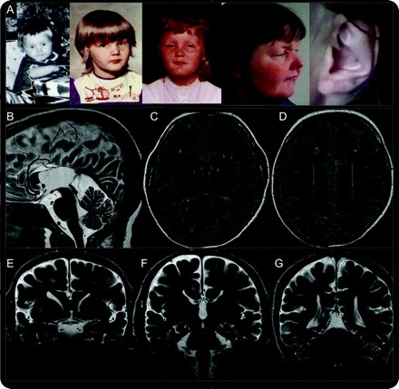
Figure Facial features and brain MRI
(A) Facial features of patient at age 1, 3, 7, and 43 years, with attention to the diagnostic clue, her enlarged and calcified external ear. Important early features include prognathism, repaired cleft palate, anteverted nares, prominent ears, and nasal roots. Early prognathism gave way to micrognathia in subsequent years. Ptosis was not apparent in early childhood. (B) Sagittal T2-weighted brain MRI demonstrates partial agenesis of the corpus callosum with “wandering” of the anterior cerebral arteries and high-riding third ventricle. (C–E) Increased fluid-attenuated inversion recovery signal is seen in patchy subcortical and diffuse periventricular distribution. (E–G) Coronal T2-weighted sequences demonstrate partial calcification of the putamen and globus pallidum and teardrop deformity of the lateral ventricles. The brainstem and cerebellum showed normal size and configuration. The patient's mother has given full informed consent for the publication of the video and images.
