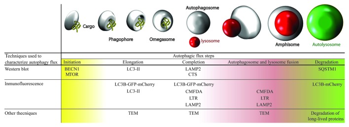
Figure 1. Table and illustration with techniques used for studying basal autophagy. Complete autophagic flux was studied in both cell types, analyzing each stage of autophagy using various techniques. We observed that G2019S LRRK2 mutant fibroblasts present higher basal autophagy levels than wild-type cells. TEM, transmission electron microscopy; CMFDA, 5-chloromethylfluorescein diacetate; LTR, LysoTracker Red.

An official website of the United States government
Here's how you know
Official websites use .gov
A
.gov website belongs to an official
government organization in the United States.
Secure .gov websites use HTTPS
A lock (
) or https:// means you've safely
connected to the .gov website. Share sensitive
information only on official, secure websites.
