Abstract
Lawsonia intracellularis is the causative agent of proliferative enteropathy. This disease affects various animal species, including nonhuman primates, has been endemic in pigs, and is an emerging concern in horses. Non-pathogenic variants obtained through multiple passages in vitro do not induce disease, but bacterial isolates at low passage induce clinical and pathological changes. We hypothesize that genes differentially expressed between pathogenic (passage 10) and non-pathogenic (passage 60) L. intracellularis isolates encode potential bacterial virulence factors. The present study used high-throughput sequencing technology to characterize the transcriptional profiling of a pathogenic and a non-pathogenic homologous L. intracellularis variant during in vitro infection. A total of 401 genes were exclusively expressed by the pathogenic variant. Plasmid-encoded genes and those involved in membrane transporter (e.g. ATP-binding cassette), adaptation and stress response (e.g. transcriptional regulators) were the categories mostly responsible for this wider transcriptional landscape. The entire gene repertoire of plasmid A was repressed in the non-pathogenic variant suggesting its relevant role in the virulence phenotype of the pathogenic variant. Of the 319 genes which were commonly expressed in both pathogenic and non-pathogenic variants, no significant difference was observed by comparing their normalized transcription levels (fold change±2; p<0.05). Unexpectedly, these genes demonstrated a positive correlation (r2 = 0.81; p<0.05), indicating the involvement of gene silencing (switching off) mechanisms to attenuate virulence properties of the pathogenic variant during multiple cell passages. Following the validation of these results by reverse transcriptase-quantitative PCR using ten selected genes, the present study represents the first report characterizing the transcriptional profile of L. intracellularis. The complexity of the virulence phenotype was demonstrated by the diversity of genes exclusively expressed in the pathogenic isolate. The results support our hypothesis and provide the basis for prospective mechanistic studies regarding specific roles of target genes involved in the pathogenesis, diagnosis and control of proliferative enteropathy.
Introduction
Lawsonia intracellularis is a fastidious intracellular bacterium and the etiologic agent of proliferative enteropathy (PE), an intestinal hyperplasic disease characterized by thickening of the mucosa of the intestine due to enterocyte proliferation [1]. Cell proliferation is directly associated with bacterial infection and replication in the intestinal epithelium [2]. As a result, mild to severe diarrhea is the major clinical sign described in infected animals [3]. Since the 1990s, PE has been endemic in swine herds and has been occasionally reported in various other species, including nonhuman primates, wild mammalians and ratite birds [4], [5]. Outbreaks among foals began to be reported on breeding farms worldwide within the last decade [6], [7]. Therefore, PE is now considered an emerging disease in horses [8].
Although PE was first reported in 1931 [9], the causative bacterium was primarily isolated only in 1993 using rat small intestinal cells (IEC-18) in strict microaerophilic environmental conditions [10]. Since then, various cell lines have supported L. intracellularis growth in vitro, including insect and avian cell lines [11]–[14]. To date, growth of the bacteria in axenic (cell-free) media has not been reported. Regardless of the cell type, the dynamics of the infection in vitro requires actively dividing cells in a microaerophilic atmosphere with the peak of infection at six to seven days post-inoculation [10], [14], [15]. McOrist et al (1995) chronologically described the dynamics of the infection and bacterial replication in intestinal porcine epithelial cells (IPEC-J2). Most events closely resembled those observed at the cellular level in infected animals, including multiplication of the bacteria freely in the cell cytoplasm. While the dynamics of the infection have been well-characterized, little is known so far about the genetic basis for the virulence, pathogenesis or physiology of L. intracellularis [16].
Spontaneous attenuated isolates obtained through multiple passages in cell culture have not been successful at inducing typical PE lesions or reversing its virulence in experimentally-infected pigs [12]. Conversely, bacterial isolates at low passage induce clinical and pathological changes typical of PE [17]. Various standard DNA-based typing techniques, such as pulsed field gel electrophoresis (PFGE), multilocus sequence typing (MLST) and variable number tandem repeat (VNTR) have shown identical genotypes in both pathogenic (low passage) and non-pathogenic (high passage) variants [18]–[20]. As a result, we believe their phenotypic properties occur at the transcriptional level. Bacterial genes differentially expressed between pathogenic (low passage) and non-pathogenic (high passage) homologous isolates have not been reported and this information may help to elucidate genes encoding the major bacterial virulence factors involved in the pathogenesis of PE.
We hypothesize that genes differentially expressed between pathogenic (passage 10) and non-pathogenic (passage 60) homologous L. intracellularis isolates encode potential bacterial virulence factors. High-throughput technology (RNA-seq) was used to characterize and compare the transcriptional profile of a pathogenic and a non-pathogenic variant. Plasmid-encoded genes, regulatory factors and ATP-binding cassette (ABC) transporters associated genes were important for contributing to the wider transcriptional landscape observed in the pathogenic isolate. Additionally, the present study provided novel information for studying specific mechanisms of target genes and their potential usefulness for the diagnosis and control of PE.
Results and Discussion
Whole-transcriptome profiling of bacterial organisms has been widely studied to understand global changes in gene expression in vitro and in vivo [21], [22]. Hybridization-based approaches have been successfully applied for years [23], but it has limited use in obligate intracellular bacteria since it needs a reference purified sample from cultivated bacteria. High-throughput technologies have overcome these limitations by providing high resolution data to describe the bacterial transcripts in different experimental conditions [24], [25]. The present study used RNA-seq to qualitatively and quantitatively characterize the transcriptional profile of pathogenic and non-pathogenic homologous L. intracellularis isolates during in vitro infection. Since this is the first comprehensive gene expression analysis regarding this organism, the findings are presented and discussed in a comparative pathogenomic approach based on the information available from other related bacterial organisms.
Mapping and Differential Expression
The sequence reads representing the RNA transcripts derived from the pathogenic and non-pathogenic homologous L. intracellularis PHE/MN1-00 isolates were mapped onto the complete genome sequence of the same bacterial strain available at the National Center for Biotechnology Information (NCBI) (accession: NC_008011). The circular L. intracellularis genome has 1,719,014 base pairs (bp) comprised of one chromosome (1,457,619 bp) and three plasmids (plasmid A: 27,048 bp, plasmid B: 39,794 bp and plasmid C: 194,553 bp). From a total of 1,391 computationally predicted genes in the annotated PHE/MN1-00 genome, 1,340 are protein coding. Combining the pathogenic and non-pathogenic transcript reads, 731 protein coding genes were mapped onto the reference DNA sequence of the L. intracellularis PHE/MN1-00 isolate. Sequence reads mapped against the bacterial genome were used to quantify the gene expression levels based on the number of reads per kilobase of coding sequence per million mapped reads (RPKM). The expression data were sufficiently reproducible by showing a positive correlation between the two biological replicates of the pathogenic (r2 = 0.86) and non-pathogenic (r2 = 0.81) variants which was obtained using a linear regression model (p<0.05). Genes with low expression levels showed lesser agreement between replicates (Figure S1). This observation was reported using Illumina® RNA-seq data from human brain RNA [26] and using ABI SOLiD™ platform in a transcriptome study of Neisseria gonorrhoeae [25]. Following the default parameters of the CuffDiff software (see Material and Methods) our study used ten sequenced reads as the minimum number of alignments in a locus needed to conduct agreement and comparison testing within and between the biological replicates.
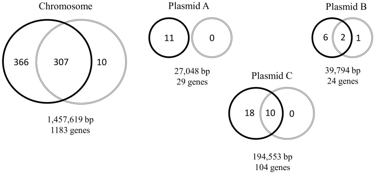
A total of 720 and 330 genes were expressed by the pathogenic and non-pathogenic variants, respectively (Figure S2). The wider transcriptional landscape observed in the pathogenic variant was characterized by 401 genes uniquely expressed by this variant. Genes with the highest transcription levels according to their biological function categories are shown in Table 1. Only 11 genes were expressed exclusively by the non-pathogenic variant (Table 2). Differential mapping and distribution of the expressed genes into the L. intracellularis chromosome and its three plasmids are summarized in Figure 1. Plasmid-encoded genes significantly contributed to this broader profile of gene expression exhibited by the pathogenic isolate. The entire transcriptional repertoire of the plasmid A was suppressed in the non-pathogenic isolate suggesting its potential contribution in the L. intracellularis virulence. Transcription factors located in bacterial chromosomes have been shown to positively regulate virulence factors in the plasmid of Shigella flexneri and Pseudomonas syringae [27], [28]. Our study also identified many regulatory factors exclusively expressed by the pathogenic L. intracellularis variant which potentially regulate plasmid-encoded genes. Specific findings related to regulatory factors are discussed later.
Table 1. Chromosomal genes showing highest transcript levels exclusively expressed by the pathogenic variant of the L. intracellularis isolate PHE/MN1-00 according to the putative biological function.
| Locus | Gene | Description | Log2 (RPKM) |
| Biological function | |||
| Cell division and macromolecule biosynthesis | |||
| LI0372 | putative cell division protein FtsB | 12.8 | |
| LI0844 | tRNA and rRNA cytosine-C5-methylases | 14.1 | |
| LI0962 | rplW | 50S ribosomal protein L23 | 13.5 |
| Energy production and conversion | |||
| LI0442 | hyaD | processing of HyaA and HyaB proteins | 10.9 |
| LI1176 | nitroreductase | 11.1 | |
| Cellular processes and small molecule biosynthesis | |||
| LI0154 | ribH | riboflavin synthase beta-chain | 12.6 |
| LI0361 | quinolinate synthetase | 12.7 | |
| LI0735 | ychB | 4-diphosphocytidyl-2-C-methyl-D-erythritol kinase | 14.5 |
| Membrane transport | |||
| LI0338 | livH | branched chain amino acid ABC transporter (permease) | 12.5 |
| LI0754 | glnH | amino acid ABC transporter substrate-binding protein | 13.9 |
| LI0995 | oprM | outer membrane efflux protein | 12.1 |
| Protein turnover and chaperones | |||
| LI0375 | pcm | protein-L-isoaspartate carboxylmethyltransferase | 12.2 |
| LI0225 | smpB | SsrA-binding protein | 11.4 |
| LI0618 | ATPases with chaperone activity, ATP-binding subunit ClpA | 11.4 | |
| Cell membrane and motility | |||
| LI0825 | Lipid A core-O-antigen ligase and related enzymes | 13.2 | |
| LI0947 | cell wall-associated hydrolases | 12.6 | |
| LI1141 | cheW | chemotaxis signal transduction protein | 11.0 |
| Adaptation and stress response | |||
| LI0035 | fur | Fe2+/Zn2+ uptake regulation proteins | 11.8 |
| LI0301 | recO | DNA repair protein RecO (recombination protein O) | 13.0 |
| LI0457 | rpoN | Sigma54-like protein | 11.7 |
| General predicted function | |||
| LI0140 | glycosyltransferase | 11.5 | |
| LI0246 | hypA | zinc finger protein | 11.6 |
| LI0880 | sfsA | DNA-binding protein, stimulates sugar fermentation | 11.4 |
| Hypothetical/Unknown function | |||
| LI0259 | hypothetical protein | 13.9 | |
| LI0917 | hypothetical protein | 12.9 | |
| LI1056 | hypothetical protein | 12.3 | |
Table 2. Genes exclusively expressed by the non-pathogenic variant of the L. intracellularis isolate PHE/MN1-00.
| Locus | Gene | Description | Log2 (RPKM) |
| Biological function | |||
| Cell division and macromolecule biosynthesis | |||
| LI0702 | maf | nucleotide-binding protein involved in septum formation | 11.7 |
| LIB024 | parA | ATPases involved in chromosome partitioning | 11.1 |
| Energy production and conversion | |||
| LI0614 | Thiol-disulfide isomerase and thioredoxins | 12.1 | |
| LI1001 | napC | cytochrome c nitrite reductase small subunit | 12.2 |
| Cellular processes and small molecule biosynthesis | |||
| LI0709 | ribF | FAD synthase involved in riboflavin metabolism | 12.2 |
| Membrane transport | |||
| LI0540 | yscN | type III secretion system ATPase | 11.3 |
| LI1163 | YscJ | type III secretion protein | 10.8 |
| Cell membrane and motility | |||
| LI0747 | flgK | flagellar hook-associated protein | 11.2 |
| Predicted and unknown function | |||
| LI0427 | hypothetical protein | 9.8 | |
| LI0554 | hypothetical protein | 10.8 | |
| LI0583 | hypothetical protein | 10.1 | |
Figure 1. Schematic representation of the L. intracellularis genome.
Distribution of genes expressed by the pathogenic (black circles) and the non-pathogenic (gray circles) variants. Overlapping zones represent genes expressed in both variants.
A correlation between infectivity and loss of DNA contents from linear and/or circular plasmids during serial passages in vitro has been well established in Borrelia burgdorferi infections [29], [30]. Since there are a large number of different plasmids found in Borreliae genomes, their stability and loss in vitro vary between and within species [31]. Although our study did not identify any transcriptional activities in the plasmid A of the non-pathogenic variant, the DNA contents of this plasmid were identical in both pathogenic and non-pathogenic variants (data not shown). Additionally, transcription levels were detected in three and ten genes present in the plasmid B and C of the non-pathogenic variant, respectively (Figure 1). These findings demonstrated the occurrence of transcriptional activities and, consequently, the presence of these two plasmids in this variant. These observations support the hypothesis that global changes in gene expression are able to drive the loss of the virulence phenotype in cultivated L. intracellularis without altering the genomic DNA.
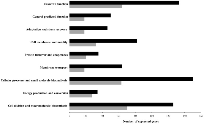
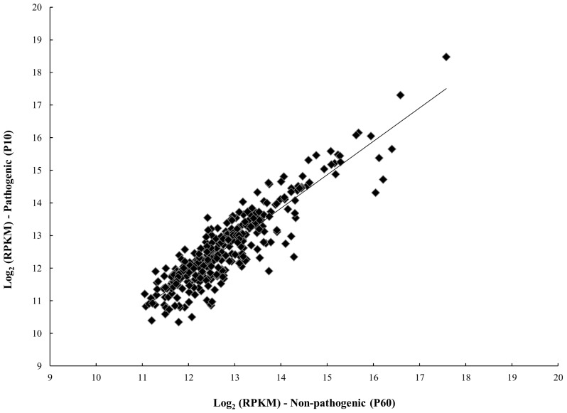
Narrowing the analysis to the putative biological gene functions, the number of genes expressed by the pathogenic variant was consistently higher throughout the functional categories (Figure 2). The transcriptional landscape was most significantly reduced in those genes involved in the membrane transport (72%), general predicted function (64%), cell membrane and motility (61%) and adaptation and stress response (61%) categories. In comparing the transcription levels of all 319 genes commonly expressed in both the pathogenic and non-pathogenic variants, there was no significant difference (fold change ±2; adjusted p-value <0.05– Table S1). When the expression levels from both variants were plotted against each other (Figure 3), log2 (RPKM) values unexpectedly showed a positive correlation (0.809) in a linear regression model. These results revealed the importance of gene silencing (switching off) mechanisms to attenuate virulence properties of L. intracellularis throughout passages in vitro.
Figure 2. Functional categories of genes expressed by the pathogenic and non-pathogenic homologous L. intracellularis isolate PHE/MN1-00.
Black and gray bars represent the number of genes expressed by the pathogenic and non-pathogenic variants, respectively.
Figure 3. Average log-transformed RPKM (log2 [RPKM]) of the 319 genes commonly expressed by the pathogenic (y-axis) and non-pathogenic (x-axis) variant.
The trend line represent a linear regression model (p-value <0.05; r2 = 0.809).
Regulation of gene expression during in vitro cultivation has been widely reported in various bacterial organisms [32]. Bordetella pertussis switches off the expression of type III secretion system (TTSS) proteins in laboratory-adapted strains [33]. One of the TTSS components, protein Bsp22, ceases to be expressed between passages three and four [34]. Interestingly, these authors also showed the reversible re-expression of this protein after contact with the host in vivo. The expression of TTSS proteins has been demonstrated during L. intracellularis infection [35]. However, our study failed to demonstrate consistent differences in the expression of TTSS proteins between pathogenic and non-pathogenic variants. Additionally, reversibility of virulence has not been reported in animals infected with laboratory-adapted L. intracellularis.
Transcriptional regulation has also been associated with mutations characterized by non-synonymous substitutions at the DNA level in Escherichia coli cultivated in glucose-limited medium for 20,000 generations [36]. Regulatory changes in gene expression following by mutations in 12 lines of E. coli were driven by the specific environment in vitro evolving an ecological specialization in laboratory-adapted strains [37]. Narrower transcriptional profiling of the non-pathogenic L. intracellularis passed 60 times in vitro was observed in our study; this could represent earlier stages of adaptation to a specialized in vitro environment. As in the case of E. coli, unnecessary functions that are costly to fitness in the in vitro condition might then be eliminated after hundreds or thousands of generations. According to ecological specialization models, organisms genetically adapted to one environment may lose fitness in other environments [37], [38]. Corroborating with this principle, the lack of reversible virulence in animals infected with laboratory-adapted L. intracellularis suggests that improving fitness in a specialized environment in vitro adversely affects performance in other complex substrates in vivo.
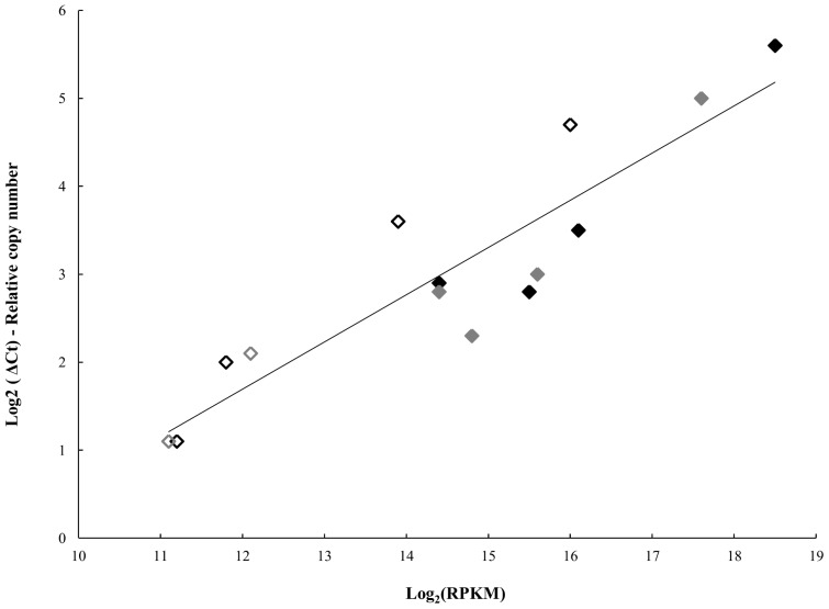
Gene expression data generated by RNA-seq were validated using quantitative reverse transcriptase PCR (qRT-PCR). The reliability of RNA-seq results was confirmed based on the relative quantification of 10 unlinked genes: four expressed in both variants, four expressed by the pathogenic and two expressed by the non-pathogenic (see Materials and Methods). The average log-transformed from two qRT-PCR replicates was plotted against the log2 (RPKM) (Figure 4). The transcript levels were positively correlated on a linear regression model (p-value <0.05; r2 = 0.827). Therefore, the RNA-seq data were consistent in quantitatively estimating the transcription levels of L. intracellularis during infection in vitro. The validation of RNA-seq data using qRT-PCR has also been previously reported in yeast [39] and other bacterial transcriptomes [24], [25], [40].
Figure 4. Correlation between RNA-seq and qRT-PCR.
Plot demonstrating the relative quantification of 10 unlinked genes by qRT-PCR (y-axis) and the transcript levels generated by RNA-seq (x-axis). Genes commonly expressed by the pathogenic (♦) and non-pathogenic (♦) variants. Genes exclusively expressed by the pathogenic (◊) and non-pathogenic (◊) variants.
Cell Division and Macromolecule Biosynthesis
Synthesis of proteins involved in cell division and ribosome biogenesis has been closely related to bacterial growth rate [41], [42]. From a total of 43 ribosomal protein-encoding genes present in the L. intracellularis reference genome, we identified 31 genes expressed by both the pathogenic and non-pathogenic variants and eight additional genes exclusively expressed by the pathogenic one. Furthermore, the operon responsible for orchestrating bacterial cell division that contained the FtsA and FtsZ genes [43] was also identified in both variants (Table S1). The expression of RNA polymerase á and â subunits has also been associated with growth rate in that their synthesis increases in response to a rich nutrient medium (nutritional shift-up) [44]. All L. intracellularis genes encoding RNA polymerase subunits previously predicted from the annotated genome were expressed in both variants. Regardless of the pathogenic or non-pathogenic phenotype, these observations demonstrated that both variants grew at the same rate and were harvested during exponential phase at time of RNA harvesting (five days post-infection). Likewise, the results corroborate previous studies which described an increase in the number of Lawsonia-infected cells from two to seven days after infection [15], [45]. Adverse effects in bacterial growth have been related to the reduced expression of RNA polymerase subunits and ribosomal proteins in Neisseria gonorrhoeae cultivated in an oxygen-limited environment [25]. The microaerophilic environment provided in our study was first described by Lawson et al (1993) and it has been shown to be an optimal atmosphere for cultivation of L. intracellularis [10], [45].
Energy Production and Conversion
Pathogenic bacteria are frequently able to couple virulence pathways with general metabolic functions such as energy production, cellular signaling and molecular biosynthesis [46]. The operon comprising F0–F1 ATP synthase subunits, which was expressed in both pathogenic and non-pathogenic L. intracellularis variants (Table S1), is essential for life in many bacterial species since it maintains pH homeostasis of the cell [47]. Concomitantly, it plays a critical role in the acid stress response, specifically in enteric bacteria which encounter low pH conditions transiting through stomach and in the phagosome of host cells [48]. Although we intended to harvest L. intracellularis freely multiplying in the cell cytoplasm (five days post-infection) which would fully represent its exponential growth phase, the identification of the F0–F1 operon in both variants suggests that a fraction of harvested intracellular bacteria was in the transient phagosome phase.
Along with these observations, the expression data also indicated the presence of free intracellular bacteria in the cell cytoplasm by detecting high transcriptional levels (Table S1) of rickettsia-like ATP/ADP translocase gene in both variants. This transmembrane protein was first identified in a few obligate intracellular bacteria, including Chlamydiales, Rickettsiales and amoebal symbionts [49], and more recently in L. intracellularis [50]. A remarkable adaptation of these organisms to an intracellular microenvironment allows them to import cytoplasmic ATP generated in their hosts into the prokaryotic cell across the bacterial cell membrane [51]. In an exchange mode, the bacterial ADP is exported back into the host cytosol. This exploitation of the host’s energy pool is also referred to as energy parasitism. In agreement with the exponential growth phase of L. intracellularis, we also identified the expression of cytochrome bd operon in the pathogenic and non-pathogenic variant (Table S1). This oxygen reductase is used in the bioenergetic pathway in a variety of bacterial organisms under O2-limited conditions [52].
Cellular Processes and Small Molecule Biosynthesis
Although L. intracellularis and other obligate intracellular bacteria depend on their hosts for certain nutrients (e.g. cytoplasmic ATP, see above), a wide spectrum of biosynthetic pathway-encoding genes are critical for fulfilling their essential functions. The 2-C-methyl-D-erythritol 4-phosphate (MEP) pathway is primarily involved in the biosynthesis of structural and functional isoprenoid molecules but the genes encoding the final two enzymes, ispG and lytB, have also been related to intracellular survival and induction of cellular immune response [53]. Six genes of the Lawsonia MEP pathway previously predicted in the Kyoto encyclopedia of gene and genomes (KEGG) database were exclusively expressed by the pathogenic variant. However, the final remaining two enzymes (ispG and lytB) were transcribed by both the pathogenic and non-pathogenic variants. We suggest that ispG and lytB may be essential genes in L. intracellularis, as previously reported in several other bacterial pathogens [53]. Based on this vital characteristic of the MEP pathway, it has recently been used as a drug target [54]. Fosmidomicyn showed the ability to inhibit the second enzyme of the MEP pathway (Dxr) and has been used in clinical trials [55]. Interestingly, our study identified expression levels of the Dxr-encoding gene (log2 [RPKM] = 10.1) exclusively in the pathogenic variant, suggesting its potential use for drug targeting.
Despite the similarities in the expression of ribosomal proteins and RNA polymerase subunits between pathogenic and non-pathogenic variants described previously, our study also identified the expression of relA gene exclusively in the pathogenic variant. This gene encodes an enzyme responsible for synthesizing guanosine tetraphosphate (ppGpp). This small signal molecule, also called stringent factor, is involved in various cellular processes and affects the control of growth rates by binding to RNA polymerase and reduces synthesis of ribosomal proteins and tRNA molecules during nutritional stress [56], [57]. Although the transcript reads of relA gene were exclusively identified in the pathogenic variant, its expression levels were relatively low (log2 [RPKM] = 9.7) suggesting that it was not sufficient to affect the expression of genes involved in macromolecule biosynthesis.
Based on the empirical observation that high-passage variants of L. intracellularis have higher growth rates than low passages in vitro, we believe that relA-dependent accumulation of ppGpp may control the bacterial growth rate at some point during the course of the infection in vitro. However, chronological transcriptional analysis is needed in order to elucidate this question. Supporting this speculation, Cooper et al (2003) demonstrated an increased fitness of laboratory-adapted E. coli (after 20,000 generations in vitro) associated with reduction in the concentration of ppGpp. These authors did not observe any mutation in the DNA sequence of relA gene. Similarly, the suppression of Lawsonia relA gene in the non-pathogenic variant was not associated with any mutations in the relA DNA sequence (data not shown).
The stringent response mediated by ppGpp also plays an important role in bacterial virulence, especially to adapt to conditions encountered in the intracellular host environment [46]. A relA mutant strain of Listeria monocytogenes showed no virulence in vivo using murine infection models [58]. However, the same study shows no difference between the wild type and ÄrelA mutant strain in their ability to escape from the phagosome and polymerize host cell actin in vitro using Caco-2 cells. Comparable phenotypic differences between in vitro and in vivo infections have also been observed in L. intracellularis. Low passage and high passage variants have similar abilities to infect cells in vitro but they demonstrated differences in pathogenic and non-pathogenic phenotypes in vivo [59].
Membrane Transport
The majority of genes encoding ABC transporters identified in the L. intracellularis genome were shown to be expressed in the pathogenic variant. From a total of 14 ABC transporter operons, 10 cassettes were specifically expressed by the pathogenic variant, including those involved in resistance to organic solvents, membrane transport of polyamines (spermidine), phosphates, nitrates, amino acids (glutamine and branched-chain), metallic cations (cobalt and nickel), lipopolysaccharides and lipoproteins. We specifically observed the highest expression levels in ABC uptake systems for amino acids (Table 1). Elevated transcription levels of the glutamine transport system (glnHPQ operon) were shown to be essential for virulence in Salmonella enterica Serovar Typhimurium [60] and Streptococcus pneumoniae [61]. Both studies demonstrated significant attenuation of the virulence in vivo using mouse models infected with mutant strains (ÄglnHPQ). Despite the attenuated phenotype, the mutant strain of S. enterica Typhimurium provided a protective immune response against challenge with wild-type. Additionally, the authors showed that glnHPQ operon is positively regulated by the sigma factor ó54 (RpoN). This regulatory factor was also exclusively expressed by the pathogenic L. intracellularis variant in our study (Table 1). The expression of ó54 and other transcription factors are discussed later in the subsection related to bacterial adaptation and stress response.
The in vitro growth kinetics of S. enterica Typhimurium and S. pneumoniae glnHPQ mutants were not affected in either broth or agar supplemented with glutamine or peptides [60], [61]. On the other hand, both studies showed reduced intracellular survival of these mutants in macrophage cell lines. There is no information regarding the intracellular survival of pathogenic or non-pathogenic L. intracellularis in infected macrophages and no impairment of intracellular survival has been reported in L. intracellularis continually cultivated using epithelial or fibroblastic-like cells [15], [45]. Although the IPEC-J2 cell culture media used in our study was not supplemented with glutamine, the glutamine ABC transporters were consistently expressed only by the pathogenic variant in the two glnHPQ operons present in the L. intracellularis genome.
The ABC transporter involving polyamine uptake (PotABCD operon) and uniquely expressed in the pathogenic variant at moderate levels (log2 [RPKM] = 11.1) has also been important for virulence in other bacterial organisms [62]. Corresponding with the glutamine ABC transporters discussed above, mutations in the PotABCD operon of S. pneumoniae showed no effect on the growth rates in vitro, but the mutant strain showed significant attenuation of virulence within murine models regardless of the inoculation route [62].
The differential mapping of other bacterial transport systems including TTSS and general secretory (Sec) pathways was not consistently different between the pathogenic and non-pathogenic variants. The role of TTSS proteins has been well established at earlier stages of infection and has been required for the invasion of various bacterial organisms [63]. Our study detected the intracellular expression of five of a total of 15 L. intracellularis genes involved in the synthesis of TTSS previously identified in the KEGG database. This unexpected intracellular expression of TTSS proteins was reported in S. enterica Typhimurium and plays a role in the survival of this organism within the Salmonella-containing vacuole [64], [65].
The expression of genes encoding structural components of TTSS (YscN, YscO and YscQ) was previously identified by RT-PCR in three L. intracellularis isolates infecting rat small intestinal cells (IEC-18) [35]. However, the study did not provide information about the number of cell passages the isolates had undergone or the time points in which the bacterial RNA was harvested from the infected cell monolayers. Our study identified two (YscQ and YscN) of the three major TTSS components cited above. The YscQ gene was expressed in both the pathogenic and non-pathogenic variant and YscN was uniquely identified in the non-pathogenic variant (Table 2). These variable results regarding the expression of TTSS proteins suggest that further studies are required in order to elucidate the role of TTSS in L. intracellularis infection in vitro.
Protein Turnover and Chaperones
Chaperone-encoded genes (DnaK, DnaJ, GroEL and GroES) were highly expressed by the pathogenic and non-pathogenic variants. This observation corroborates the up-regulation of this gene category identified in a variety of bacterial organisms growing inside eukaryotic cells [66]. Furthermore, bacterial chaperones are known to be essential for overcoming bacterial stress by ensuring the proper folding of proteins. Three additional genes (CbpA, CrpE and ClpA) involving the synthesis of chaperone molecules were exclusively expressed by the pathogenic variant and their specific biological functions remain to be determined.
Cell Membrane and Motility
A broader diversity of genes encoding cell wall molecules were observed in the pathogenic L. intracellularis variant, suggesting more extensive options for remodeling of the bacterial envelope compared with the non-pathogenic variant. This characteristic is important for altering the bacterial cell surface during different steps of the infectious process [66]. The wider variety of this gene category in the pathogenic variant was predominantly comprised of genes encoding glycosyltransferase enzymes which have been implicated in posttranslational processing of proteins responsible for modifying the lipopolysaccharide composition. Modifications of the glycosylation composition of the cell wall were depicted in Campylobacter jejuni during its intestinal life cycle. These events were then considered part of the strategy used by the bacterium to resist and evade the host immune responses [67].
In the mapping of genes involved in the flagellar assembly pathway, there were no consistent differences between the pathogenic and non-pathogenic variants. From a total of 30 predicted genes in the KEGG database, ten were expressed in both variants (Table S1), eight were exclusively identified in the pathogenic and one was unique from the non-pathogenic variant (Table 2). Our study detected similar expression levels of the FliC gene in both variants (Table S1). The flagellin encoded by this gene has been well studied regarding its ability to induce an immune response [68]. S. enterica Typhimurium expressed this protein in the membrane-bound compartment and it was then translocated into the host cytoplasm to be detected by cytosolic Nod-like receptors which are able to mount an innate immune response [69]. Studies characterizing the immune response of cells infected with pathogenic and non-pathogenic L. intracellularis would be important to detect specific host factors associated with these two variants.
Adaptation and Stress Response
The number of genes encoding proteins involved in adaptation and stress response was one of the gene categories with significant reduction (61%) in the non-pathogenic variant (Figure 2). Ten of the 28 genes uniquely expressed by the pathogenic variant in this category were transcriptional regulatory factors. These molecules regulate bacterial gene expression by acting globally or at specific DNA regions to active or repress transcription or modulate DNA topology. The ó70 is the “housekeeping” sigma factor and has been required for cell growth in the majority of bacterial organisms [70]. In agreement with this, we observed similar expression levels of ó70 in the pathogenic and non-pathogenic variants (Table S1). Conversely, the global regulator ó54 was uniquely expressed in the pathogenic variant (Table 1) and has been reported to control the transcription of virulence genes in a variety of bacteria species [70]. Specifically in S. enterica Typhimurium, the role of ó54 is to coordinate the transcription of the glutamine ABC transporter, glnHPQ operon, during the intracellular life cycle. The coincidental and exclusive co-expression of ó54 and glnHPQ by the pathogenic variant supports the hypothesis that this sigma factor may also coordinate the glutamine uptake operon in L. intracellularis to ensure an adequate supply of this crucial amino acid during its intracellular life. Additionally, the consistently higher number of ABC transporter-encoding genes expressed by the pathogenic, but not the non-pathogenic, variant begs the question whether ó54 is also able to positively regulate other ABC transporters (e.g. polyamines and branched-chain amino acid transporters). The obligate intracellular nature of L. intracellularis has imposed considerable limitations in elucidating this and other specific regulatory mechanisms. To date, the construction of recombinant plasmids appears to be an alternative model for overcoming these barriers.
A gene encoding the major transcription factor involved in the iron homeostasis (fur regulator) was uniquely expressed by the pathogenic variant (Table 1) and its transcription levels were confirmed by qRT-PCR (Figure 4; Table S2). The fur regulator can act as either a repressor or an activator in a variety of bacterial organisms [71]. The wider gene expression profile identified in the pathogenic variant of L. intracellularis suggests the role of fur as an activator in our experimental conditions. Supporting this hypothesis, fur-activated genes have been reported in other enteric pathogens (e.g. S. enterica Typhimurium) and bacteria that replicate freely in the host cytoplasm (e.g. L. monocytogenes) [71], [72]. Although this transcription factor is typically related to the iron metabolism in response to iron availability, its role in the stress response and virulence in vivo has also been well established [71]. For instance, fur mutants of L. monocytogenes and C. jejuni showed reduced virulence in experimental models [73], [74]. Furthermore, the in vitro growth rates in a fur mutant strain of Desulfovibrio vulgaris, one of the closest bacterial species genetically related to L. intracellularis [75], [76], were not affected.
In addition to its role in virulence, fur acts in the regulation of genes involved in oxidative stress, such as superoxide dismutase (sod). However, the unique sod gene previously annotated in the L. intracellularis genome (sodC gene) was expressed at high levels by both pathogenic and non-pathogenic variants (Table S1). Its transcription levels were validated by qRT-PCR (Figure 4; Table S2). This finding indicates that a fur-independent mechanism potentially regulates sodC expression in vitro. In agreement with this speculation, the literature describes fur regulation of sodA (Mn2+-containing sod) and sodB (Fe2+-containing sod) but not sodC (Cu-Zn2+-containing sod) [71]. In addition, fur mutation had no effect on the regulation of sodC in E. coli [77]. Regardless of specific regulatory mechanisms, the expression of the sodC gene is critical for intracellular survival of pathogenic bacteria by catalyzing reactive host-derived superoxide radicals (O2 −) to hydrogen peroxide (H2O2) and oxygen (O2) [78]. In our study, this mechanism also appears to be essential for both pathogenic and non-pathogenic L. intracellularis during in vitro infection. The gene (rubA) encoding rubredoxin 2 (Rb-2) was computationally predicted in the L. intracellularis genome and has been described to complement the catalytic activity of sodC against oxidative stress in Desulfovibrio species. Our study identified rubA as the second most commonly expressed gene by both pathogenic and non-pathogenic variants (Table S1). According to a model proposed by Lumppio et al (2001), the superoxide dismutase acts in the periplasm fraction and the Rb-2 neutralizes superoxide radicals in the bacterial cytoplasm [79].
Predicted/unknown Function
Genes encoding hypothetical proteins comprise approximately 27.2% of the 1340 protein encoding genes previously predicted in the reference DNA sequence of the L. intracellularis PHE/MN1-00 isolate. This significant number becomes more evident within the plasmid A (55.2%), plasmid B (58.3%) and plasmid C (63.5%). Our study identified four genes encoding hypothetical proteins in the ten most highly expressed genes commonly identified in the pathogenic and non-pathogenic variants. Among all commonly expressed genes, the LI0447 gene demonstrated the highest transcription levels (Table S1) which was confirmed by qRT-PCR (Figure 4; Table S2). The protein encoded by the LI0447 gene was computationally predicted to be a transmembrane protein with 50.7% identity at the amino acid level to other hypothetical proteins identified in the Mannheimia haemolytica genome (MHA_1476– accession: ZP_04978003.1). An autotransporter protein (LatA) was recently characterized as a prominent antigen during infection of IEC-18 cells with the L. intracellularis isolate LR189/5/83 [80]. However, the authors did not specify the number of in vitro passages that bacterial isolate had. Our study identified the gene encoding this protein, referred to as LI0649, in both the pathogenic and non-pathogenic variants (Table S1). Regarding the genes uniquely expressed by the pathogenic variant, the hypothetical protein encoded by the LI0259 gene was the third most highly expressed chromosomal gene (Table 1). This protein (accession: YP_594636) has a conserved putative domain of 41 amino acids found in a wide range of bacteria and is noted as a regulatory factor included in the FmdB family.
From a total of 35 plasmid-encoded genes uniquely expressed by the pathogenic variant (Figure 1), 19 genes were predicted to encode hypothetical proteins. Genes showing the highest transcription levels in the plasmids are summarized in Table 3. The LIC056 gene was the most highly expressed gene identified uniquely in the pathogenic variant and its transcription levels were confirmed by qRT-PCR (Figure 4; Table S2). This gene encodes a predicted transmembrane protein containing a conserved autotransporter beta-domain. This class of proteins has been implied to mediate secretion by translocation of bacterial proteins across the outer membrane. Another intriguing observation was the expression of the LIC091 gene exclusively in the pathogenic variant (Table 3). This gene has been predicted to encode one of the largest proteins among all bacterial organisms containing 8746 amino acids. Eight different protein families have been proposed for this molecule including a transmembrane protein with 99.5% of its amino acid sequence belonging to an extracellular domain. Our study provides evidence regarding the potential role of genes encoding hypothetical proteins during L. intracellularis infection in vitro, but their specific biological functions remain to be elucidated. Additionally, the substantial number of these genes previously identified in the L. intracellularis genome and now associated with its transcriptional profiling suggests that this organism may adopt unique mechanisms of survival and pathogenesis among bacterial pathogens.
Table 3. Plasmid-encoded genes with highest transcript levels exclusively expressed by the pathogenic variant of the L. intracellularis isolate PHE/MN1-00.
| Locus | Gene | Description | Log2 (RPKM) |
| Biological function | |||
| Plasmid A | |||
| LIA022 | cell wall biosynthesis glycosyltransferase | 12.1 | |
| LIA025 | plasmid stabilization system protein | 11.7 | |
| LIA017 | bchE | Fe-S oxidoreductase | 11.2 |
| Plasmid B | |||
| LIB002 | PbsX family transcriptional regulator | 13.5 | |
| LIB017 | hypothetical protein | 12.1 | |
| LIB019 | hypothetical protein | 13.3 | |
| Plasmid C | |||
| LIC047 | hypothetical protein | 12.8 | |
| LIC056 | hypothetical protein | 16 | |
| LIC079 | hypothetical protein | 11.4 | |
| LIC091 | hypothetical protein | 11.4 | |
Conclusions
The present study is the first report characterizing the transcriptional profile of L. intracellularis and comparing the gene expression levels of a pathogenic and a non-pathogenic homologous isolate. The wider transcriptional landscape identified in the pathogenic variant was consistent throughout the gene categories and had significant contributions of plasmid-encoded genes. High expression levels of genes encoding ABC transporters and specific transcriptional regulators were uniquely identified in the pathogenic variant and suggest specific metabolic adaptation of L. intracellularis, including substrate acquisition that allows its efficient proliferation in the infected host. The dynamics of the genetic changes in laboratory-adapted bacterial organisms have developed a new ecological specialization which results in different bacterial phenotypes [37]. In our study, the lack of selective pressure during multiple cell passages in vitro might be the reason for the narrower transcriptional profile observed in the non-pathogenic variant and loss of pathogenicity in vivo by gene silencing (switching off) mechanisms.
The diversity of genes exclusively expressed in the pathogenic variant and repressed in the non-pathogenic, including those involved in membrane transport, adaptation and stress response, justifies the complexity of the virulence phenotype which was attenuated probably due to a combination of mechanisms. In this regard, the wide gene expression analysis in our study was important to globally characterize both pathogenic and non-pathogenic phenotypes and to provide the basis for future mechanistic studies. Finally, the results support our hypothesis and open a new research field for studying target genes involved in the pathogenesis, diagnosis and control of PE.
Materials and Methods
Cell Culture and Infection in vitro
The intestinal piglet epithelial cell line IPEC-J2 is a non-transformed columnar cell type derived from neonatal piglet mid-jejunum [81]. The cells were maintained in T75 cell culture flasks with Dulbecco’s MEM/F12 nutrient mix (1∶1) supplemented with 5% Fetal Bovine Serum, 5 ng/ml Epidermal Growth Factor (Sigma-Aldrich) and 5 ng/ml Insulin-Transferrin-Selenium mixture (BD Biosciences) without antibiotics at 37°C in a humidified atmosphere of 5% CO2, as previously described [15].
L. intracellularis isolate PHE/MN1-00 (ATCC PTA-3457) previously isolated from a pig with the hemorrhagic form of PE was used for passage 6 (pathogenic variant) and 56 (non-pathogenic variant) in cell culture. The pathogenic and non-pathogenic properties of these two variants were confirmed by experimental inoculation in pigs, performed in a previous study [59]. Pure culture of the bacteria was thawed and grown in IPEC-J2 for three continuous passages in order to allow the bacteria to recover from frozen storage. Cell culture flasks T75 containing 30% confluent IPEC-J2 cell monolayer were infected with 103 pathogenic (passage 10) and non-pathogenic (passage 60) L. intracellularis organisms derived from the isolate (PHE/MN1-00). Inoculated cultures were placed in an anaerobic chamber, which was evacuated to 500 mmHg and refilled with hydrogen gas. Infected cultures were then incubated for seven days in a Tri-gas incubator with 83.2% nitrogen gas, 8.8% carbon dioxide, 8% oxygen gas and a temperature of 37°C [10].
The infection was monitored daily by counting the number of heavily infected cells (HIC) using immunoperoxidase staining with polyclonal antibody specific for L. intracellularis, as previously described [82]. A parallel infection was also monitored using 16-well tissue culture plates, as described in our previous study [45]. The inoculated doses in this parallel monitoring system were proportional to those used in the T75 flasks according to the number of IPEC-J2 cells previously passed on the day before the infection. The monitoring was achieved by counting the number of HIC daily in eight wells (replicates) infected with the pathogenic and eight with the non-pathogenic variant. Cells containing 30 or more intracellular bacteria were considered to be HIC [83], [84]. Additionally, quantitative PCR (qPCR) based on the copy numbers of aspartate ammonia lyase gene of L. intracellularis was performed daily as a second parameter of monitoring, as described elsewhere [45]. Direct counting of infected cells and qPCR were used to confirm the exponential phase of the bacterial growth and to standardize the amount of L. intracellularis used as starting material for RNA isolation. All procedures used in the present study were approved by the Institutional Sponsored Projects Administration of the University of Minnesota.
RNA Isolation and Enrichment
On the fourth continuous passage, the infection was passed using two replicates from each variant (pathogenic and non-pathogenic) and a total of four infected monolayers were harvested. A negative control using non-infected IPEC-J2 cells was conducted by treating the monolayers with sterile complete media. On day five post-inoculation the supernatants were removed and the infected monolayers were washed with RNAprotect® Bacterial Reagent (Qiagen). The infected cells were scraped and passed through a 20-gauge needle five times. Total RNA were extracted using RNeasy® Mini Kit (Qiagen) with an additional step for removing residual DNA using DNase I (Qiagen). Total RNA were assessed by a NanoDrop ND-8000 Spectrophotometer and Agilent Bioanalyzer for quality and integrity. Only samples with RNA Integrity Numbers (RIN) ≥8.0 were used in subsequent mRNA purification steps.
The bacterial mRNA was enriched from the total extracted RNA (mixture of cells and bacterial RNA) by subtractive hybridization using MicrobEnrich™ and MicrobExpress™ kits (Ambion Inc). Briefly, oligonucleotides specific to the mammalian RNAs (18S rRNA, 28S rRNA and polyadenylated mRNAs) and to the bacterial ribosomal RNA (16S and 23S) were hybridized and captured by magnetic beads. Equal amount of input RNA was used following the manufacturer’s instructions for both pathogenic and non-pathogenic L. intracellularis variants. Internal controls provided in both kits were performed in all enrichments. The effectiveness of rRNA depletions was evaluated using an Agilent Bioanalyzer 2100 (mRNA Nano Series Assay).
Library Preparation and Sequencing
The library preparation and sequencing were conducted in the core facility of the Biomedical Genomics Center at the University of Minnesota. Briefly, 100 ng of the enriched mRNAs from both pathogenic and non-pathogenic L. intracellularis variants were fragmented and the first and second strand sDNAs were synthetized and ends repaired. The cDNA template was enriched by PCR and validated using High Sensitivity Chip on the Agilent2100 Bioanalyzer. Following quantification of the cDNA generated for the library using PicoGreen Assay, the samples were clustered and loaded on the Illumina® Genome Analyzer GA IIx platform which generated on average 6,190,522 single reads with 76 bp. Base calling and quality filtering were performed following the manufacturer’s instructions (Illumina® GA Pipeline).
Analysis and Mapping the Sequence Reads
Data analysis including quality control, trimming and mapping were performed in the Galaxy platform [85]. Initially, FastQC tool was applied on the raw sequence data followed by FastQ Trimmer [86]. Using these tools, ten base pairs were trimmed from the 5¢end and six from the 3¢end. As a result, trimmed sequences containing 60 bp were used in the gene expression analysis. The sequence reads showed more than 25 phred quality score [87] and were then mapped onto the L. intracellularis isolate PHE/MN1-00 reference genome obtained from the NCBI database using Bowtie short read aligner [88], with no more than two mismatches.
The number of reads that were mapped within each annotated coding sequence (CDS) was calculated in order to estimate the level of transcription for each gene. The Cufflinks tool was used to estimate the relative abundances of the transcript reads in each gene [89]. For comparison of the expression levels between the pathogenic and non-pathogenic L. intracellularis variants, the read counts were normalized based on the number of reads per kilobase of coding sequence per million mapped reads (RPKM).
Differential Expression Analysis
Following the quantitative analysis of expressed genes, the differential expression between pathogenic and non-pathogenic variants was assessed using the CuffDiff tool [89]. The total read count was determined for each gene by combining data from the two replicate sequencing runs. RPKM values were expressed in log2 (RPKM) to allow for the statistical comparison of transcription levels. As a result, log2-fold change in abundance of each transcript was obtained by log2 (RPKM [pathogenic]/RPKM [non-pathogenic]). P-values were calculated and adjusted for multiple comparisons using false discovery rate (FDR) [90]. Significant differential expression was determined in genes with FDR-adjusted p-values <0.05 and fold change ±2 in the comparison of transcription levels between pathogenic and non-pathogenic variants.
Quantitative Reverse Transcriptase PCR
The validation of the expression data identified by RNA-seq was performed by qRT-PCR from a specific set of genes: LI0005 (superoxide dismutase); LI0035 (fur regulator); LI0447 (hypothetical protein); LI0614 (thioredoxin); LI0902 (outer membrane protein); LIA017 (Fe-S oxidoreductase); LIC056 (hypothetical protein); LIB024 (chromosome partitioning ATPase); LI0825 (lipid A core-O-antigen ligase) and LI0959 (30S ribosomal protein S10). Specific primers were designed using Roche Universal Probe Library (UPL) generating a UPL probe number (Table S2). RNA samples were synthesized to first-strand cDNA using SuperScript® II RT (Invitrogen, Carlsbad, CA). Duplicate qRT-PCR reactions from each primer probe set were validated by five serial dilutions of cDNA on the ABI7900HT instrument (Applied Biosystems, Foster City, CA). After validation, quantitative PCR was performed in duplicates using 15 ng of cDNA per sample with the following conditions: 60°C for 2 min, 95°C for 5 min and 45 cycles (95°C/10 sec and 60° for 1 min). Averages of relative transcriptional levels were calculated and log2 transformed in order to be compared to the RNA-seq expression levels. Linear regression model was used to evaluate the correlation between average RPKM and qRT-PCR data.
Supporting Information
Reproducibility of biological replicates. RPKM of replicate 1 plotted on the y-axis and replicate 2 on the x-axis. Each spot represents a single gene. Black circles represent genes expressed by the pathogenic isolate (PHE/MN1-00 at passage 10) and the linear regression (solid trendline −r2 = 0.862). Gray squares represent genes expressed by the non-pathogenic isolate (PHE/MN1-00 at passage 60) and the linear regression (dashed trend line −r2 = 0.813).
(TIF)
RPKM representing the transcription levels (y-axis) and the number of mapped genes (x-axis) onto the L. intracellularis reference genome. (A) Pathogenic variant showing 720 expressed genes. (B) Non-pathogenic variant showing 330 expressed genes. The locus tags of the four highest expressed genes are described.
(TIF)
Comparison between the transcription levels of genes commonly expressed by the pathogenic and non-pathogenic homologous L. intracellularis isolate PHE/MN1-00.
(XLS)
Primers used for validation of RNA-seq expression data by quantitative reverse transcriptase PCR assay.
(XLS)
Acknowledgments
We thank Molly Kelley for technical lab assistance and the Minnesota Supercomputing Institute at the University of Minnesota for technical data analysis assistance.
Funding Statement
Funding provided by Swine Disease Eradication Center of the College of Veterinary Medicine – University of Minnesota, Emerging and Zoonotic Diseases Mentored Funding Research of the College of Veterinary Medicine – University of Minnesota and Brazilian government agency “Conselho Nacional de Desenvolvimento Científico e Tecnológico”. The funders had no role in study design, data collection and analysis, decision to publish, or preparation of the manuscript.
References
- 1.Gebhart CJ, Guedes RMC (2010) Lawsonia intracellularis. In: Gyles CL, Prescott JF, Songer JG, Thoen CO, editors. Pathogenesis of bacterial infections in animals. Fourth ed. Ames, Iowa, USA: Blackwell Publishing. 503–509.
- 2. McOrist S, Roberts L, Jasni S, Rowland AC, Lawson GH, et al. (1996) Developed and resolving lesions in porcine proliferative enteropathy: possible pathogenetic mechanisms. J Comp Pathol 115: 35–45. [DOI] [PubMed] [Google Scholar]
- 3. Lawson GH, Gebhart CJ (2000) Proliferative enteropathy. J Comp Pathol 122: 77–100. [DOI] [PubMed] [Google Scholar]
- 4. Cooper DM, Swanson DL, Gebhart CJ (1997) Diagnosis of proliferative enteritis in frozen and formalin-fixed, paraffin-embedded tissues from a hamster, horse, deer and ostrich using a Lawsonia intracellularis-specific multiplex PCR assay. Vet Microbiol 54: 47–62. [DOI] [PubMed] [Google Scholar]
- 5. Lafortune M, Wellehan JF, Jacobson ER, Troutman JM, Gebhart CJ, et al. (2004) Proliferative enteritis associated with Lawsonia intracellularis in a Japanese macaque (Macaca fuscata). J Zoo Wildl Med 35: 549–552. [DOI] [PubMed] [Google Scholar]
- 6. McGurrin MK, Vengust M, Arroyo LG, Baird JD (2007) An outbreak of Lawsonia intracellularis infection in a standardbred herd in Ontario. Can Vet J 48: 927–930. [PMC free article] [PubMed] [Google Scholar]
- 7. Pusterla N, Jackson R, Wilson R, Collier J, Mapes S, et al. (2009) Temporal detection of Lawsonia intracellularis using serology and real-time PCR in Thoroughbred horses residing on a farm endemic for equine proliferative enteropathy. Vet Microbiol 136: 173–176. [DOI] [PubMed] [Google Scholar]
- 8. Pusterla N, Gebhart C (2009) Equine Proliferative Enteropathy caused by Lawsonia intracellularis. Equine Veterinary Education 21: 415–419. [DOI] [PMC free article] [PubMed] [Google Scholar]
- 9. Biester HE, Schwarte LH (1931) Intestinal Adenoma in Swine. Am J Pathol 7: 175–185.176. [PMC free article] [PubMed] [Google Scholar]
- 10. Lawson GH, McOrist S, Jasni S, Mackie RA (1993) Intracellular bacteria of porcine proliferative enteropathy: cultivation and maintenance in vitro. J Clin Microbiol 31: 1136–1142. [DOI] [PMC free article] [PubMed] [Google Scholar]
- 11. McOrist S, Mackie RA, Lawson GH, Smith DG (1997) In-vitro interactions of Lawsonia intracellularis with cultured enterocytes. Vet Microbiol 54: 385–392. [DOI] [PubMed] [Google Scholar]
- 12.Knittel JP, Roof MB (1999) Lawsonia intracellularis cultivation, anti-Lawsonia intracellularis vaccines and diagnostic agents. US Patent. C12N 1/20 ed. United States: NOBL Laboratories Inc. Sioux Center, Iowa.
- 13. Guedes RM, Gebhart CJ (2003) Preparation and characterization of polyclonal and monoclonal antibodies against Lawsonia intracellularis. J Vet Diagn Invest 15: 438–446. [DOI] [PubMed] [Google Scholar]
- 14.Evans J, Gebhart CJ, Huether MJ, Krishnan R, Nitzel GP, et al. (2011) Methods of culturing Lawsonia intracellularis. US Patent. C12N 1/20 ed. United States: Pfizer Inc., Madison, NJ.
- 15. McOrist S, Jasni S, Mackie RA, Berschneider HM, Rowland AC, et al. (1995) Entry of the bacterium ileal symbiont intracellularis into cultured enterocytes and its subsequent release. Res Vet Sci 59: 255–260. [DOI] [PubMed] [Google Scholar]
- 16. Jacobson M, Fellström C, Jensen-Waern M (2010) Porcine proliferative enteropathy: an important disease with questions remaining to be solved. Vet J 184: 264–268. [DOI] [PubMed] [Google Scholar]
- 17. Guedes RM, Gebhart CJ (2003) Onset and duration of fecal shedding, cell-mediated and humoral immune responses in pigs after challenge with a pathogenic isolate or attenuated vaccine strain of Lawsonia intracellularis. Vet Microbiol 91: 135–145. [DOI] [PubMed] [Google Scholar]
- 18.Oliveira AR, Gebhart CJ. Differentiation of Lawsonia intracellularis isolates from different animal species using Pulsed field gel electrophoresis (PFGE); 2008; Saint Paul.
- 19.Kelley MF, Gebhart CJ, Oliveira S. Multilocus sequencing typing system for Lawsonia intracellularis; 2010; Chicago, IL. CRWAD.
- 20. Beckler DC, Kapur V, Gebhart CJ. Molecular epidemiologic typing of Lawsonia intracellularis . In: CRWAD, editor; 2004; Chicago IL: 124. [Google Scholar]
- 21. Wehrly TD, Chong A, Virtaneva K, Sturdevant DE, Child R, et al. (2009) Intracellular biology and virulence determinants of Francisella tularensis revealed by transcriptional profiling inside macrophages. Cell Microbiol 11: 1128–1150. [DOI] [PMC free article] [PubMed] [Google Scholar]
- 22. Virtaneva K, Porcella SF, Graham MR, Ireland RM, Johnson CA, et al. (2005) Longitudinal analysis of the group A Streptococcus transcriptome in experimental pharyngitis in cynomolgus macaques. Proc Natl Acad Sci U S A 102: 9014–9019. [DOI] [PMC free article] [PubMed] [Google Scholar]
- 23. Behr MA, Wilson MA, Gill WP, Salamon H (1999) Schoolnik GK, et al (1999) Comparative genomics of BCG vaccines by whole-genome DNA microarray. Science 284: 1520–1523. [DOI] [PubMed] [Google Scholar]
- 24. Yoder-Himes DR, Chain PS, Zhu Y, Wurtzel O, Rubin EM, et al. (2009) Mapping the Burkholderia cenocepacia niche response via high-throughput sequencing. Proc Natl Acad Sci U S A 106: 3976–3981. [DOI] [PMC free article] [PubMed] [Google Scholar]
- 25. Isabella VM, Clark VL (2011) Deep sequencing-based analysis of the anaerobic stimulon in Neisseria gonorrhoeae. BMC Genomics 12: 51. [DOI] [PMC free article] [PubMed] [Google Scholar]
- 26. Bullard JH, Purdom E, Hansen KD, Dudoit S (2010) Evaluation of statistical methods for normalization and differential expression in mRNA-Seq experiments. BMC Bioinformatics 11: 94. [DOI] [PMC free article] [PubMed] [Google Scholar]
- 27. Zhu L, Liu X, Zheng X, Bu X, Zhao G, et al. (2010) Global analysis of a plasmid-cured Shigella flexneri strain: new insights into the interaction between the chromosome and a virulence plasmid. J Proteome Res 9: 843–854. [DOI] [PubMed] [Google Scholar]
- 28. Alarcón-Chaidez FJ, Keith L, Zhao Y, Bender CL (2003) RpoN (sigma(54)) is required for plasmid-encoded coronatine biosynthesis in Pseudomonas syringae. Plasmid 49: 106–117. [DOI] [PubMed] [Google Scholar]
- 29. Purser JE, Norris SJ (2000) Correlation between plasmid content and infectivity in Borrelia burgdorferi. Proc Natl Acad Sci U S A 97: 13865–13870. [DOI] [PMC free article] [PubMed] [Google Scholar]
- 30. Biškup UG, Strle F, Ružić-Sabljić E (2011) Loss of plasmids of Borrelia burgdorferi sensu lato during prolonged in vitro cultivation. Plasmid 66: 1–6. [DOI] [PubMed] [Google Scholar]
- 31. Glöckner G, Schulte-Spechtel U, Schilhabel M, Felder M, Sühnel J, et al. (2006) Comparative genome analysis: selection pressure on the Borrelia vls cassettes is essential for infectivity. BMC Genomics 7: 211. [DOI] [PMC free article] [PubMed] [Google Scholar]
- 32. Fux CA, Shirtliff M, Stoodley P, Costerton JW (2005) Can laboratory reference strains mirror “real-world” pathogenesis? Trends Microbiol 13: 58–63. [DOI] [PubMed] [Google Scholar]
- 33. Fennelly NK, Sisti F, Higgins SC, Ross PJ, van der Heide H, et al. (2008) Bordetella pertussis expresses a functional type III secretion system that subverts protective innate and adaptive immune responses. Infect Immun 76: 1257–1266. [DOI] [PMC free article] [PubMed] [Google Scholar]
- 34. Gaillard ME, Bottero D, Castuma CE, Basile LA, Hozbor D (2011) Laboratory adaptation of Bordetella pertussis is associated with the loss of type three secretion system functionality. Infect Immun 79: 3677–3682. [DOI] [PMC free article] [PubMed] [Google Scholar]
- 35. Alberdi MP, Watson E, McAllister GE, Harris JD, Paxton EA, et al. (2009) Expression by Lawsonia intracellularis of type III secretion system components during infection. Vet Microbiol 139: 298–303. [DOI] [PubMed] [Google Scholar]
- 36. Cooper TF, Rozen DE, Lenski RE (2003) Parallel changes in gene expression after 20,000 generations of evolution in Escherichiacoli. Proc Natl Acad Sci U S A 100: 1072–1077. [DOI] [PMC free article] [PubMed] [Google Scholar]
- 37. Cooper VS, Lenski RE (2000) The population genetics of ecological specialization in evolving Escherichia coli populations. Nature 407: 736–739. [DOI] [PubMed] [Google Scholar]
- 38. Futuyma D, Moreno G (1988) The evolution of ecological specialization. Annual Review of Ecology, Evolution and Systematics 19: 207–233. [Google Scholar]
- 39. Nagalakshmi U, Wang Z, Waern K, Shou C, Raha D, et al. (2008) The transcriptional landscape of the yeast genome defined by RNA sequencing. Science 320: 1344–1349. [DOI] [PMC free article] [PubMed] [Google Scholar]
- 40. Oliver HF, Orsi RH, Ponnala L, Keich U, Wang W, et al. (2009) Deep RNA sequencing of L. monocytogenes reveals overlapping and extensive stationary phase and sigma B-dependent transcriptomes, including multiple highly transcribed noncoding RNAs. BMC Genomics 10: 641. [DOI] [PMC free article] [PubMed] [Google Scholar]
- 41. Weart RB, Levin PA (2003) Growth rate-dependent regulation of medial FtsZ ring formation. J Bacteriol 185: 2826–2834. [DOI] [PMC free article] [PubMed] [Google Scholar]
- 42. Nomura M, Gourse R, Baughman G (1984) Regulation of the synthesis of ribosomes and ribosomal components. Annu Rev Biochem 53: 75–117. [DOI] [PubMed] [Google Scholar]
- 43. Adams DW, Errington J (2009) Bacterial cell division: assembly, maintenance and disassembly of the Z ring. Nat Rev Microbiol 7: 642–653. [DOI] [PubMed] [Google Scholar]
- 44. Shepherd N, Churchward G, Bremer H (1980) Synthesis and function of ribonucleic acid polymerase and ribosomes in Escherichia coli B/r after a nutritional shift-up. J Bacteriol 143: 1332–1344. [DOI] [PMC free article] [PubMed] [Google Scholar]
- 45. Vannucci FA, Wattanaphansak S, Gebhart CJ (2012) An Alternative Method for Cultivation of Lawsonia intracellularis. J Clin Microbiol 50: 1070–1072. [DOI] [PMC free article] [PubMed] [Google Scholar]
- 46. Dalebroux ZD, Svensson SL, Gaynor EC, Swanson MS (2010) ppGpp conjures bacterial virulence. Microbiol Mol Biol Rev 74: 171–199. [DOI] [PMC free article] [PubMed] [Google Scholar]
- 47. Ryan S, Hill C, Gahan CG (2008) Acid stress responses in Listeria monocytogenes. Adv Appl Microbiol 65: 67–91. [DOI] [PubMed] [Google Scholar]
- 48. Gahan CG, Hill C (1999) The relationship between acid stress responses and virulence in Salmonella typhimurium and Listeria monocytogenes. Int J Food Microbiol 50: 93–100. [DOI] [PubMed] [Google Scholar]
- 49. Schmitz-Esser S, Linka N, Collingro A, Beier CL, Neuhaus HE, et al. (2004) ATP/ADP translocases: a common feature of obligate intracellular amoebal symbionts related to Chlamydiae and Rickettsiae. J Bacteriol 186: 683–691. [DOI] [PMC free article] [PubMed] [Google Scholar]
- 50. Schmitz-Esser S, Haferkamp I, Knab S, Penz T, Ast M, et al. (2008) Lawsonia intracellularis contains a gene encoding a functional rickettsia-like ATP/ADP translocase for host exploitation. J Bacteriol 190: 5746–5752. [DOI] [PMC free article] [PubMed] [Google Scholar]
- 51. Winkler HH, Neuhaus HE (1999) Non-mitochondrial ATP transport. Trends Biochem Sci 24: 64–68. [DOI] [PubMed] [Google Scholar]
- 52. Borisov VB, Gennis RB, Hemp J, Verkhovsky MI (2011) The cytochrome bd respiratory oxygen reductases. Biochim Biophys Acta 1807: 1398–1413. [DOI] [PMC free article] [PubMed] [Google Scholar]
- 53.Heuston S, Begley M, Gahan CG, Hill C (2012) Isoprenoid biosynthesis in bacterial pathogens. Microbiology. [DOI] [PubMed]
- 54. Obiol-Pardo C, Rubio-Martinez J, Imperial S (2011) The methylerythritol phosphate (MEP) pathway for isoprenoid biosynthesis as a target for the development of new drugs against tuberculosis. Curr Med Chem 18: 1325–1338. [DOI] [PubMed] [Google Scholar]
- 55. Oyakhirome S, Issifou S, Pongratz P, Barondi F, Ramharter M, et al. (2007) Randomized controlled trial of fosmidomycin-clindamycin versus sulfadoxine-pyrimethamine in the treatment of Plasmodium falciparum malaria. Antimicrob Agents Chemother 51: 1869–1871. [DOI] [PMC free article] [PubMed] [Google Scholar]
- 56. Potrykus K, Murphy H, Philippe N, Cashel M (2011) ppGpp is the major source of growth rate control in E. coli. Environ Microbiol 13: 563–575. [DOI] [PMC free article] [PubMed] [Google Scholar]
- 57. Jain V, Kumar M, Chatterji D (2006) ppGpp: stringent response and survival. J Microbiol 44: 1–10. [PubMed] [Google Scholar]
- 58. Bennett HJ, Pearce DM, Glenn S, Taylor CM, Kuhn M, et al. (2007) Characterization of relA and codY mutants of Listeria monocytogenes: identification of the CodY regulon and its role in virulence. Mol Microbiol 63: 1453–1467. [DOI] [PubMed] [Google Scholar]
- 59.Vannucci FA, Beckler D, Pusterla N, Mapes SM, Gebhart CJ (2012) Attenuation of virulence of Lawsonia intracellularis after in vitro passages and its effects on the experimental reproductionof porcine proliferative enteropathy. Veterinary Microbiology (In press). [DOI] [PubMed]
- 60. Klose KE, Mekalanos JJ (1997) Simultaneous prevention of glutamine synthesis and high-affinity transport attenuates Salmonella typhimurium virulence. Infect Immun 65: 587–596. [DOI] [PMC free article] [PubMed] [Google Scholar]
- 61. Härtel T, Klein M, Koedel U, Rohde M, Petruschka L, et al. (2011) Impact of glutamine transporters on pneumococcal fitness under infection-related conditions. Infect Immun 79: 44–58. [DOI] [PMC free article] [PubMed] [Google Scholar]
- 62. Ware D, Jiang Y, Lin W, Swiatlo E (2006) Involvement of potD in Streptococcus pneumoniae polyamine transport and pathogenesis. Infect Immun 74: 352–361. [DOI] [PMC free article] [PubMed] [Google Scholar]
- 63. Knodler LA, Steele-Mortimer O (2003) Taking possession: biogenesis of the Salmonella-containing vacuole. Traffic 4: 587–599. [DOI] [PubMed] [Google Scholar]
- 64. Hensel M, Shea JE, Waterman SR, Mundy R, Nikolaus T, et al. (1998) Genes encoding putative effector proteins of the type III secretion system of Salmonella pathogenicity island 2 are required for bacterial virulence and proliferation in macrophages. Mol Microbiol 30: 163–174. [DOI] [PubMed] [Google Scholar]
- 65. Hautefort I, Thompson A, Eriksson-Ygberg S, Parker ML, Lucchini S, et al. (2008) During infection of epithelial cells Salmonella enterica serovar Typhimurium undergoes a time-dependent transcriptional adaptation that results in simultaneous expression of three type 3 secretion systems. Cell Microbiol 10: 958–984. [DOI] [PMC free article] [PubMed] [Google Scholar]
- 66. La MV, Raoult D, Renesto P (2008) Regulation of whole bacterial pathogen transcription within infected hosts. FEMS Microbiol Rev 32: 440–460. [DOI] [PubMed] [Google Scholar]
- 67. Stintzi A, Marlow D, Palyada K, Naikare H, Panciera R, et al. (2005) Use of genome-wide expression profiling and mutagenesis to study the intestinal lifestyle of Campylobacter jejuni. Infect Immun 73: 1797–1810. [DOI] [PMC free article] [PubMed] [Google Scholar]
- 68. Bobat S, Flores-Langarica A, Hitchcock J, Marshall JL, Kingsley RA, et al. (2011) Soluble flagellin, FliC, induces an Ag-specific Th2 response, yet promotes T-bet-regulated Th1 clearance of Salmonella typhimurium infection. Eur J Immunol 41: 1606–1618. [DOI] [PubMed] [Google Scholar]
- 69. Sun YH, Rolán HG, Tsolis RM (2007) Injection of flagellin into the host cell cytosol by Salmonella enterica serotype Typhimurium. J Biol Chem 282: 33897–33901. [DOI] [PubMed] [Google Scholar]
- 70. Kazmierczak MJ, Wiedmann M, Boor KJ (2005) Alternative sigma factors and their roles in bacterial virulence. Microbiol Mol Biol Rev 69: 527–543. [DOI] [PMC free article] [PubMed] [Google Scholar]
- 71. Carpenter BM, Whitmire JM, Merrell DS (2009) This is not your mother's repressor: the complex role of fur in pathogenesis. Infect Immun 77: 2590–2601. [DOI] [PMC free article] [PubMed] [Google Scholar]
- 72. McLaughlin HP, Hill C, Gahan CG (2011) The impact of iron on Listeria monocytogenes; inside and outside the host. Curr Opin Biotechnol 22: 194–199. [DOI] [PubMed] [Google Scholar]
- 73. Rea RB, Gahan CG, Hill C (2004) Disruption of putative regulatory loci in Listeria monocytogenes demonstrates a significant role for Fur and PerR in virulence. Infect Immun 72: 717–727. [DOI] [PMC free article] [PubMed] [Google Scholar]
- 74. Palyada K, Threadgill D, Stintzi A (2004) Iron acquisition and regulation in Campylobacter jejuni. J Bacteriol 186: 4714–4729. [DOI] [PMC free article] [PubMed] [Google Scholar]
- 75. Bender KS, Yen HC, Hemme CL, Yang Z, He Z, et al. (2007) Analysis of a ferric uptake regulator (Fur) mutant of Desulfovibrio vulgaris Hildenborough. Appl Environ Microbiol 73: 5389–5400. [DOI] [PMC free article] [PubMed] [Google Scholar]
- 76. McOrist S, Gebhart CJ, Boid R, Barns SM (1995) Characterization of Lawsonia intracellularis gen. nov., sp. nov., the obligately intracellular bacterium of porcine proliferative enteropathy. Int J Syst Bacteriol 45: 820–825. [DOI] [PubMed] [Google Scholar]
- 77. Lynch M, Kuramitsu H (2000) Expression and role of superoxide dismutases (SOD) in pathogenic bacteria. Microbes Infect 2: 1245–1255. [DOI] [PubMed] [Google Scholar]
- 78. McCord JM, Fridovich I (1969) Superoxide dismutase. An enzymic function for erythrocuprein (hemocuprein). J Biol Chem 244: 6049–6055. [PubMed] [Google Scholar]
- 79. Lumppio HL, Shenvi NV, Summers AO, Voordouw G, Kurtz DM (2001) Rubrerythrin and rubredoxin oxidoreductase in Desulfovibrio vulgaris: a novel oxidative stress protection system. J Bacteriol 183: 101–108. [DOI] [PMC free article] [PubMed] [Google Scholar]
- 80.Watson E, Clark EM, Alberdi MP, Inglis NF, Porter M, et al.. (2011) A novel Lawsonia intracellularis autotransporter protein is a prominent antigen. Clin Vaccine Immunol. [DOI] [PMC free article] [PubMed]
- 81. Berschneider HM (1989) Development of normal cultured small intestinal epithelial cell lines which transport Na and Cl. Gastroenterology 96: A41. [Google Scholar]
- 82. Guedes RM, Gebhart CJ, Deen J, Winkelman NL (2002) Validation of an immunoperoxidase monolayer assay as a serologic test for porcine proliferative enteropathy. J Vet Diagn Invest 14: 528–530. [DOI] [PubMed] [Google Scholar]
- 83. McOrist S, Mackie RA, Lawson GH (1995) Antimicrobial susceptibility of ileal symbiont intracellularis isolated from pigs with proliferative enteropathy. J Clin Microbiol 33: 1314–1317. [DOI] [PMC free article] [PubMed] [Google Scholar]
- 84. Wattanaphansak S, Singer RS, Gebhart CJ (2009) In vitro antimicrobial activity against 10 North American and European Lawsonia intracellularis isolates. Vet Microbiol 134: 305–310. [DOI] [PubMed] [Google Scholar]
- 85. Giardine B, Riemer C, Hardison RC, Burhans R, Elnitski L, et al. (2005) Galaxy: a platform for interactive large-scale genome analysis. Genome Res 15: 1451–1455. [DOI] [PMC free article] [PubMed] [Google Scholar]
- 86. Blankenberg D, Gordon A, Von Kuster G, Coraor N, Taylor J, et al. (2010) Manipulation of FASTQ data with Galaxy. Bioinformatics 26: 1783–1785. [DOI] [PMC free article] [PubMed] [Google Scholar]
- 87. Ewing B, Hillier L, Wendl MC, Green P (1998) Base-calling of automated sequencer traces using phred. I. Accuracy assessment. Genome Res 8: 175–185. [DOI] [PubMed] [Google Scholar]
- 88. Langmead B, Trapnell C, Pop M, Salzberg SL (2009) Ultrafast and memory-efficient alignment of short DNA sequences to the human genome. Genome Biol 10: R25. [DOI] [PMC free article] [PubMed] [Google Scholar]
- 89. Trapnell C, Williams BA, Pertea G, Mortazavi A, Kwan G, et al. (2010) Transcript assembly and quantification by RNA-Seq reveals unannotated transcripts and isoform switching during cell differentiation. Nat Biotechnol 28: 511–515. [DOI] [PMC free article] [PubMed] [Google Scholar]
- 90. Benjamini Y, Hochberg Y (1995) Controlling the False Discovery Rate: A Practical and Powerful Approach to Multiple Testing. Journal of the Royal Statistical Society Series B 57: 289–300. [Google Scholar]
Associated Data
This section collects any data citations, data availability statements, or supplementary materials included in this article.
Supplementary Materials
Reproducibility of biological replicates. RPKM of replicate 1 plotted on the y-axis and replicate 2 on the x-axis. Each spot represents a single gene. Black circles represent genes expressed by the pathogenic isolate (PHE/MN1-00 at passage 10) and the linear regression (solid trendline −r2 = 0.862). Gray squares represent genes expressed by the non-pathogenic isolate (PHE/MN1-00 at passage 60) and the linear regression (dashed trend line −r2 = 0.813).
(TIF)
RPKM representing the transcription levels (y-axis) and the number of mapped genes (x-axis) onto the L. intracellularis reference genome. (A) Pathogenic variant showing 720 expressed genes. (B) Non-pathogenic variant showing 330 expressed genes. The locus tags of the four highest expressed genes are described.
(TIF)
Comparison between the transcription levels of genes commonly expressed by the pathogenic and non-pathogenic homologous L. intracellularis isolate PHE/MN1-00.
(XLS)
Primers used for validation of RNA-seq expression data by quantitative reverse transcriptase PCR assay.
(XLS)


