

Abstract
Purpose
To obtain a precise estimate of ovarian follicle density and variation in the number of follicles at several gestational ages during human fetal development.
Methods
Twelve necropsied ovaries from 9 fetuses (gestational age: 24 to 36 weeks) and 3 neonates (who died within the first hours of life) were studied. Ovaries were fixed with 4 % formaldehyde and embedded in paraffin. Serial, 7 mm thick sections of the ovaries were cut and evaluated at every 50 cuts. Follicles were counted in 10 regions (each measuring 625 μm2) of the ovarian cortex and the number of follicles per mm³ was calculated.
Results
The number of follicles per 0.25 mm² ranged from 10.9 (± 4.8) in a neonate to 34.7 (± 10.6) also in a neonate. Among fetuses, follicle density was lowest at 36 weeks of gestation (11.1 ± 6.2) and highest at 26 weeks (32 ± 8.9). The total number of follicles ranged from 500,000 at the age of 22 weeks to > 1,000,000 at the age of 39 weeks.
Conclusion
Our results show a peak in the number of follicles during intrauterine life at approximately 26 weeks, followed by a rapid reduction in this number before birth, providing a step forward towards the understanding of primordial follicular assembly in humans and, ultimately, the identification of the determinants of reproductive capacity.
Keywords: Follicular density, Human ovaries, Fetal development
Introduction
Understanding the dynamics of ovarian follicular development in humans, particularly primordial follicle assembly during embryonic development, has direct implications for improving reproductive productivity, understanding female infertility and predicting the reproductive lifespan of women. For example, reproductive age and senescence in women are chiefly determined by the ovarian pool of non-growing follicles (NGFs) at birth and the processes underlying the depletion of this pool until menopause.
Data on the number of germ cells in the ovaries of fetuses and neonates is, however, still scarce and controversial. First, data collection is often constrained by ethical and practical issues. Additionally, although previous studies have investigated the various developmental stages of the human ovary [1–3, 6], methodological shortcomings have hindered progress in the area. For example, the adoption of different approaches to determine gestational age and estimate follicle numbers has often limited the comparison of results. Moreover, since follicles have different sizes and are distributed unevenly in the human ovary, it would be necessary to severe the entire organ and calculate the number of follicles in each section to obtain a precise estimate of their total number and density at various stages of embryonic development.
The total number of primordial follicles is determined early in life, predominantly during the second trimester of gestation. Oogenesis starts in the early stages of embryonic development. When migrating from the yolk sac to the gonadal ridge [5] germ cells proliferate by mitosis and differentiate into oogonia [9]. Histological and ultra-structural evidence suggests that the differentiation of oogonia into oocytes occurs at around 11–12 weeks of intrauterine life [1, 7, 10]. Between 11.5 and 15 weeks of gestation approximately half of the oocytes are surrounded by an incomplete layer of follicular cells. Germ cell numbers reach a peak around 16–20 weeks of gestation, when a higher number of follicle atresia is also observed. Between the 19th and 22nd week, oogonial division rates begin to decline. Indeed, oogonial proliferation is restricted to antenatal development or ceases shortly after birth, when most germ cells (approximately 70 %) are in primordial follicles.
Accordingly, previous research [1] has shown that the number of germ cells in human ovaries increases from 600,000 2 months after conception to a peak of 6,800,000 in the fifth month, then declining to 2,000,000 at birth (of which 50 % are atretic). Similarly, only 30 % of the normal oocytes of the newborn have been shown to survive until the age of 7 years. Another study [3] reported an average of 266,000 follicles in the ovaries of five newborns. Recently, an increase in follicle numbers up to 34 weeks of gestation has also been reported [4].
The aim of this study was to obtain a precise estimate of follicular density and variation in the number of follicles during fetal development by determining the number of follicles in serial sections of the ovaries of a larger sample of human fetuses. Our findings provide a step further towards the understanding of the dynamics of follicular assembly during embryonic development and the size of the ovarian pool of follicles at birth.
Material and methods
A total of 14 necropsied ovaries were studied over a period of 18 months (January 2005 to July 2006). Of this, 12 ovaries (from 9 fetuses and 3 neonates) were selected for analysis, and 2 excluded due to the presence of morphological abnormalities. The age of fetuses was determined by chronology and ultrasound, and ranged from 24 to 36 weeks. The three newborns died in the early hours of life, after 39 weeks of gestation.
Ovarian samples were fixed in a 4 % buffered formaldehyde solution soon after necropsy to prevent tissue autolysis. After dehydration, samples were embedded in paraffin. Serial, 7 mm tick sections of the ovaries were cut, and at every 50 cuts the material was stained with hematoxylin-eosin (HE) and examined under an (400x) optical microscope (Nikon, Japan).
For each slide, the follicles of ten regions of the ovarian cortex were counted, where each region comprised an area of 625 μm2. The presence of a nucleus in the surface was used as a marker for establishing the limit of each area. The mean number of follicles and its standard deviation (SD) was calculated for each ovary.
Follicular density was calculated using the method previously described by Gougeon and Chainy [8], which prevents that a follicle is counted more than once. The total number of follicles in 1 mm³ was then calculated using the formula: Nt = (No x St x t)/do where Nt is the number in follicles, No is the average of follicles observed in 1 mm², St is the total number of cuts in 1 mm³ (correction factor for gestational age = 143), t is the thickness of the cut (7 mm) and do is the average diameter of the core of primordial follicles (16.1 ± 6.1 mm), which represents approximately 95 % of all follicles in fetuses. Follicular density was then estimated for fetuses of different gestational ages. Results are expressed as means and standard deviations. The study was approved by the research ethics committee of the Universidade Federal de Minas Gerais (COEP/UFMG) and all couples (parents) signed an Informed Consent Form.
Results
The analyses of the ovarian cortex revealed a high predominance of germ cells and primordial follicles (Fig. 1), with only few primary follicles observed. No further stage of follicular development was observed. The number of follicles per 0.25 mm2 ranged from 10.9 ± 4.8 (in a neonate) to 34.7 ± 10.6 (also in a neonate). Among fetuses, follicle density was lowest at 36 weeks of gestation (11.1 ± 6.2 follicles per 0.25 mm2) and highest at 26 weeks (32 ± 8.9). Table 1 shows estimated follicular density at each gestational age. Table 2 shows the total number of follicles per fetus based on the ovarian volume described by Baker [1] and that the cortex volume corresponds to 90 % of the ovary [3, 11]. The mean number of follicles according to the gestational age is shown in Fig. 2.
Fig. 1.
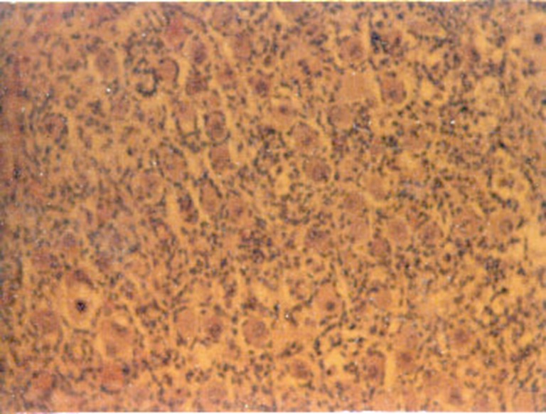
Ovarian cortex of a 39 week old fetus
Table 1.
Follicular density in ovaries of human fetuses of different gestational ages
| Gestational age (weeks) | Follicles/0.25 mm2 | Area (mm2) | Follicles counted | Follicles/mm3 |
|---|---|---|---|---|
| 22 | 18.8 ± 6.5 | 20 | 1,880 | 18,701 |
| 23 | 17.1 ± 5.8 | 25 | 1,712 | 17,010 |
| 24 | 13.1 ± 6.2 | 27.5 | 1,444 | 13,031 |
| 24 | 20.2 ± 7.2 | 22.5 | 1,617 | 20,094 |
| 26 | 32 ± 8.9 | 25 | 3,205 | 31,833 |
| 28 | 25.3 ± 9.6 | 20 | 2,025 | 25,168 |
| 31 | 18.2 ± 6.1 | 15 | 1,096 | 18,105 |
| 33 | 11.2 ± 5.1 | 22.5 | 1,004 | 11,141 |
| 36 | 11.1 ± 6.2 | 30 | 1,326 | 11,042 |
| 39 | 12.6 ± 6.5 | 30 | 1,512 | 12,534 |
| 39 | 34.7 ± 10.3 | 27.5 | 3,814 | 34,518 |
| 39 | 10.9 ± 4.8 | 32.5 | 1,406 | 10,843 |
Numbers are mean ± SD
Table 2.
Total number of follicles in ovaries of human fetuses of different gestational ages
| Gestational age (weeks) | Ovarian volume1 | Cortex volume1 | Follicles/ovary | Total (both ovaries) |
|---|---|---|---|---|
| 22 | 14.6 | 13.14 | 245,743 | 491,486 |
| 23 | 37.8 | 30.24 | 578,706 | 1,157,413 |
| 24 | 37.8 | 30.24 | 443,336 | 886,673 |
| 24 | 37.8 | 30.24 | 683,618 | 1,367,237 |
| 26 | 37.8 | 30.24 | 1,082,960 | 2,165,920 |
| 28 | 27.5 | 22 | 622,908 | 1,245,816 |
| 31 | 27.5 | 22 | 448,099 | 896,199 |
| 33 | 27.8 | 22.24 | 278,761 | 557,523 |
| 36 | 41.1 | 33.12 | 411,428 | 822,856 |
| 39 | 46.1 | 36.9 | 520,046 | 1,040,092 |
| 39 | 46.1 | 36.9 | 1,432,191 | 2,864,383 |
| 39 | 46.1 | 36.9 | 449,881 | 899,762 |
1Ovarian and cortex volumes were obtained from the literature
Fig. 2.

Mean number of ovarian follicles according to the gestational age during human fetal development
Discussion
To our knowledge, this is the first study to determine follicular density in human fetuses of several gestational ages using direct methods. Our results show that follicle numbers peak at 26 weeks of gestation during fetal development. In the specific case of neonates (39 weeks of gestation), the present findings are similar to those reported in an early study [1], revealing a mean number of 1,601,412 follicles per ovary (range: 899,762 to 2,864,383).
Although previously available estimates of primordial follicular numbers are still used as a reference, these estimates were based on indirect methods. Additionally, early estimates of the number of oocytes at birth (estimated at 2,000,000) [1] were not consistent with those from recent studies (from 100,000 to 600,000, estimated indirectly) or from a study from the early 50s’ [2], which estimated the number of follicles in neonatal ovaries to be on average 360,000 by counting the follicles directly and correcting the estimates by the thickness of the cuts.
Given the difficulties to calculate the volume of the ovarian cortex, previously published values were used. Therefore, we adopted a methodology that assumes a relatively uniform distribution of follicles in the cortex of fetal ovaries. Indeed, standard errors ranged from 1.07 to 0.425 in our series, hence a relatively small variation that meets the assumption of a uniform distribution.
The comparison of the present results with previously published estimates [1] reveal substantial differences in the number of follicles at each gestational age, most likely due to the methodological differences between these studies. Importantly, the present findings reveal a peak in the number of follicles during intrauterine life, followed by a rapid reduction in this number before birth. As the loss of most follicles occurs after recruitment begins, these results indicate the highest rates of follicular recruitment also occur at the same period.
In summary, by describing the variation in ovarian follicular density during embryonic development using a direct counting method, our results provide an important contribution to the understanding of primordial follicular assembly in humans and, ultimately, the determinants of reproductive capacity.
Footnotes
Capsule
Necropsied ovaries from fetuses and neonates were studied and a peak in the number of follicles was observed at approximately 26 weeks, followed by a rapid reduction in this number before birth.
References
- 1.Baker TG. A quantitative and citological study of germ cells in human ovaries. Proc R Soc Lond. 1963;158:417–420. doi: 10.1098/rspb.1963.0055. [DOI] [PubMed] [Google Scholar]
- 2.Block E. Quantitative morphological investigations of the follicular system in women; methods of quantitative determinations. Acta Anat. 1951;12:267–270. doi: 10.1159/000140549. [DOI] [PubMed] [Google Scholar]
- 3.Forabosco A, Sforza C, Pol A, Vizzotto L, Marzona L, Ferrario VF. Morphometric study of the human neonatal ovary. Anat Rec. 1991;231:201–205. doi: 10.1002/ar.1092310208. [DOI] [PubMed] [Google Scholar]
- 4.Forabosco A, Sforza C. Establishment of ovarian reserve: a quantitative morphometric study of the developing human ovary. Fertil Steril. 2007;88:675–683. doi: 10.1016/j.fertnstert.2006.11.191. [DOI] [PubMed] [Google Scholar]
- 5.Godin I, Wylie CC. TGF beta1 inhibits proliferation and has a chemotropic effect on mouse primordial germ cells in culture. Development. 1991;113:1451–1457. doi: 10.1242/dev.113.4.1451. [DOI] [PubMed] [Google Scholar]
- 6.Gondos B, Bhiraleus P, Hobel CJ. Ultrastructural observations on germ cells in human fetal ovaries. Am J Obstet Gynecol. 1971;110:644–648. doi: 10.1016/0002-9378(71)90245-6. [DOI] [PubMed] [Google Scholar]
- 7.Gondos B, Westergaard L, Byskov A. Initiation of oogenesis in human fetal ovary: ultrastuctural and squash preparation study. Am J Obstet Gynecol. 1986;155:189–192. doi: 10.1016/0002-9378(86)90109-2. [DOI] [PubMed] [Google Scholar]
- 8.Gougeon A, Chainy GBN. Morphometric studies of small follicles in ovaries of women at different ages. J Reprod Fertil. 1987;81:433–438. doi: 10.1530/jrf.0.0810433. [DOI] [PubMed] [Google Scholar]
- 9.Hilscher W. The genetic control and germ cell kinetics of female and male germ line in mammals including man. Hum Reprod. 1991;6:1416–1425. doi: 10.1093/oxfordjournals.humrep.a137281. [DOI] [PubMed] [Google Scholar]
- 10.Kurilo LF. Oogenesis in antenatal development in man. Hum Genet. 1981;57:1–8. doi: 10.1007/BF00271175. [DOI] [PubMed] [Google Scholar]
- 11.Sforza C, Forabosco A. A morphometric approach to the study of human ovarian organogenesis. Ital J Anat Embryol. 1998;103(Suppl 1):51–62. [PubMed] [Google Scholar]