Abstract
Gene targeting in most of human somatic cell lines has been labor-intensive because of low homologous recombination efficiency. The development of an experimental system that permits a facile evaluation of gene targeting efficiency in human somatic cell lines is the first step towards the improvement of this technology and its application to a broad range of cell lines. In this study, we utilized phosphatidylinositol glycan anchor biosynthesis class A (PIGA), a gene essential for the synthesis of glycosylphosphatidyl inositol (GPI) anchors, as a reporter of gene targeting events in human somatic cell lines. Targeted disruption of PIGA was quantitatively detected with FLAER, a reagent that specifically binds to GPI anchors. Using this PIGA-based reporter system, we successfully detected adeno-associated virus (AAV)-mediated gene targeting events both with and without promoter-trap enrichment of gene-targeted cell population. The PIGA-based reporter system was also capable of reproducing previous findings that an AAV-mediated gene targeting achieves a remarkably higher ratio of homologous versus random integration (H/R ratio) of targeting vectors than a plasmid-mediated gene targeting. The PIGA-based system also detected an approximately 2-fold increase in the H/R ratio achieved by a small negative selection cassette introduced at the end of the AAV-based targeting vector with a promoter-trap system. Thus, our PIGA-based system is useful for monitoring AAV-mediated gene targeting and will assist in improving gene targeting technology in human somatic cell lines.
Introduction
Gene targeting is a powerful technology to explore gene functions in eukaryotes by allowing the manipulation of endogenous genes via homologous recombination (HR). Although gene targeting in mammalian cells has been mainly conducted in murine embryonic stem (ES) cells for the purpose of creating knockout or knock-in mouse models, human somatic cells have also been subjected to gene targeting [1]. Gene-targeted human cell lines are useful to study human gene functions and diseases caused by genetic alterations, such as cancer [2], [3]. These gene-targeted cell lines are also believed to be promising drug screening platforms used for the development and validation of molecularly targeted therapies [4], [5].
Several significant improvements in gene targeting technology have been made in previous studies, including the use of adeno-associated virus (AAV)-based targeting vectors [6], [7], a promoter-trap strategy with the aid of internal ribosomal entry sites (IRES) [8], and a systematic PCR screening method for gene-targeted cell clones [9]. Despite these improvements, gene targeting in most of human somatic cell lines remains to be difficult probably because of low HR efficiencies as demonstrated in murine somatic cells [10]. The vast majority of targeting vectors introduced into human somatic cells are integrated randomly, rather than via HR, into the genome. To further improve this technology and make it readily applicable to a wide range of human cell lines, it is advantageous to have an experimental system monitoring gene targeting efficiencies with a simple procedure. In particular, monitoring the frequency of homologous versus random integration (H/R ratio) of targeting vectors is informative, as this ratio is a major determinant of difficulty in conducting gene targeting.
The phosphatidylinositol glycan anchor biosynthesis class A (PIGA) gene on the X chromosome encodes a protein that is required for glycosylphosphatidyl inositol (GPI) anchor biosynthesis [11], [12]. An inactivating mutation in the PIGA gene results in the loss of GPI anchors in the cells carrying a single X chromosome, such as diploid cells of male origin. The loss of GPI anchors then leads to the release of extracellular proteins that are otherwise linked to cell membranes by GPI anchors. Cells without GPI anchors are readily detectable with negative staining by reagents that bind to GPI anchors or by antibodies targeted against GPI-anchored proteins [13], [14]. Thus, the frequency of PIGA inactivation can be monitored by simple cell staining followed by fluorescence flow cytometry. This unique property of PIGA has been exploited to estimate the incidence of genetic mutations occurring spontaneously or elicited by mutagens during cell culture or in animal models [15]–[18]. PIGA was also disrupted by zinc-finger nucleases (ZFNs) in human ES cells and induced pluripotent stem cells to provide a proof of principle for the genome editing mediated by ZFNs [19]. In the current study, we used PIGA to develop an assay that evaluates gene targeting efficiencies represented by H/R ratios in human somatic cell lines. Using this system, we also attempted to modify the structure of an AAV-based targeting vector and thereby improve the gene targeting efficiency in order to facilitate the application of this technology to various human somatic cell lines.
Results
Quantitative measurement of PIGA gene targeting frequency
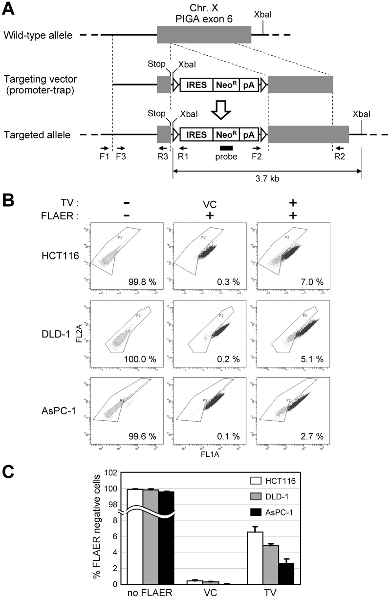
We initially constructed a targeting vector that disrupts PIGA exon 6 as shown in Fig. 1A. This targeting vector has 5′ and 3′ homology arms, both of which are approximately 1 kb in length. This targeting vector also has a neomycin resistance gene (NeoR) downstream of an IRES that allows for the enrichment of gene-targeted clones by promoter-trap strategy. In addition, the vector has two loxP sites encompassing the NeoR expression cassette so that NeoR can be excised by Cre-loxP recombination, which allows NeoR to be reused for further genetic manipulation. An artificial stop codon within exon 6 was incorporated at the proximal end of the 5′ arm to ensure PIGA disruption. This targeting vector was created based on an AAV backbone, as AAV-based targeting vectors reportedly achieve more than 1,000-times higher gene targeting frequencies than conventional plasmid-based targeting vectors [6], and have been employed in many recent studies to overcome low gene targeting efficiencies in human somatic cell lines [5], [20]–[28].
Figure 1. PIGA gene targeting in human somatic cell lines.
(A) A diagram of the promoter-trap PIGA targeting vector. Gray boxes indicate PIGA exon 6, while thick lines indicate PIGA intron 5 and intergenic genomic region. Thin dotted lines indicate homology between the endogenous gene locus and the targeting vector. F1 to F3 and R1 to R3 indicate PCR primers used in the experiment shown in Fig. 2A and B. A filled rectangle indicates the location of the probe used in Southern blot analysis shown in Fig. 2C. XbaI restriction enzyme sites are marked in the diagram. An arrow at the bottom shows the distance between two XbaI sites at the targeted PIGA gene locus. Triangles: loxP; Stop: stop codon (TAA); IRES: internal ribosomal entry site; NeoR: neomycin resistance gene; pA: polyadenylation site. (B and C) Human somatic cell lines were infected with the promoter-trap PIGA targeting vector or a vector control (VC), and processed for FLAER staining and then flow cytometric analyses. Shown are the representative dot plots (B) and a graphic representation of the entire results (mean ± s.e.m.; n = 3) (C). In (B), the FLAER-negative ratio for each experiment is noted in the dot plot. TV: promoter-trap PIGA targeting vector.
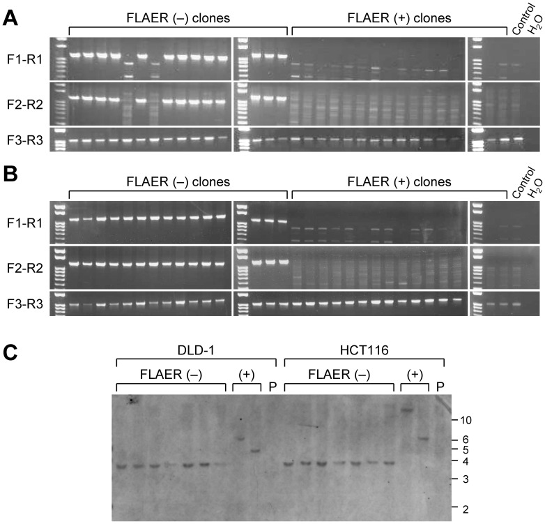
The constructed AAV-based PIGA targeting vector was used to infect two colon cancer cell lines HCT116 and DLD-1 and a pancreatic cancer cell line AsPC-1, all of which are of male origin. The infected cells were selected with G418 to assess the H/R ratio of the targeting vector, and stained with an Alexa 488-conjugated inactive aerolysin variant, FLAER, which specifically binds to GPI-anchors [29]. Cells were then analyzed by fluorescence flow cytometry to determine the percentage of FLAER-negative cells. As expected, virtually all the cells not processed for FLAER staining did not have an Alexa 488 signal, whereas the cells that were processed for FLAER staining but that had been infected with an unrelated AAV vector driving NeoR gene expression (vector control; VC) were mostly FLAER-positive (Fig. 1B and C). In contrast, FLAER-negative populations accounted for 2.7–7.0% of the cells that were subjected to both PIGA gene targeting and FLAER staining, suggesting that gene targeting successfully inactivated PIGA. We subsequently sorted both FLAER-negative and -positive populations from HCT116 and DLD-1 cells infected with the PIGA targeting vector, and established 55 and 15 single cell clones from FLAER-negative and -positive populations, respectively, for both cell lines. The resulting cell clones were examined with a pair of analytical PCR reactions encompassing the 5′ and 3′ homology arms of the targeting vector, respectively. These PCR reactions demonstrated that 51 of 55 (93%; HCT116) and 54 of 55 (98%; DLD-1) FLAER-negative clones underwent PIGA gene targeting, while all 15 FLAER-positive clones were apparently formed by the random integration of the targeting vector into the genome (Fig. 2A and B). We also confirmed the DNA sequence around the targeted genomic locus at the PIGA exon 6 in 15 DLD-1-derived FLAER-negative cell clones (data not shown). We further analyzed a subset of FLAER-negative and -positive cell clones derived from HCT116 and DLD-1 by Southern blotting, and thereby confirmed that the PIGA gene had been targeted as expected in FLAER-negative cell clones (Fig. 2C). These data collectively indicate that the negative FLAER staining in our system truly represents the abrogation of PIGA by gene targeting.
Figure 2. Confirmatory assays examining the PIGA gene locus in FLAER-negative and -positive cell clones.
(A and B) Representative results of PCR examining FLAER-negative and -positive single cell clones derived from HCT116 (A) and DLD-1 (B) infected with the promoter-trap PIGA targeting vector. PCR was conducted with primer pairs F1-R1, F2-R2 and F3-R3 that yielded PCR products of 1,140 bp, 1,184 bp, and 1,031 bp in size, respectively. For the location of primers, see Fig. 1A. Control: parental cell lines. (C) Southern blot analysis of FLAER-negative and -positive single cell clones. Genomic DNA was digested with XbaI, separated on a gel, and hybridized with a probe shown in Fig. 1A. The positions of the DNA size standards are shown to the right in kilobase. P: parental cell lines.
Disruption of PIGA does not affect the proliferation of HCT116 and DLD-1 cells
PIGA encodes a protein with important cellular function. If the disruption of PIGA affects cell proliferation, flow cytometric analysis of the cells transduced with PIGA targeting vector after a period of antibiotic selection may incorrectly estimate gene targeting efficiency. To clarify whether PIGA-deficient cells have growth advantage or disadvantage against PIGA-proficient cells, we next compared the growth indices of FLAER-negative bulk cell populations and single cell clones derived from them with those of the FLAER-positive bulk cell populations, single cell clones derived from them, and parental cell lines. As shown in Fig. 3, no significant difference in cell growth was observed between PIGA-deficient and -proficient cells both in HCT116 and DLD-1 series.
Figure 3. Growth indices of PIGA-targeted and non-targeted cell populations.
Cells derived from HCT116 (left) and DLD-1 (right) were propagated in 96-well tissue culture plates, and cell numbers assayed at Day 3, Day 5, and Day 7 were shown relative to those at Day 1. In each cluster of bar graphs, open bars represent a FLAER-negative bulk cell population and PIGA-targeted (confirmed by Southern blotting) single cell clones #1, #2 and #3 (from left to right); gray bars represent a FLAER-positive bulk cell population and PIGA-non-targeted (determined by analytical PCR) single cell clones #1, #2 and #3 (from left to right); a filled bar represents the parental cell line (mean ± s.d.; n = 6).
Gene targeting in human somatic cell lines with various experimental conditions
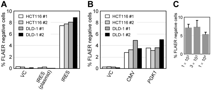
We subsequently investigated whether our PIGA-based system can reproduce a previous finding that an AAV-based targeting vector achieves several orders of magnitude greater targeting efficiency than a plasmid-based targeting vector that, except for its backbone, has an identical structure [6]. Since HCT116 and DLD-1 were found to be more amenable to PIGA gene targeting than AsPC-1 in our system (Fig. 1B and C), we used these two cell lines in most of the experiments hereafter performed. We first transfected (as a plasmid) or infected (as an AAV) our PIGA targeting vector into the human cell lines, selected the cells with G418, and then compared the percentages of FLAER-negative cells between the plasmid and AAV groups. We detected no increase of FLAER-negative cells by the transfection of PIGA targeting vector; however, the infection of the cells with PIGA targeting vector resulted in an increase of FLAER-negative cell populations to 7.4–8.9% (p <0.0001, versus the plasmid-based targeting vector; Fig. 4A). These data suggest that, in the PIGA gene targeting, an AAV-based targeting vector promotes a higher gene targeting frequency than a plasmid-based vector consistent with previous studies [6], [8], [30]. However, our PIGA-based system may not be sufficiently sensitive to detect low gene targeting frequencies achieved by plasmid-based targeting vectors in human somatic cell lines.
Figure 4. Monitoring of the gene targeting efficiencies in various experimental conditions using the PIGA-based reporter system.
(A) HCT116 and DLD-1 cells were transfected (as a plasmid) or infected (as an AAV) with the promoter-trap PIGA targeting vector, and the ratios of FLAER-negative cell populations were determined in duplicate. In X-axis labels, VC, IRES (plasmid) and IRES indicate a vector control, and the plasmid and AAV versions of promoter-trap PIGA targeting vector, respectively. (B) HCT116 and DLD-1 cells were infected with PIGA targeting vectors in which the NeoR gene was driven by the cytomegalovirus (CMV) promoter or the promoter of human phosphoglycerate kinase 1 (PGK1) gene, and the ratios of FLAER-negative cell populations were determined in duplicate. (C) HCT116 was infected with the indicated multiplicity of infection of the AAV-based promoter-trap PIGA targeting vector, and the ratios of FLAER-negative cell populations were determined (mean ± s.e.m.; n = 3).
We next investigated whether our PIGA-based system can detect AAV-mediated gene targeting performed without a promoter-trap system. To this end, we created a pair of new PIGA targeting vectors by replacing IRES within the above-described promoter-trap PIGA targeting vector with either the cytomegalovirus (CMV) immediate early promoter or the promoter of human phosphoglycerate kinase 1 (PGK1) gene. AAV was produced with these constructs and then used for the infection of the HCT116 and DLD-1 cell lines. Flow cytometric analysis of the cell lines after G418 selection exhibited an increase of FLAER-negative cell populations to 2.8–5.1% (p = 0.0005 for CMV versus VC, and p = 0.0004 for PGK1 versus VC; Fig. 4B), suggesting that our PIGA-based system is capable of detecting gene targeting events elicited by AAV-based non-promoter-trap targeting vectors (Fig. 4B).
We also examined whether the multiplicity of infection (MOI) of the PIGA targeting vector has an impact on the H/R ratio in our system. To this end, HCT116 cells were infected with the promoter-trap PIGA targeting vector at MOIs between 1×10³ and 1×10⁴, and processed for the quantification of FLAER-negative cell populations as described above. As a result, no significant change in the H/R ratio was observed within this range of MOIs (Fig. 4C). It has been shown that both homologous and random integration of AAV-based targeting vectors increase as the MOI increases [6], [31], [32]. A previous study also showed that, unlike our results, the H/R ratio of an AAV-based targeting vector increases in an MOI-dependent manner [32], even though this increase may be compromised because of the counterbalance between two parameters, i.e., homologous and random integration. The inconsistency between the results of this study and ours may be due to the different experimental systems that were used to determine gene targeting frequencies (GFP-based versus PIGA-based systems) or the different types of cells employed for gene targeting (293 cells versus colon cancer cell lines). However, a possibility remains that limited sensitivity in determining H/R ratio in our system might account for the absence of the correlation between MOI and H/R ratio in our experimental results.
Positive-negative selection (PNS) in an AAV-based targeting vector
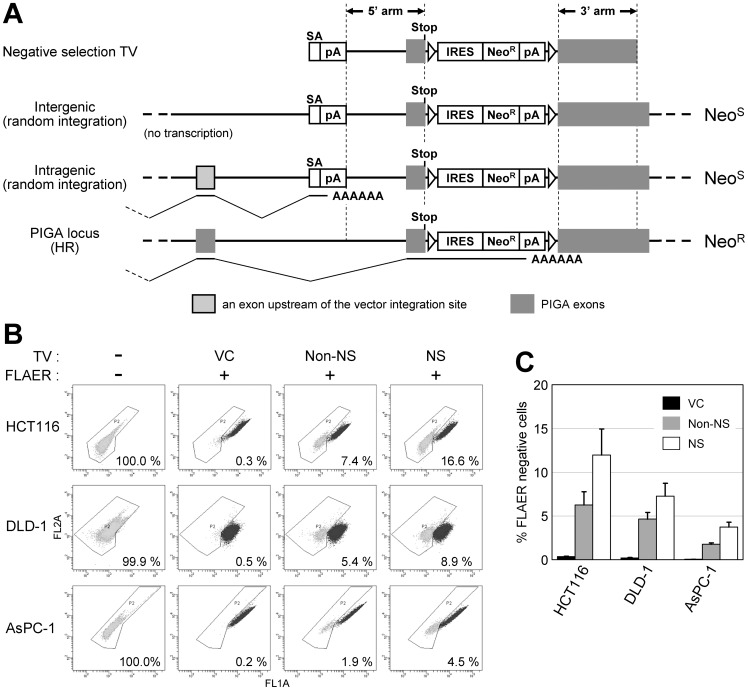
We next utilized this PIGA-based reporter system in an attempt to employ PNS in the context of AAV-mediated gene targeting. PNS is a useful strategy to enrich gene-targeted cell populations during plasmid-based gene targeting [33] and has been primarily used in murine ES cells. Negative selection (NS) is accomplished by inserting a genetic cassette that contains a gene conferring cytotoxicity or drug sensitivity, such as herpes simplex virus thymidine kinase [33] and diphtheria toxin A fragment [34], at the distal end of either of the homology arms. To the best of our knowledge, PNS has not been utilized in AAV-based targeting vectors because AAV vectors have a strict size limit of approximately 5 kb in total, and NS cassettes described above are too large to be used in AAV-based targeting vectors. Thus, in place of a large gene for NS, we tested a small cassette comprising a splice acceptor derived from human BCL2 intron 2/exon 3 boundary followed by a bovine growth hormone (BGH) polyadenylation site (polyA) inserted at the distal end of the 5′ arm in the PIGA targeting vector (Fig. 5A). This cassette is 524 bp long in total and is thus sufficiently small to place within an AAV vector. When a targeting vector carrying this cassette, along with an IRES-based promoter-trap system, is randomly integrated into an intragenic region of the genome, this cassette will trap at least a part of transcription machinery at the disrupted endogenous gene locus, and thereby reduce NeoR gene expression. In contrast, when the targeting vector is integrated into the PIGA gene locus via HR, this cassette will be removed upon integration, and thus the NeoR gene will be transcribed by the endogenous PIGA promoter without interruption. Thus, this small cassette in principle will provide NS when used with a promoter-trap system. To our knowledge, this is the first report officially assessing the impact of this type of NS cassette on gene targeting efficiency.
Figure 5. Negative selection (NS) in AAV-mediated gene targeting.
(A) A diagram describing the NS conferred by a small cassette consisting of splice acceptor (SA) and a polyadenylation site (pA). “Negative selection TV” depicts a promoter-trap PIGA targeting vector with a NS cassette. Other notations on the left indicate the location and the manner (in parenthesis) of vector integration. Bold and thin lines depict introns and anticipated splicing in the locus, respectively. Noted to the right are the theoretical neomycin sensitive (NeoS) or resistant (NeoR) statuses of the cells resulting from the integration of vectors. HR: homologous recombination. (B and C) Cell lines were infected with indicated PIGA targeting vectors or a vector control (VC), and processed for FLAER staining and then flow cytometric analyses. Shown are the representative dot plots (B) and a graphic representation of the entire results (mean ± s.e.m.; n = 3) (C). In (B), the FLAER-negative ratio for each experiment is noted in the dot plot. Non-NS: a promoter-trap PIGA targeting vector without the NS cassette (depicted in Fig. 1A).
We assayed the difference in gene targeting efficiencies achieved by a promoter-trap PIGA targeting vector between in the presence and absence of this NS cassette in the vector. HCT116, DLD-1, and AsPC-1 cells were infected with these AAV-based targeting vectors, selected with G418, stained with FLAER, and then processed for flow cytometry to quantify FLAER-negative cells. These assays demonstrated a 1.6- to 2.1-fold enrichment of gene-targeted clones by the use of the NS cassette (p<0.05; Fig. 5B and C). In accord with this enrichment, the numbers of G418 resistant colonies derived from the infected cells were decreased by half to one-third with the use of the NS cassette (data not shown).
We further investigated whether the enrichment of gene-targeted clones by this NS cassette enables our PIGA-based system to detect gene targeting events elicited by plasmid-based targeting vectors. HCT116 and DLD-1 cells were transfected (as a plasmid) with the PIGA targeting vector carrying the NS cassette and a promoter-trap system, selected with G418, and then analyzed by fluorescence flow cytometry. This analysis exhibited no increase in the ratio of FLAER-negative cells by the transfection of the above-mentioned PIGA targeting vector versus VC (data not shown), again suggesting the difficulty in detecting plasmid-mediated gene targeting in human somatic cell lines using our PIGA-based system.
We also designed another NS cassette comprising a splice acceptor site (111 bp) obtained from pIRESneo3 and a SV40 early messenger RNA polyA (238 bp). This newly designed NS cassette was incorporated at the distal end of the 5′ arm in the promoter-trap PIGA targeting vector in place of the above-mentioned NS cassette. HCT116 and DLD-1 cells were infected with the resultant AAV-based targeting vector, and the percentages of FLAER-negative cell populations were compared with those of the cells infected with a promoter-trap PIGA targeting vector without a NS cassette. This analysis demonstrated no significant difference in FLAER-negative ratios depending on the presence or absence of the newly designed NS cassette (data not shown), suggesting no appreciable capacity of this NS cassette to enrich gene-targeted cell clones.
These experiments investigating small NS cassettes collectively indicate that the introduction of a small NS cassette comprising a splice acceptor and a polyA sites into a promoter-trap targeting vector may moderately elevate the H/R ratio of the targeting vector in human somatic cell lines. However, it is likely that only a limited selection of splice acceptor and polyA sites are capable of elevating H/R ratio. In addition, this increase of H/R ratio may only be detectable with AAV-mediated and not with plasmid-mediated gene targeting in the experimental setting used in this study.
Discussion
In the current study, we developed a molecular system that exploits a unique property of the PIGA gene to evaluate gene targeting efficiencies in human somatic cell lines. The PIGA-based system was capable of monitoring AAV-mediated gene targeting in both promoter-trap and non-promoter-trap systems. In previous studies, exogenously introduced marker genes, including GFP and NeoR, have frequently been utilized to measure gene targeting frequencies [6], [35]. However, it is often found that the marker genes driven by strong, ectopic promoters are initially expressed constitutively, but eventually reduce their expression to an undetectable level, probably because of epigenetic changes in promoter regions. In contrast, endogenous marker genes, which are regulated by their native promoters, allow for stable monitoring systems that operate in more physiological settings.
The hypoxanthine phosphoribosyltransferase 1 (HPRT1) gene is one of the most commonly used endogenous markers [6], [36], [37]. Owing to its localization on the X chromosome, the abrogation of an HPRT1 allele in cells bearing a single X chromosome leads to the loss of the HPRT1 protein, which results in resistance to 6-Thioguanine (6-TG) and lethality in hypoxanthine-aminopterin-thymidine (HAT) medium. Thus, cells undergoing targeted disruption of HPRT1 can be quantified by colony formation assays in the presence of 6-TG, while, those undergoing targeted gene correction of mutant HPRT1 can be identified by cell culture in HAT medium. Other endogenous genes on the X chromosome, such as IL2RG, have also been utilized to determine gene targeting efficiencies by PCR-restriction fragment length polymorphism analyses upon ZFN-mediated gene targeting [38]. Compared with these systems, our PIGA-based system permits a more high-throughput and convenient measurement of gene targeting frequencies with the aid of fluorescence flow cytometry. Additionally, the PIGA-based system offers an option to assay gene targeting frequencies via colony counting after selection of the cells with aerolysin, a toxin that binds to GPI anchors and produces pore-forming channels in cell membranes [39]. Because PIGA is also located on the X chromosome, however, PIGA-based system can be applied only to cells that carry single X chromosomes, making the cell lines of female origin and many of aneuploid cell lines ineligible for this reporter system. In addition, endogenous marker genes, including HPRT1 and PIGA, may have spontaneous inactivating gene alterations in a subset of cell populations. It is also possible that genes involved in the same biological pathways with HPRT1 and PIGA are biallelically inactivated by spontaneous genetic or epigenetic changes. These events may constitute a background of the reporter systems.
Using the PIGA-based system, we evaluated the capacity of small NS cassettes inserted at the end of a targeting vector to improve the efficiency of AAV-mediated gene targeting with promoter-trap strategy, and found approximately 2-fold enrichment of gene-targeted cell clones with one of the NS cassettes. Thus, although this system needs to be further characterized, the PNS strategy described here will be potentially useful in facilitating AAV-mediated gene targeting in human somatic cell lines. However, the enrichment of gene-targeted clones achieved by our PNS strategy was less effective than anticipated. In plasmid-based targeting vectors, similarly, PNS using large NS marker genes has usually achieved only 2–5-fold enrichments of gene-targeted clones. These modest enrichments have been attributed to damage to the NS cassettes upon the random integration of the targeting vectors [40]. However, this hypothesis may not account for our results with an AAV-based targeting vector, since the terminal erosion of AAV vectors upon random integration does not generally proceed over inverted terminal repeats (ITRs); thus, any components within targeting vectors will remain intact in most cases [41]–[43]. We speculate that the observed modest enrichment in this study may be due to splicing around the NS cassette as experienced in insertion mutagenesis [44]–[47].
A variety of technologies including AAV-mediated gene targeting have been developed to achieve genome editing in human cell lines [35], [38], [48], [49]. Among these, AAV-mediated gene targeting has the virtue of being simple, open, cost-sensitive, and sufficiently effective in conducting gene targeting. Moreover, it does not actively promote DNA double strand breaks, thus likely producing a relatively low frequency of off-target DNA cleavages and resulting promiscuous genetic alterations in the genome. Therefore, AAV-mediated gene targeting remains to be an important alternative to conduct genome editing in human somatic cell lines [5], [20]–[28]. This study has shown that the efficiency of AAV-mediated gene targeting can be monitored with a simple procedure using our PIGA-based reporter system. This reporter system will be useful to improve the technology of AAV-mediated gene targeting by testing newly designed targeting vectors, optimizing experimental procedures for gene targeting, and screening for genes and drugs that have impact on gene targeting efficiencies. It may also assist in performing comparative evaluation of different human somatic cell lines and thereby identifying cell lines amenable to gene targeting. Such technical improvements will eventually lead to the development of highly efficient technologies of AAV-mediated gene targeting readily applicable to various human somatic cell lines.
Materials and Methods
Vector construction
To construct a promoter-trap PIGA targeting vector, 5′ and 3′ homology arms, which are both 1.0 kb in size, were amplified by PCR, digested with XbaI and XhoI, respectively, and inserted into pSEPT [30]. To assemble NS cassettes, a fragment consisting of 236 bp of intron 2 and 59 bp of exon 3 from human BCL2 [50] was PCR-amplified and digested with SacI and NheI. A fragment of 226 bp from BGH polyA was also PCR-amplified and digested with NheI and MluI. Meanwhile, SacI, NheI and MluI restriction enzyme sites were introduced upstream of the 5′ homology arm in the promoter-trap PIGA targeting vector, and were then used to incorporate the splice acceptor and BGH polyA sites produced as described above. For the other NS cassette, a splice acceptor site from pIRESneo3 was PCR-amplified, digested with SacI, and ligated at the distal end of the 5′ homology arm in the promoter-trap PIGA targeting vector. The resulting plasmid was cleaved with NheI and MluI, and ligated with a XbaI-MluI fragment from pIRESneo3 which contains SV40 early messenger RNA polyA.
To create a PIGA targeting vector in which the CMV promoter drives NeoR, the promoter-trap PIGA targeting vector was cleaved by BamHI and ClaI to remove the promoter-trap module. Meanwhile, pCIneo (Promega) was first digested with HindIII, and the longer fragment was self-ligated and cleaved with BglII and ClaI. A 1.7 kb fragment containing the CMV promoter, NeoR, and a polyA was then isolated and ligated to the BamHI-ClaI site of the cleaved promoter-trap PIGA targeting vector.
For the construction of a PIGA targeting vector in which human PGK1 promoter drives NeoR, the promoter-trap PIGA targeting vector was cleaved by BamHI and HindIII, and the longest fragment containing 5′ and 3′ homology arms was recovered. Meanwhile, human PGK1 promoter was PCR-amplified, digested with BamHI and HindIII, and then ligated to the above-mentioned 5′ and 3′ homology arms. The resulting plasmid was then cleaved with HindIII and ClaI and ligated to a HindIII-ClaI fragment of pCIneo containing the NeoR gene and a polyA.
Constructs were confirmed by DNA sequencing and then used to produce AAV particles with 293T cells and the AAV helper-free system (Agilent Technologies) as previously described [51]. PCR was carried out with the primers listed in Table S1.
PIGA gene targeting assay
The HCT116, DLD-1, and AsPC-1 cell lines were cultured in Dulbecco's modified Eagle&s medium (DMEM; Wako) supplemented with 5% fetal bovine serum (Biowest) and 1% penicillin/streptomycin (Wako). The infection of cells with the AAV-based targeting vectors was carried out as previously described [51] at an MOI of 1×10⁴ unless otherwise noted. The MOI of AAV was determined by its copy number, which was quantified by real-time PCR using StepOnePlus (Applied Biosystems). Initially, the concentration of the solution of plasmid-based PIGA targeting vector was assayed with NanoDrop (NanoDrop Technologies) and used to determine its copy number in reference to its molecular weight. This plasmid prep was then diluted and used as a control in real-time PCR. For antibiotic selection, 0.4 mg/ml and 1 mg/ml of G418 (Invitrogen) were used for HCT116 and DLD-1 cells, respectively. After 1 to 2 weeks of G418 selection in 75 cm2 flasks, the bulk population of cells was dissociated and stained with FLAER (Pinewood Scientific Services) as per the manufacturer's instructions. FLAER was used at a final concentration of 5 nM. Flow cytometric analyses were carried out with FACSCanto II (BD Biosciences). Cell sorting was conducted with FACSVantage SE (BD Biosciences).
To compare the AAV- and plasmid-based targeting vectors, cells seeded in 75 cm2 flasks at 30% confluency were transfected with 7.5 μg/flask of the plasmid-based promoter-trap PIGA targeting vector using TransIT-LT1 (Mirus Bio) as per the manufacturer's instructions, and were then selected with G418. The resulting bulk cells were stained with FLAER and analyzed by flow cytometry as described above. Data were compared with those obtained in parallel through the infection of the AAV-based promoter-trap PIGA targeting vector.
PCR
To confirm the homologous integration of the PIGA targeting vector by PCR, genomic DNA (gDNA) was extracted from single cell clones using PureLink Genomic DNA Mini Kit (Invitrogen) and used as templates. PCR was performed with Platinum Taq (Invitrogen), the primers shown in Table S1, and a GeneAmp 9700 thermal cycler (Applied Biosystems).
Southern blotting
Southern blotting was carried out using gDNA extracted with a standard phenol/chloroform extraction and ethanol precipitation protocol. Five micrograms of gDNA was digested with XbaI, electrophoresed on 0.7% agarose gels, blotted onto Hybond-N+ membranes (GE Healthcare), and probed with an approximately 300-bp internal sequence of NeoR. Labeling of the Probe was carried out with AlkPhos Direct Labelling and Detection System with CDP-Star kit (GE Healthcare).
Cell proliferation assay
Cells derived from HCT116 and DLD-1 parental cell lines were seeded in 96-well tissue culture plates at a density of 1,000 cells and 500 cells per well, respectively. Cell numbers were quantified every other day from Day 1 (the day after seeding) to Day 7 using crystal violet staining. For staining, cells in the plates were first washed with phosphate buffered saline (PBS), fixed with 10% buffered formalin (Sigma–Aldrich), stained with 0.2% crystal violet (Sigma–Aldrich), washed with PBS three times, and air-dried. Crystal violet was then dissolved into 10% acetic acid, and absorbance at wavelength 590 nm was measured by VersaMax Microplate Reader (Molecular Devices). For background subtraction, the averaged absorbance of blank wells (medium only) in a tissue culture plate was subtracted from the measurement of each well in the same plate.
Statistics
The impact of MOI on gene targeting efficiency was analyzed by a one-way factorial analysis of variance (ANOVA). All the other statistical analyses in this study were carried out with a two-way factorial ANOVA. Intercooled Stata (Stata) was used for the analyses.
Acknowledgments
We thank Dr. Fred Bunz (Johns Hopkins University) for providing pSEPT.
Funding Statement
This work was supported in part by Grants-in-Aid for Scientific Research from the Japan Society for the Promotion of Science (#22700888 to SK, #21591225 to YH, and #22500999 to HK), a Strategic Research Foundation Grant-aided Project for Private Universities from the Ministry of Education, Culture, Sports, Science and Technology (MEXT), Japan (S1101027 to SK, YH, and HK), Takeda Science Foundation (to HK) and AIKEIKAI foundation (to SK). The funders had no role in study design, data collection and analysis, decision to publish, or preparation of the manuscript.
References
- 1. Smithies O, Gregg RG, Boggs SS, Koralewski MA, Kucherlapati RS (1985) Insertion of DNA sequences into the human chromosomal beta-globin locus by homologous recombination. Nature 317: 230–234. [DOI] [PubMed] [Google Scholar]
- 2. Shirasawa S, Furuse M, Yokoyama N, Sasazuki T (1993) Altered growth of human colon cancer cell lines disrupted at activated Ki-ras. Science 260: 85–88. [DOI] [PubMed] [Google Scholar]
- 3. Sedivy JM, Vogelstein B, Liber HL, Hendrickson EA, Rosmarin A (1999) Gene targeting in human cells without isogenic DNA. Science 283: 9a. [Google Scholar]
- 4. Di Nicolantonio F, Arena S, Gallicchio M, Zecchin D, Martini M, et al. (2008) Replacement of normal with mutant alleles in the genome of normal human cells unveils mutation-specific drug responses. Proc Natl Acad Sci U S A 105: 20864–20869. [DOI] [PMC free article] [PubMed] [Google Scholar]
- 5. Di Nicolantonio F, Arena S, Tabernero J, Grosso S, Molinari F, et al. (2010) Deregulation of the PI3K and KRAS signaling pathways in human cancer cells determines their response to everolimus. J Clin Invest 120: 2858–2866. [DOI] [PMC free article] [PubMed] [Google Scholar]
- 6. Russell DW, Hirata RK (1998) Human gene targeting by viral vectors. Nat Genet 18: 325–330. [DOI] [PMC free article] [PubMed] [Google Scholar]
- 7. Khan IF, Hirata RK, Russell DW (2011) AAV-mediated gene targeting methods for human cells. Nat Protoc 6: 482–501. [DOI] [PMC free article] [PubMed] [Google Scholar]
- 8. Hirata R, Chamberlain J, Dong R, Russell DW (2002) Targeted transgene insertion into human chromosomes by adeno-associated virus vectors. Nat Biotechnol 20: 735–738. [DOI] [PubMed] [Google Scholar]
- 9. Konishi H, Lauring J, Garay JP, Karakas B, Abukhdeir AM, et al. (2007) A PCR-based high-throughput screen with multiround sample pooling: application to somatic cell gene targeting. Nat Protoc 2: 2865–2874. [DOI] [PubMed] [Google Scholar]
- 10. Arbones ML, Austin HA, Capon DJ, Greenburg G (1994) Gene targeting in normal somatic cells: inactivation of the interferon-gamma receptor in myoblasts. Nat Genet 6: 90–97. [DOI] [PubMed] [Google Scholar]
- 11. Miyata T, Takeda J, Iida Y, Yamada N, Inoue N, et al. (1993) The cloning of PIG-A, a component in the early step of GPI-anchor biosynthesis. Science 259: 1318–1320. [DOI] [PubMed] [Google Scholar]
- 12. Takeda J, Miyata T, Kawagoe K, Iida Y, Endo Y, et al. (1993) Deficiency of the GPI anchor caused by a somatic mutation of the PIG-A gene in paroxysmal nocturnal hemoglobinuria. Cell 73: 703–711. [DOI] [PubMed] [Google Scholar]
- 13. Parker C, Omine M, Richards S, Nishimura J, Bessler M, et al. (2005) Diagnosis and management of paroxysmal nocturnal hemoglobinuria. Blood 106: 3699–3709. [DOI] [PMC free article] [PubMed] [Google Scholar]
- 14. Brodsky RA (2009) How I treat paroxysmal nocturnal hemoglobinuria. Blood 113: 6522–6527. [DOI] [PMC free article] [PubMed] [Google Scholar]
- 15. Chen R, Eshleman JR, Brodsky RA, Medof ME (2001) Glycophosphatidylinositol-anchored protein deficiency as a marker of mutator phenotypes in cancer. Cancer Res 61: 654–658. [PubMed] [Google Scholar]
- 16. Araten DJ, Golde DW, Zhang RH, Thaler HT, Gargiulo L, et al. (2005) A quantitative measurement of the human somatic mutation rate. Cancer Res 65: 8111–8117. [DOI] [PubMed] [Google Scholar]
- 17. Miura D, Dobrovolsky VN, Kasahara Y, Katsuura Y, Heflich RH (2008) Development of an in vivo gene mutation assay using the endogenous Pig-A gene: I. Flow cytometric detection of CD59-negative peripheral red blood cells and CD48-negative spleen T-cells from the rat. Environ Mol Mutagen 49: 614–621. [DOI] [PubMed] [Google Scholar]
- 18. Dobo KL, Fiedler RD, Gunther WC, Thiffeault CJ, Cammerer Z, et al. (2011) Defining EMS and ENU dose-response relationships using the Pig-a mutation assay in rats. Mutat Res 725: 13–21. [DOI] [PMC free article] [PubMed] [Google Scholar]
- 19. Zou J, Maeder ML, Mali P, Pruett-Miller SM, Thibodeau-Beganny S, et al. (2009) Gene targeting of a disease-related gene in human induced pluripotent stem and embryonic stem cells. Cell Stem Cell 5: 97–110. [DOI] [PMC free article] [PubMed] [Google Scholar]
- 20. Konishi H, Karakas B, Abukhdeir AM, Lauring J, Gustin JP, et al. (2007) Knock-in of mutant K-ras in nontumorigenic human epithelial cells as a new model for studying K-ras mediated transformation. Cancer Res 67: 8460–8467. [DOI] [PubMed] [Google Scholar]
- 21. Hucl T, Rago C, Gallmeier E, Brody JR, Gorospe M, et al. (2008) A syngeneic variance library for functional annotation of human variation: application to BRCA2. Cancer Res 68: 5023–5030. [DOI] [PMC free article] [PubMed] [Google Scholar]
- 22. Gustin JP, Karakas B, Weiss MB, Abukhdeir AM, Lauring J, et al. (2009) Knockin of mutant PIK3CA activates multiple oncogenic pathways. Proc Natl Acad Sci U S A 106: 2835–2840. [DOI] [PMC free article] [PubMed] [Google Scholar]
- 23. Yun J, Rago C, Cheong I, Pagliarini R, Angenendt P, et al. (2009) Glucose deprivation contributes to the development of KRAS pathway mutations in tumor cells. Science 325: 1555–1559. [DOI] [PMC free article] [PubMed] [Google Scholar]
- 24. Vitolo MI, Weiss MB, Szmacinski M, Tahir K, Waldman T, et al. (2009) Deletion of PTEN promotes tumorigenic signaling, resistance to anoikis, and altered response to chemotherapeutic agents in human mammary epithelial cells. Cancer Res 69: 8275–8283. [DOI] [PMC free article] [PubMed] [Google Scholar]
- 25. Chung JH, Bunz F (2010) Cdk2 is required for p53-independent G2/M checkpoint control. PLoS Genet 6: e1000863. [DOI] [PMC free article] [PubMed] [Google Scholar]
- 26. Fattah F, Lee EH, Weisensel N, Wang Y, Lichter N, et al. (2010) Ku regulates the non-homologous end joining pathway choice of DNA double-strand break repair in human somatic cells. PLoS Genet 6: e1000855. [DOI] [PMC free article] [PubMed] [Google Scholar]
- 27. Solomon DA, Kim T, Diaz-Martinez LA, Fair J, Elkahloun AG, et al. (2011) Mutational inactivation of STAG2 causes aneuploidy in human cancer. Science 333: 1039–1043. [DOI] [PMC free article] [PubMed] [Google Scholar]
- 28. Konishi H, Mohseni M, Tamaki A, Garay JP, Croessmann S, et al. (2011) Mutation of a single allele of the cancer susceptibility gene BRCA1 leads to genomic instability in human breast epithelial cells. Proc Natl Acad Sci U S A 108: 17773–17778. [DOI] [PMC free article] [PubMed] [Google Scholar]
- 29. Brodsky RA, Mukhina GL, Li S, Nelson KL, Chiurazzi PL, et al. (2000) Improved detection and characterization of paroxysmal nocturnal hemoglobinuria using fluorescent aerolysin. Am J Clin Pathol 114: 459–466. [DOI] [PMC free article] [PubMed] [Google Scholar]
- 30. Topaloglu O, Hurley PJ, Yildirim O, Civin CI, Bunz F (2005) Improved methods for the generation of human gene knockout and knockin cell lines. Nucleic Acids Res 33: e158. [DOI] [PMC free article] [PubMed] [Google Scholar]
- 31. Hirata RK, Russell DW (2000) Design and packaging of adeno-associated virus gene targeting vectors. J Virol 74: 4612–4620. [DOI] [PMC free article] [PubMed] [Google Scholar]
- 32. Porteus MH, Cathomen T, Weitzman MD, Baltimore D (2003) Efficient gene targeting mediated by adeno-associated virus and DNA double-strand breaks. Mol Cell Biol 23: 3558–3565. [DOI] [PMC free article] [PubMed] [Google Scholar]
- 33. Mansour SL, Thomas KR, Capecchi MR (1988) Disruption of the proto-oncogene int-2 in mouse embryo-derived stem cells: a general strategy for targeting mutations to non-selectable genes. Nature 336: 348–352. [DOI] [PubMed] [Google Scholar]
- 34. Yagi T, Ikawa Y, Yoshida K, Shigetani Y, Takeda N, et al. (1990) Homologous recombination at c-fyn locus of mouse embryonic stem cells with use of diphtheria toxin A-fragment gene in negative selection. Proc Natl Acad Sci U S A 87: 9918–9922. [DOI] [PMC free article] [PubMed] [Google Scholar]
- 35. Porteus MH, Baltimore D (2003) Chimeric nucleases stimulate gene targeting in human cells. Science 300: 763. [DOI] [PubMed] [Google Scholar]
- 36. Suzuki K, Mitsui K, Aizawa E, Hasegawa K, Kawase E, et al. (2008) Highly efficient transient gene expression and gene targeting in primate embryonic stem cells with helper-dependent adenoviral vectors. Proc Natl Acad Sci U S A 105: 13781–13786. [DOI] [PMC free article] [PubMed] [Google Scholar]
- 37. Iiizumi S, Kurosawa A, So S, Ishii Y, Chikaraishi Y, et al. (2008) Impact of non-homologous end-joining deficiency on random and targeted DNA integration: implications for gene targeting. Nucleic Acids Res 36: 6333–6342. [DOI] [PMC free article] [PubMed] [Google Scholar]
- 38. Urnov FD, Miller JC, Lee YL, Beausejour CM, Rock JM, et al. (2005) Highly efficient endogenous human gene correction using designed zinc-finger nucleases. Nature 435: 646–651. [DOI] [PubMed] [Google Scholar]
- 39. Diep DB, Nelson KL, Raja SM, Pleshak EN, Buckley JT (1998) Glycosylphosphatidylinositol anchors of membrane glycoproteins are binding determinants for the channel-forming toxin aerolysin. J Biol Chem 273: 2355–2360. [DOI] [PubMed] [Google Scholar]
- 40. Sedivy JM, Dutriaux A (1999) Gene targeting and somatic cell genetics–a rebirth or a coming of age? Trends Genet 15: 88–90. [DOI] [PubMed] [Google Scholar]
- 41. Rutledge EA, Russell DW (1997) Adeno-associated virus vector integration junctions. J Virol 71: 8429–8436. [DOI] [PMC free article] [PubMed] [Google Scholar]
- 42. Miller DG, Petek LM, Russell DW (2004) Adeno-associated virus vectors integrate at chromosome breakage sites. Nat Genet 36: 767–773. [DOI] [PubMed] [Google Scholar]
- 43. Miller DG, Trobridge GD, Petek LM, Jacobs MA, Kaul R, et al. (2005) Large-scale analysis of adeno-associated virus vector integration sites in normal human cells. J Virol 79: 11434–11442. [DOI] [PMC free article] [PubMed] [Google Scholar]
- 44. Voss AK, Thomas T, Gruss P (1998) Efficiency assessment of the gene trap approach. Dev Dyn 212: 171–180. [DOI] [PubMed] [Google Scholar]
- 45. McClive P, Pall G, Newton K, Lee M, Mullins J, et al. (1998) Gene trap integrations expressed in the developing heart: insertion site affects splicing of the PT1-ATG vector. Dev Dyn 212: 267–276. [DOI] [PubMed] [Google Scholar]
- 46. Elmore CL, Wu X, Leclerc D, Watson ED, Bottiglieri T, et al. (2007) Metabolic derangement of methionine and folate metabolism in mice deficient in methionine synthase reductase. Mol Genet Metab 91: 85–97. [DOI] [PMC free article] [PubMed] [Google Scholar]
- 47. Debrand E, El Jai Y, Spence L, Bate N, Praekelt U, et al. (2009) Talin 2 is a large and complex gene encoding multiple transcripts and protein isoforms. FEBS J 276: 1610–1628. [DOI] [PMC free article] [PubMed] [Google Scholar]
- 48. Christian M, Cermak T, Doyle EL, Schmidt C, Zhang F, et al. (2010) Targeting DNA double-strand breaks with TAL effector nucleases. Genetics 186: 757–761. [DOI] [PMC free article] [PubMed] [Google Scholar]
- 49. Miller JC, Tan S, Qiao G, Barlow KA, Wang J, et al. (2011) A TALE nuclease architecture for efficient genome editing. Nat Biotechnol 29: 143–148. [DOI] [PubMed] [Google Scholar]
- 50. Ishida Y, Leder P (1999) RET: a poly A-trap retrovirus vector for reversible disruption and expression monitoring of genes in living cells. Nucleic Acids Res 27: e35. [DOI] [PMC free article] [PubMed] [Google Scholar]
- 51. Rago C, Vogelstein B, Bunz F (2007) Genetic knockouts and knockins in human somatic cells. Nat Protoc 2: 2734–2746. [DOI] [PubMed] [Google Scholar]


