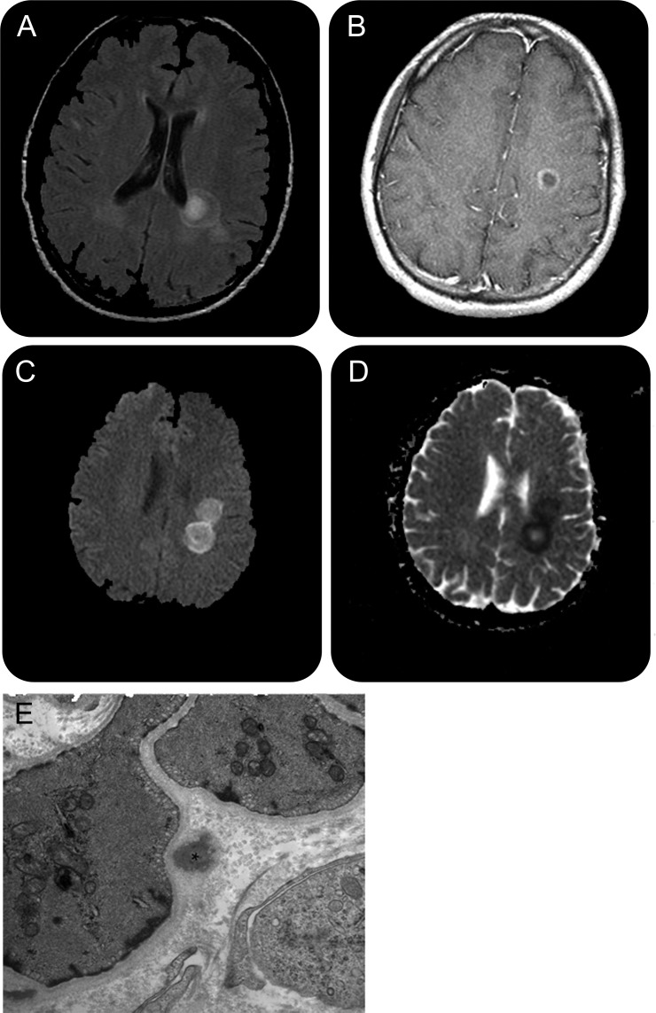
A 26-year-old man of Portuguese descent with no significant past medical history presents with subacute onset of right-sided hemiparesis and aphasia, with marked expressive aphasia, word-finding difficulty, and rare paraphrasic errors. Examination demonstrated right central facial weakness, 2–3/5 strength in the right arm and leg, and dysmetria in the right upper and lower extremities. Reflexes were hyperactive on the right with a right-sided Babinski response. MRI of the brain showed multiple bilateral concentric ring-like structures in the centrum semiovale and the corona radiata on T2 imaging (figure,A), consistent with the pattern of Balo concentric sclerosis. There was associated restricted diffusion in 3 lesions and incomplete ring enhancement in 1 lesion. CSF analysis showed 30 leukocytes (79% lymphocytes, 4% monocytes), erythrocyte count was 850, and oligoclonal bands were absent. He was treated with 1 g of IV methylprednisolone daily for 5 days, with little improvement. He then underwent 5 plasmapheresis exchanges, with significant improvement of motor, sensory, speech deficits, and gait. Forty-five days after discharge, his examination showed 4+/5 strength in the right intrinsic muscles of the hand and a right-sided positional tremor. Gait and tandem gait were normal. He was placed on β-interferon-1a 44 μg subcutaneously 3 times a week, for treatment of presumed clinically isolated syndrome, with good response. One year later, his neurologic examination was essentially normal.
Figure. Brain MRI (A–D) and electron microscopy photomicrograph of skin biopsy (E).
(A–D) Concentric rings on brain MRI with restricted diffusion. (A) Axial brain MRI shows a lesion adjacent to the left lateral ventricle, which demonstrates a ring and central core hyperintensity on T2 fluid-attenuated inversion recovery image. (B) Axial T1 image of a gadolinium ring-enhancing lesion adjacent to the left lateral ventricle. (C) Axial diffusion-weighted image (DWI) shows high signal within 2 lesions. (D) Axial apparent diffusion coefficient map confirms the presence of restricted diffusion of lesions seen on DWI in (C). (E) Electron microscopy photomicrograph of skin biopsy: skin punch biopsy of the right deltoid was performed. A dermal arteriole with one definite granular osmiophilic material (GOM) deposit marked with an asterisk. The GOM deposit is in close apposition to the pinocytic vesicles of the vascular smooth muscle cell plasma membrane (magnification ×25,000).
There was no family history of demyelinating or autoimmune disorders. His mother has a significant history of headaches and early-onset strokes, and was found to be a carrier of the Notch3 mutation consistent with cerebral autosomal dominant arteriopathy with subcortical infarcts and leukoencephalopathy (CADASIL). His sister had a stroke in her 30s, and a history of migraine headaches, but has not undergone medical evaluation.
Testing for CADASIL in the patient showed the Notch3 mutation associated with CADASIL (transition C>T, nucleotide position 1750, codon 558). Skin biopsy showed electron-dense extracellular material in close apposition to the smooth muscle of the vascular media1 (figure, B).
Discussion.
This patient's clinical presentation, neuroimaging findings, disease course, and response to therapy are consistent with Balo concentric sclerosis. However, the presence of the Notch3 mutation associated with CADASIL in this patient raises questions about disease pathogenesis in this particular patient, and of possible relationships between the 2 disorders.
Balo concentric sclerosis is rare, and has been associated with demyelinating diseases including multiple sclerosis (MS) and neuromyelitis optica.2 It is thought to represent the host tissue response to injury, and is associated with restricted diffusion on MRI.3 Of note, restricted diffusion on MRI has also been described in more “typical” MS lesions.4 A recent neuropathologic analysis of brain tissue from 14 patients with concentric lesions consistent with Balo found that all active concentric lesions followed the type III pattern of demyelination,5 which was not seen in any of the 18 MS cases reviewed.6 The type III pattern shares common features with hypoxia-like tissue injury. Lesions with a type III pattern as well as Balo lesions are characterized by preferential loss of MAG, with initial relative preservation of PLP and MOG. Oligodendrocytes display occasional nuclear shrinkage and fragmentation, and expressed CNPase in the cytoplasm. Additionally, expression of molecules involved in tissue preconditioning, including hypoxia-inducible factor 1-α, heat-shock protein 70, and D-110 epitope, were increased at the edge of active concentric lesions. These molecules were mainly expressed in oligodendrocytes, astrocytes, microglia, and cortical neurons. CADASIL mutation has been shown to be absent in patients with typical MS.7
Although these findings may be entirely coincidental, the presence of CADASIL Notch3 mutation in our patient with a clinical presentation of Balo concentric sclerosis raises the following hypotheses regarding pathogenesis.
Hypothesis 1. Balo is caused by mutations predisposing to hypoxic tissue injury, which includes the CADASIL mutation: Balo concentric sclerosis is due to mutations which affect vascular smooth muscle and lead to hypoxic-like injury in tissue. Such mutations may include the Notch3 mutation associated with CADASIL.
Hypothesis 2. CADASIL can present as Balo: CADASIL may present as Balo in some patients who are genetically predisposed to limited tissue injury, resulting in areas of preserved myelin.
Hypothesis 3. Vascular risk factors may modify demyelinating presentation: Notch3 mutation and other vascular risk factors could influence the presentation of a primary demyelinating disease, thus changing its phenotype toward the concentric Balo pattern.
Based on this case observation, we suggest systematic testing for the CADASIL mutation in patients with a demyelinating presentation consistent with Balo concentric sclerosis or significant restricted diffusion on MRI. Further genetic and biological studies are required to investigate the relationship between these 2 conditions.
Footnotes
Author contributions: T. Chitnis contributed to design and drafting for intellectual contents. T. Hollmann contributed to intellectual contents.
Disclosure: Dr. Chitnis has served as a consultant for Biogen Idec, Teva Pharmaceutical Industries Ltd., Novartis, Sanofi-Aventis, and EMD Serono, Inc.; receives research support from EMD Serono, Inc. the NIH/NINDS, and the National Multiple Sclerosis Society; and her spouse is employed by Novartis. Dr. Hollmann reports no disclosures.
References
- 1. Goebel HH, Meyermann R, Rosin R, Schlote W. Characteristic morphologic manifestation of CADASIL, cerebral autosomal-dominant arteriopathy with subcortical infarcts and leukoencephalopathy, in skeletal muscle and skin. Muscle Nerve 1997; 20: 625– 627 [DOI] [PubMed] [Google Scholar]
- 2. Graber JJ, Kister I, Geyer H, Khaund M, Herbert J. Neuromyelitis optica and concentric rings of Balo in the brainstem. Arch Neurol 2009; 66: 274– 275 [DOI] [PubMed] [Google Scholar]
- 3. Kavanagh EC, Heran MK, Fenton DM, Lapointe JS, Nugent RA, Graeb DA. Diffusion-weighted imaging findings in Balo concentric sclerosis. Br J Radiol 2006; 79: e28– e31 [DOI] [PubMed] [Google Scholar]
- 4. Balashov KE, Aung LL, Dhib-Jalbut S, Keller IA. Acute multiple sclerosis lesion: conversion of restricted diffusion due to vasogenic edema. J Neuroimaging 2011; 21: 202– 204 [DOI] [PMC free article] [PubMed] [Google Scholar]
- 5. Lucchinetti C, Bruck W, Parisi J, Scheithauer B, Rodriguez M, Lassmann H. Heterogeneity of multiple sclerosis lesions: implications for the pathogenesis of demyelination. Ann Neurol 2000; 47: 707– 717 [DOI] [PubMed] [Google Scholar]
- 6. Stadelmann C, Ludwin S, Tabira T, et al. Tissue preconditioning may explain concentric lesions in Balo's type of multiple sclerosis. Brain 2005; 128: 979– 987 [DOI] [PubMed] [Google Scholar]
- 7. Broadley SA, Sawcer SJ, Chataway SJ, et al. No association between multiple sclerosis and the Notch3 gene responsible for cerebral autosomal dominant arteriopathy with subcortical infarcts and leukoencephalopathy (CADASIL). J Neurol Neurosurg Psychiatry 2001; 71: 97– 99 [DOI] [PMC free article] [PubMed] [Google Scholar]


