Abstract
Objective:
Retention after orthodontic treatment is still an important part of the treatment. Splints are considered as an alternative for removable retainers. The main goal of this study was to assess splinting biomechanically.
Materials and Methods:
Three dimensional finite element models (3D) were designed of a mandibular anterior segment which included six anterior teeth with their supporting tissues (model 1 as control) and with a bonded lingual fixed retainer in the two other models. The wire cross section was round (0.016”) in model 2 and rectangular (0.016” × 0.022”) in model 3. The models were designed in Solid Works 2006 and analyzed in ANSYS Workbench Ver. 11.0 SolidWorks Incisors were loaded with a vertical force of 187 N. PDL stress and tooth displacements were evaluated.
Results:
The numeric findings showed an increase after splinting in the central incisors [2.42 MPa to 4.57 MPa (round) and 16.66 (rectangular) MPa] in biting with four incisors. Biting with two incisors decreased the stress after splinting [2.42 MPa to 1.7 MPa (round wire) and 1.77 MPa (rectangular wire)]. In lateral movement, all teeth showed an increased stress except for the working side canine.
Conclusion:
Splinted cases (with round or rectangular wires) can benefit from stress redistribution when biting small food particles and in lateral movement.
Keywords: Lower Anterior Teeth, Retention, Orthodontic Treatment, Splinting, Finite element method
INTRODUCTION
Retention after orthodontic treatment has been recommended by several authors and researchers. It can be stated as holding the teeth in optimal esthetic and functional positions. A proper retention regimen to prevent relapse after the end of an orthodontic treatment is frequently overlooked by orthodontists. Adequate retention is needed to let PDL and supracrestal fibers reorganize, to allow remodeling of the alveolar bone and to manage differential growth of the jaws, while managing soft tissue pressure [1]. The extent of the changes happening after the end of active treatment is not predictable [2]. These changes may be partly dependent on the type of malocclusion and the type of the mechano-therapy used [3]. Removable retainers were usually prescribed for the lower arch after the end of fixed orthodontic treatment.
Presence of an acceptable undercut space in lower molars is a prerequisite to provide needed retention of the removable retainer.4 However, more patients require a fixed type of retention like a bonded retainer on the lingual part of the lower incisors and canines to eliminate the risk of relapse due to a less than adequate collaboration.
Development of acid etching and bonding techniques made it possible for bonded retainer devices to gain popularity. Fixed lingual retainers in the mandibular arch were first used in the 1970s [5] and have since been considered as an accepted part of orthodontic treatment procedures to prevent relapse [6,7].
The first goal of orthodontists to suggest splinting in anterior teeth of patients after treatment is to solve the problem of cooperation leading to frequent relapse of crowding in the lower anterior segment.
According to Keim, up to one third of orthodontists use fixed lingual retainers after treatment and relapse reduction has been reported.8 The early bonded retainers were made of conventional round or rectangular orthodontic wires [9–12].
The elegance and efficiency of the lingual bonded retainers in the mandible, lack of any need for patient cooperation, complete invisibility and finding a way to bypass molars without undercut is their advantage over removable retainers. Initially, Zachrisson proposed using a multi-strand wire for producing a canine-canine retainer (Flexible Spiral Wire) [13].
This retainer was bonded only to the lower canines with thick wire (0.03 inch diameter). In 1983, a fixed retainer was introduced by Zachrisson which bonded to all the anterior teeth with a thin wire (0.0215 inch diameter) (14) providing a solution to the problem of central and lateral incisors’ undesirable movement encountered in the previous design. Different types of fixed bonded retainers have been introduced that vary in wire material, size and diameter [15,16]. Some use different types of composite [11,12,15,16] or resin fiberglass strips [17] or may have mesh pads. Numerous types of fixed retainers have been introduced which include 1-mandibular canine-to-canine retainer, 2-direct contact splinting, 3-flexible spiral wire (FSW), 4-retainers for individual teeth.
In a review on the retention process for fixing tooth location, the success of bonding retainers in maintaining anterior mandibular teeth was validated using numerous randomized clinical trial studies [18].
It is worth noting that bonded retainers are also used as a labial or lingual periodontal splint for stabilizing hypermobile teeth due to horizontal bone loss [19–23]. This type of splint is also applicable after trauma, periodontal surgery and occlusal trauma.
Negative effects of using these types of retainers and splints have been the matter of controversy for many years. It seems to be a rather unpleasant strategy from a periodontist’s point of view. However, long-term studies have indicated that although these devices make the bonding area vulnerable to calculus and plaque accumulation around the wire, periodontal health may be maintained if oral hygiene is adequate [24–27].
Enamel decalcification was not reported except for very rare cases [28].
No reports have been found that look at the effect of long-term bonding of anterior teeth on their ability to respond to occlusal stresses and the effects on the periodontal ligament.
Finite element method, as a numeric method of finding accurate answers to different questions was introduced less than a century ago in aerospace industry and soon found its way through biologic sciences. This method has proven its efficiency in different question scales [29–37].
Considering the popular usage of fixed retainers, the purpose of this study was to quantify the changes in stress distribution and displacement manner by bonding fixed retainers on normal alveolar bone after the end of orthodontic treatment by the finite element method.
MATERIALS AND METHODS
Three dimension (3D) finite element models were designed from a mandibular anterior segment which included six anterior teeth based on the average dimensions and supporting structures. Each model consisted of a spongy core surrounded by 1 mm-thick cortical layer. A simplified 0.25 mm-thick periodontal ligament layer (PDL) was also modeled based on the root-form geometry of the teeth.
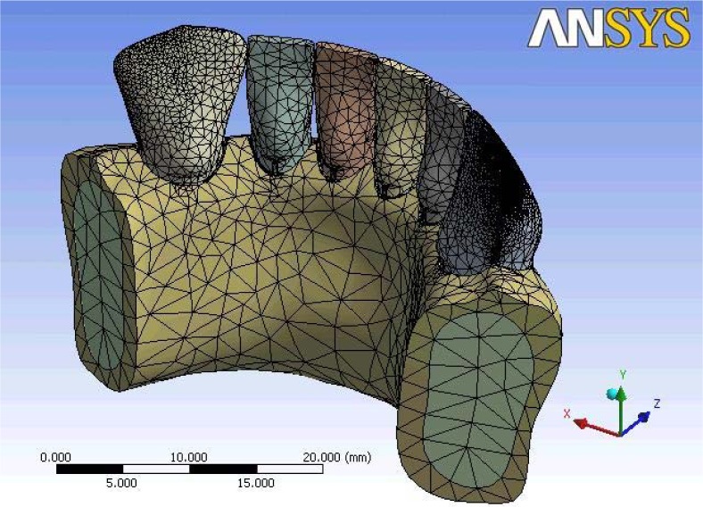
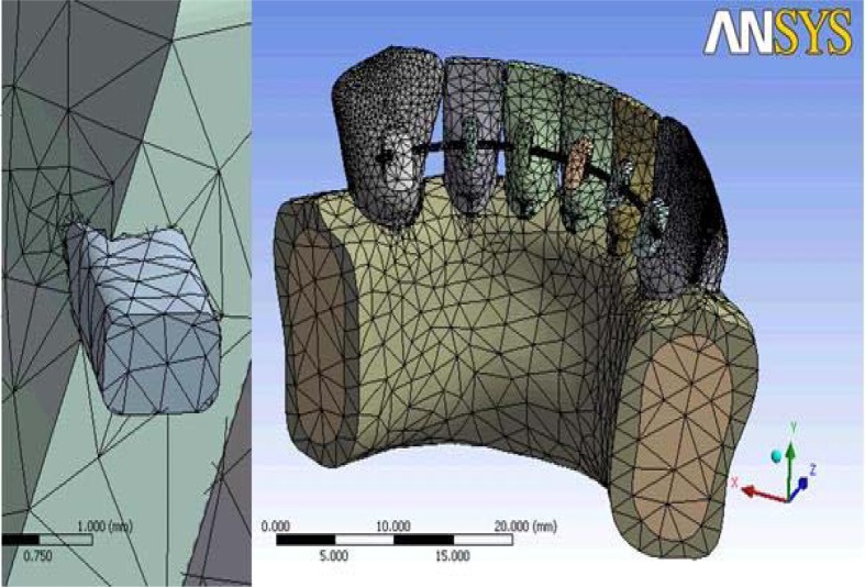
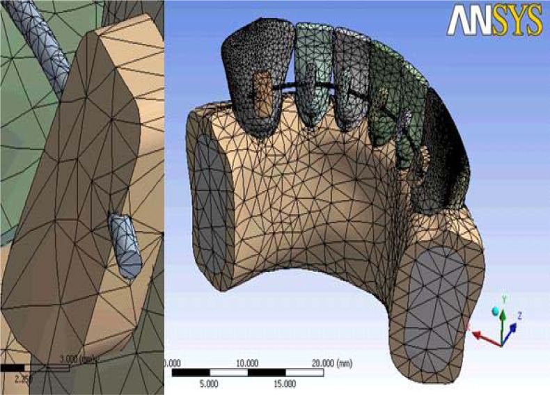
A bonded fixed retainer was designed in the lingual surface of the anterior teeth in two models and attached with composite. The models were similar except for the cross section of the retainer wire in the second (=0.4 mm diameter) and third model (= 0.4 mm × 0.55 mm) (Figure 1a–c). The first model served as control and did not have a bonded retainer. SolidWorks 2006 (300 Baker Ave. Concord, Massachusetts 01742, USA) was selected for the modeling phase. The next phase was to transfer the models for calculation to the ANSYS Workbench Ver. 11.0 (ANSYS Inc. Soutpointe, 275 Technology drive, Cononsburg PA 15317, USA). All the vital tissues were presumed elastic, homogeneous and isotropic. The corresponding elastic properties such as Young’s modulus and Poisson’s ratio were applied (Table I). Models were meshed with 95262 nodes; 84578 10-node-quadratic tetrahedron body elements, 34363 contact elements.
Fig 1a.
The non-splint model meshed
Fig 1c.
The rectangular wire splint model
Table 1.
Mechanical Properties of the Materials
| Young’s Modulus (MPa) | Poisson’s Ratio | |
|---|---|---|
| Tooth | 20300 | 0.26 |
| PDL | 0.667 | 0.49 |
| Spongy Bone | 13400 | 0.38 |
| Cortical Bone | 34000 | 0.26 |
| Composite | 61000 | 0.24 |
| SS Wire | 179000 | 0.33 |
| SS (Tube) | 205000 | 0.3 |
All nodes at the distal extremes of the models on the right and left were restrained so that all rigid body motions were prevented. A vertical force of 187 N was applied at each incisal edge of the central incisors according to previous studies [38,39].
Three functions were defined which were biting on two incisors, biting on four incisors and the canine function in mandibular lateral movement. The average von Mises stress in the PDLs and the displacement of the mesio-inciso-labial point angle of the right central incisor were considered to assess the effects of splinting.
RESULTS
Numeric findings are divided according to the approaches defined.
Stress (Table 2)
Table 2.
The Von Mises (Mpa) Stress Findings in Six Anterior Teeth PDL
| Left Canine | L2 | L1 | R1 | R2 | Right Canine | ||
|---|---|---|---|---|---|---|---|
| Biting on Four Incisors | Non-Splint | 0.06248 | 2.78 | 2.42 | 2.42 | 2.78 | 0.06248 |
| Round Splint | 0.169 | 2.075 | 4.57 | 4.57 | 2.075 | 0.169 | |
| Rectangular Splint | 0.189 | 2.53 | 16.66 | 16.66 | 2.263 | 0.183 | |
| Biting on Two Incisors | Non-Splint | 0.0349 | 0.0108 | 2.42 | 2.42 | 0.0108 | 0.0349 |
| Round-Splint | 0.05315 | 0.6965 | 1.7059 | 1.7059 | 0.6965 | 0.05315 | |
| Rectangular-Splint | 0.082 | 0.62605 | 1.7711 | 1.7711 | 0.62605 | 0.082 | |
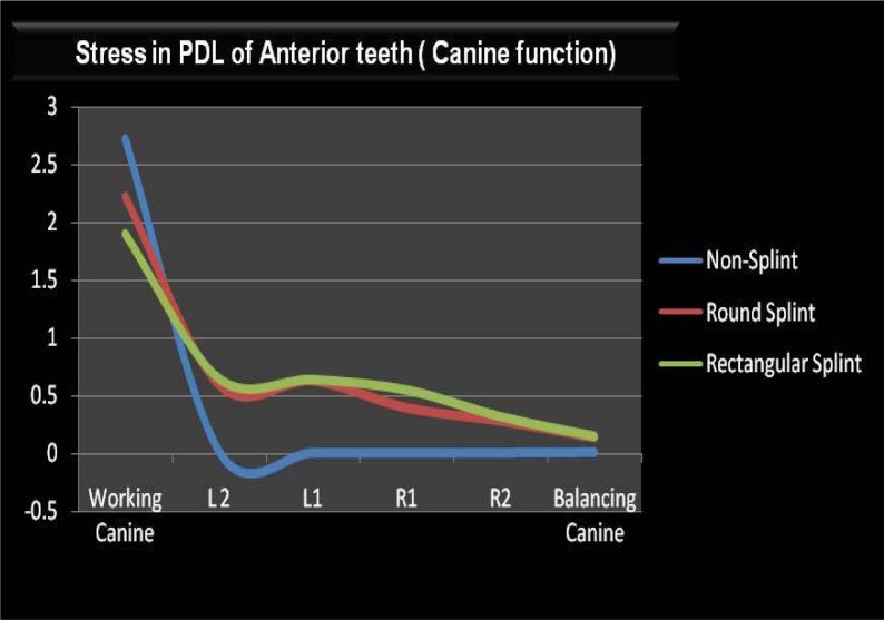
| Canine Function | Non-Splint | *2.7184 | 0.016 | 0.0032 | 0.0045 | 0.0038 | 0.00815 |
| Round Splint | 2.2245 | 0.594 | 0.6286 | 0.3938 | 0.2812 | 0.1362 | |
| Rectangular Splint | 1.9 | 0.649 | 0.64 | 0.55 | 0.32 | 0.1504 |
= The working side Canine
- Two teeth biting: The numerical findings are symmetrical to the midline in section “a” and “b”.
-
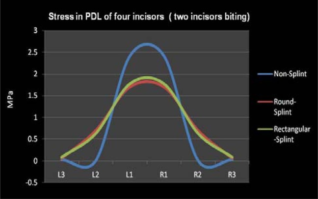
a1-Periodontal ligament stress in the non-splinted model (Table 2)In the non-splinted model the stresses are 2.42, 0.0108, and 0.0349 MPa for the central, lateral incisor and canine, respectively (Figure 2a).
-
a2-Periodontal ligament stress in the round wire splint:Findings are 1.7059 MPa for the central incisor which decreases to 0.6965 MPa in the lateral incisor and 0.05315 MPa in the canine (Figure 2a).
-
a3-Periodontal ligament stress in the rectangular wire splint:This splint caused 1.7711 MPa stress in the central incisor, 0.6261 MPa in the lateral incisor and 0.082 MPa stress in the canine (Figure 2a).
-
- Four teeth biting:
-
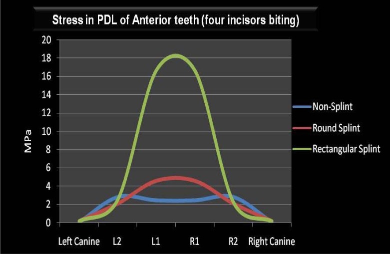
b1-Periodontal ligament stress in the non-splint model (Table 2)In the non-splint model, the stresses are 2.42, 2.78 and 0.06248 MPa for the central, lateral incisor and canine, respectively (Figure 2b).
-
b2-Periodontal ligament stress in the round wire splint:Findings are 4.57MPa for the central incisor which decreases to 2.075 MPa in the lateral incisor and 0.0169 MPa in the canine (Figure 2b).
-
b3-Periodontal ligament stress in the rectangular wire splint:This splint caused 16.66 MPa stress in the central incisor, 2.53 MPa in the lateral incisor and 0.189 MPa stress in the canine (Figure 2b).
-
- Canine function (in latero-trusive movement)
-
c1-Periodontal ligament stress in the non-splint model (Table 2)In the non-splint model, the stresses started with 2.7182 MPa on the working-side canine, decreasing to 0.016 MPa on the working-side lateral incisor and 0.0032 MPa on the working-side central incisor, and 0.0045 MPa, 0.0038 MPa, and 0.00815 MPa on the balancing-side central incisor through balancing side canine, respectively (Figure 2c).
-
c2-Periodontal ligament stress in the round wire splint:In the round wire splint model, the working-side canine stress was 2.2245 MPa, decreasing to 0.594 MPa on the working-side lateral incisor, 0.6286 MPa on the working-side central incisor, and 0.3938 MPa, 0.2812 MPa, and 0.1362 MPa on the balancing side central incisor through balancing side canine, respectively (Figure 2c).
-
c3-Periodontal ligament stress in the rectangular wire splint:In the rectangular splint model, the stress started with 1.9 MPa on the working-side canine, decreasing to 0.649 MPa on the working-side lateral incisor, 0.64 MPa on the working-side central incisor, and 0.55 MPa, 0.32 MPa, and 0.1504 MPa on the balancing-side central incisor through balancing-side canine, respectively. (Figure 2c)
-
Fig 2a.
Stress findings in four-incisor biting
Fig 2b.
Stress findings in two-incisor biting
Fig 2c.
Stress findings in lateral movement of the mandible
Displacement:
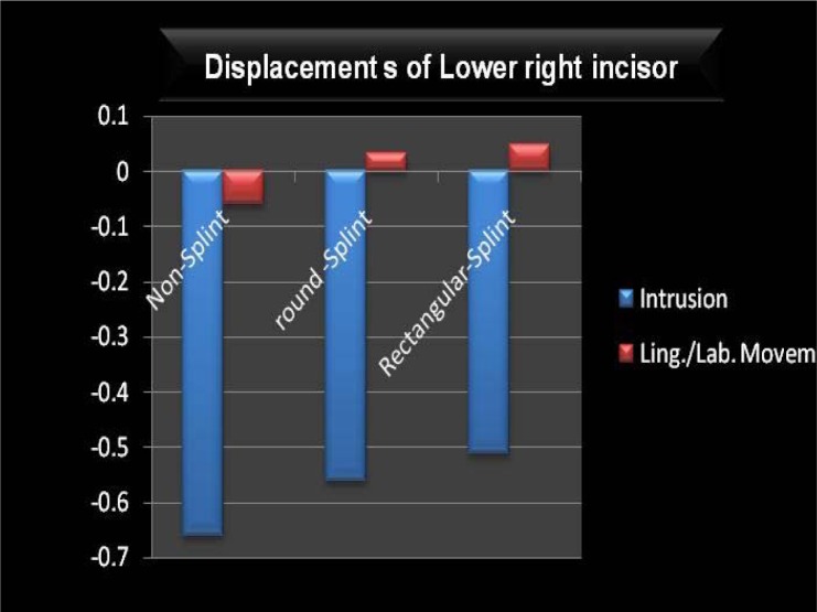
The displacements were derived from the mesio-inciso-labial point angle of the lower right incisor (Figure 3).
Fig 3.
Displacement findings in mesio-incisal line angle of the lower right incisor
Non-splint model:
The central incisor moved lingually (−0.059 mm) with intrusion (−0.659 mm).
Round wire splint:
Displacements were labial (0.0337 mm) with intrusion (−0.56 mm).
Rectangular wire splint:
In this splint, displacements were labial (0.049 mm) with intrusion (−0.511 mm).
DISCUSSION
Several studies have focused on the effect of various wire types and sizes in fixed retainers and recently fiber reinforced materials have been used widely [40–42].
Fixed retainers are advantageous in many ways, most important of which is to compensate for the lack of patient cooperation. The recommendation of this kind of retainer is based on this advantage. In the recent decade, benefit of fixed retainers in relapse control is well-known for orthodontists [43–45]. The main question to be answered at this phase is whether this retainer type has biomechanical disadvantages or not. There are benefits in its use for retention discipline, but never have its biomechanical aspects been assessed numerically.
When a large piece of food is bitten (by four incisors), splinting cannot be of benefit from the stress distribution point of view. In this situation, splinting increased the stress on the central incisors by 88% in round wire and 688% in rectangular wire splints which is against their primary defined role. This can be assumed as a disadvantage of splinting in the retention phase. Splinting may be considered advantageous when a small piece of food is bitten (by two incisors) that decreases PDL stress similarly with round and rectangular wires (29.5%).
On the other hand, in mandibular lateral movement, there are certain disadvantages in splinting teeth together. A decrease (18.5% in round wire and 30% in rectangular wire) in canine stress and an increase in stress (an average of 57 times in round wire splint and 67 times in rectangular wire splint) over the other teeth were noticed.
These effects need to be assessed in long time duration. The only judgment at this phase is to inform a modification in the stress state of the teeth in mandibular lateral movement. It seems necessary, as mentioned earlier, to design a long term study to clarify the effects of this stress modification. The displacement pattern of the central incisor was also shown to be modified after splinting. To avoid lengthening the results, the displacement of the incisors in a two-incisor biting pattern was reported. Decreasing the amount of intrusion of the anterior teeth after splinting can protect it. An interesting finding in displacement pattern was a change of the displacement direction from lingual in the non-splint model to a labial one in the splinted. It is obvious that initial displacement depends on the relation of the point of force application and the center of resistance of the teeth. A labial displacement of the incisors before splinting is expected when dealing with an increased incisal mandibular plane angle (IMPA). Lack of a telescopic movement in wire/composite connection can cause this modification. Although stress redistribution is favorable, the displacements warn us about retaining the splint for a long duration in patients with reduced labial bone thickness.
A clinical trial regarding the health of periodontal tissues before and after splinting while including teeth inclination can provide additional information about advantages and/or disadvantages of splinting teeth after orthodontic treatment. Splinting teeth together after the termination of an orthodontic treatment has two different aspects to be noticed; one is preventing teeth from unwanted movements after the end of orthodontic treatment (a positive role which is provided somewhat acceptably) and the second one is to modify the PDL stress distribution in various functions (which needs further studies to clarify its effects).
CONCLUSION
According to the results of this FEM study, the orthodontic patients with splint in their retention period will benefit from the stress redistribution provided by the splint in biting small food particles. This benefit was not detected in biting with four incisors. Round wire was shown to bring almost the same results as a rectangular wire. The results needed further clinical studies to be completely interpreted in canine function.
Fig 1b.
The round wire splint model
Acknowledgments
The authors appreciate the cooperation and assistance of the dental research center, Tehran University of Medical Sciences.
REFERENCES
- 1.Reidel RA. A review of the retention problem. Angle Orthod. 1960 Oct;30:179–99. doi: 10.1043/0003-3219(1960)030<0179:AROTRP>2.0.CO;2. [DOI] [PubMed] [Google Scholar]
- 2.Nanda RS, Nanda SK. Considerations of dentofacial growth in long-term retention and stability: is active retention needed? Am J Orthod Dentofacial Orthop. 1992 Apr;101(4):297–302. doi: 10.1016/S0889-5406(05)80321-X. [DOI] [PubMed] [Google Scholar]
- 3.Angle EH. Malocclusion of the teeth. 7th ed. Philadelphia: White Dental Manufacturing; 1907. [Google Scholar]
- 4.Proffit WR, Fields HW, Sarver DM. Contemporary orthodontics. 4th ed. Philadelphia: Mosby; 2007. Ch 11. [Google Scholar]
- 5.Kneirim RW. Invisible lower cuspid to cuspid retainer. Angle Orthod. 1973 Apr;43(2):218–20. doi: 10.1043/0003-3219(1973)043<0218:ILCTCR>2.0.CO;2. [DOI] [PubMed] [Google Scholar]
- 6.Little RM, Riedel RA, Artun J. An evaluatuin of changes in mandibular anterior alignment from 10 to 20 years post-retention. Am J Orthod Dentofacial Orthop. 1988 May;93(5):423–8. doi: 10.1016/0889-5406(88)90102-3. [DOI] [PubMed] [Google Scholar]
- 7.Orchin JD. Permanent lingual bonded retainer. J Clin Orthod. 1990 Apr;24(4):229–31. [PubMed] [Google Scholar]
- 8.Keim RG, Gottlieb EL, Nelson AH, Vogels DS., 3rd 2002 JCO study of orthodontic diagnosis and and treatment procedures. Part I. Results and trends. J Clin Orthod. 2002 Oct;36(10):553–68. [PubMed] [Google Scholar]
- 9.Rubenstein BM. A direct bond maxillary retainer. J Clin Orthod. 1976 Jan;10(1):43. [PubMed] [Google Scholar]
- 10.Carter RN. Simplified direct-bonded retainer. J Clin Orthod. 1978 Mar;12(3):221. [PubMed] [Google Scholar]
- 11.Lubit EC. The bonded lingual retainer. J Clin Orthod. 1979 May;13(5):311–3. [PubMed] [Google Scholar]
- 12.Lee RT. The lower incisor bonded retainer in clinical practice: a three year study. Br J Orthod. 1981 Jan;Apr;871(1)(4):15–8. 440–8. doi: 10.1179/bjo.8.1.15. [DOI] [PubMed] [Google Scholar]
- 13.Zachrisson BU. Clinical experience with direct-bonded retainers. Am J Orthod. 1977 doi: 10.1016/0002-9416(77)90247-0. [DOI] [PubMed] [Google Scholar]
- 14.Zachrisson BU. The bonded lingual retainers and multiples spacing of anterior teeth. Swed Dent J Suppl. 1982;15:247–55. [PubMed] [Google Scholar]
- 15.Eade P. A modified direct bond lingual retainer technique. Br J Orthod. 1980 Jul;7(3):125–6. doi: 10.1179/bjo.7.3.125. [DOI] [PubMed] [Google Scholar]
- 16.Bearn DR. Bonded orthodontic retainer: a review. Am J Orthod Dentofacial Orthop. 1995 Aug;108(2):207–13. doi: 10.1016/s0889-5406(95)70085-4. [DOI] [PubMed] [Google Scholar]
- 17.Diamond M. Resin fiberglass bonded retainer. J Clin Orthod. 1987 Mar;21(3):182–3. [PubMed] [Google Scholar]
- 18.Littlewood SJ, Millett DT, Doubleday B, Bearn DR, Worthington HV. Retention procedures for stabilizing tooth position after treatment with orthodontic braces. Cochrane Database Syst Rev. 2006 Jan 25;(1):CD002283. doi: 10.1002/14651858.CD002283.pub3. [DOI] [PubMed] [Google Scholar]
- 19.Greenfield DS, Nathanson D. Periodontal splinting with wire and composite resin. A revised approach. J Periodontal. 1980 Aug;51(8):465–8. doi: 10.1902/jop.1980.51.8.465. [DOI] [PubMed] [Google Scholar]
- 20.Ciancio SG, Nisengard RJ. Resins in periodontal splinting. Dent Clin North Am. 1975 Apr;19(2):235–42. [PubMed] [Google Scholar]
- 21.Stoller NH, Green PA. A comparison of a composite restorative material and wire ligation as methods of stabilizing excessively mobile mandibular anterior teeth. J Periodontol. 1981 Aug;52(8):451–4. doi: 10.1902/jop.1981.52.8.451. [DOI] [PubMed] [Google Scholar]
- 22.Rosenberg S. A new method of stabilization of periodontally involved teeth. J Periodontol. 1980 Aug;51(8):469–73. doi: 10.1902/jop.1980.51.8.469. [DOI] [PubMed] [Google Scholar]
- 23.Saravanamuttu R. Post-orthodontic splinting of periodontally-involved teeth. Br J Orthod. 1990 Feb;17(1):29–32. doi: 10.1179/bjo.17.1.29. [DOI] [PubMed] [Google Scholar]
- 24.Gaare D, Rolla G, Aryadi FJ, van der Ouderaa F. Improvement of gingival health by tooth brushing in individuals with large amounts of calculus. J Clin Periodontol. 1990 Jan;17(1):38–41. doi: 10.1111/j.1600-051x.1990.tb01045.x. [DOI] [PubMed] [Google Scholar]
- 25.White DJ. Dental calculus: recent insights into occurrence formation, prevention, removal and oral health effects of supragingival and subgingival deposits. Eur J Oral Sci. 1997 Oct;105(5 Pt 2):508–22. doi: 10.1111/j.1600-0722.1997.tb00238.x. [DOI] [PubMed] [Google Scholar]
- 26.Heier EE, De Smit AA, Wijgaerts IA, Adriaens PA. Periodontal implications of bonded versus removable retainers. Am J Orthod Dentofacial Orthod. 1997 Dec;112(6):607–16. doi: 10.1016/s0889-5406(97)70225-7. [DOI] [PubMed] [Google Scholar]
- 27.Zachrisson BU. Clinical implications of recent orthodontic-periodontic research findings. Semin Orthod. 1996 Mar;2(1):4–12. doi: 10.1016/s1073-8746(96)80034-x. [DOI] [PubMed] [Google Scholar]
- 28.Artun J. Caries and periodontal reactions associated with long-term use of different types of bonded lingual retainers. Am J Orthod. 1984 Aug;86(2):112–8. doi: 10.1016/0002-9416(84)90302-6. [DOI] [PubMed] [Google Scholar]
- 29.Geramy A. Alveolar bone resorption and the center of resistance modification (3-D analysis by means of the finite element method) Am J Orthod Dentofacial Orthop. 2000 Apr;117(4):399–405. doi: 10.1016/s0889-5406(00)70159-4. [DOI] [PubMed] [Google Scholar]
- 30.Geramy A. Stresses around a miniscrew. 3D analysis with the finite element method (FEM) Aust Orthod J. 2009 Nov;25(2):104–9. [PubMed] [Google Scholar]
- 31.Geramy A. Optimization of unilateral overjet management: three dimensional analysis by the finite element method. Angle Orthod. 2002 Dec;75(6):585–92. doi: 10.1043/0003-3219(2002)072<0585:OOUOMT>2.0.CO;2. [DOI] [PubMed] [Google Scholar]
- 32.Geramy A, Morgano SM. Finite element analysis of three designs of an implant-supported molar crown. J Prosthetic Dent. 2004 Nov;92(5):434–40. doi: 10.1016/j.prosdent.2004.08.011. [DOI] [PubMed] [Google Scholar]
- 33.Geramy A, Ghadirian H. Comparison of methods used to correct a lingually tilted mandibular molar: 3-D analysis using the finite element method (FEM) Aust Orthod J. 2008 Nov;24(2):96–101. [PubMed] [Google Scholar]
- 34.Geramy A, Faghihi S. Secondary trauma from occlusion: three dimensional analysis using the finite element method. Quintessence Int. 2004 Nov-Dec;35(10):835–43. [PubMed] [Google Scholar]
- 35.Hassan Ahangari A, Geramy A, Valian A. Ferrule design and stress distribution in endodontically treated upper central incisors: 3D finite element analysis. J Dent Tehran Uni Med Sci. 2008;5(3):105–10. [Google Scholar]
- 36.Geramy A. Apical third morphology and Intrusion force application: 3D finite element analysis. J Dent Tehran Uni Med Sci. 2007;4(3):130–4. [Google Scholar]
- 37.Geramy A, Ommati-Shabestary GH, Eghlima L. Influence of the angle of cervical convergence on stresses to the PDL of abutments: A 3D analysis using finite element method. J Dent Tehran Uni Med Sci. 2007;4(1):15–20. [Google Scholar]
- 38.Hsu ML, Chen FC, Kao HC, Cheng CK. Influence of off-axis loading of an anterior maxillary implant: a 3-dimensional finite element analysis. Int J Oral Maxillofac Implants. 2007 Mar;22(2):301–9. [PubMed] [Google Scholar]
- 39.Clelland NL, Lee JK, Bimbenet OC, Brantley WA. A three-dimensional finite element stress analysis of angled abutments for an implant placed in the anterior maxilla. J Prosthodont. 1995 Jun;4(2):95–100. doi: 10.1111/j.1532-849x.1995.tb00323.x. [DOI] [PubMed] [Google Scholar]
- 40.Artun J, Zachrisson B. Improving the handling properties of a composite resin for direct bonding. Am J Orthod. 1982 Apr;81(4):269–76. doi: 10.1016/0002-9416(82)90212-3. [DOI] [PubMed] [Google Scholar]
- 41.Artun J, Spadafora AT, Shapiro PA. A 3-year follow up study of various types of orthodontic canine-to-canine retainers. Eur J Orthod. 1997 Oct;19(5):501–9. doi: 10.1093/ejo/19.5.501. [DOI] [PubMed] [Google Scholar]
- 42.Geserick M, Ball J, Wichelhaus A. Bonding fiber-reinforced lingual retainers with color-reactivating flowablecomposite. J Clin Orthod. 2004 Oct;38(10):560–2. [PubMed] [Google Scholar]
- 43.Renkema AM, Al-Assad S, Bronkhorst E, Weindel S, Katsaros C, Lisson JA. Effectiveness of lingual retainers bonded to the canines in preventing mandibular incisor relapse. Am J Orthod Dentofacial Orthop. 2008 Aug;134(2):179e1–8. doi: 10.1016/j.ajodo.2008.06.003. [DOI] [PubMed] [Google Scholar]
- 44.Cerny R. The reliability of bonded lingual retainers. Aust Orthod J. 2007 May;23(1):24–9. [PubMed] [Google Scholar]
- 45.Dahl EH, Zachrisson BU. Long-term experience with direct-bonded lingual retainers. J Clin Orthod. 1991 Oct;25(10):619–30. [PubMed] [Google Scholar]


