Abstract
Smile expresses a feeling of joy, success, sensuality, affection, and courtesy, and reveals self-confidence and kindness. The harmony of the smile is determined not only by the shape, the position, and the color of the teeth, but also by the gingival tissues. Although melanin pigmentation of the gingiva is completely benign and does not present a medical problem, complaints of “black gums” are common, particularly in patients having a very high smile line. Thus, perio-esthetic treatment modalities strive to achieve a harmonious inter-relationship of the pink with white, which is imperative of all treatment procedures. For depigmentation of gingival, different treatment modalities have been reported, such as bur abrasion, scraping, partial thickness flap, cryotherapy, electrosurgery, and laser. In the present case series, scraping, electrosurgery, and diode laser have been tried for depigmentation, which are simple, effective, and yield good results, along with good patient satisfaction.
KEY WORDS: Depigmentation, electrocautery, laser, repigmentation, scraping
“Esthetics” is the science of beauty, which is the particular detail of an animate or inanimate object that makes it appealing to the eye. Esthetic or cosmetic dentistry strives to merge function and beauty with the values and individual needs of every patient. These advances now allow the practitioner to achieve a periodontal environment that complements and enhances the creation of optimal dental esthetics. Gingival health and appearance are the essential components of an attractive smile. Oral melanin pigmentation is considered to be multifactorial, physiological/pathological.[1] Melanin, a brown pigment, is the most common natural pigment contributing to endogenous pigmentation of gingiva. It is a non–hemoglobin-derived pigment formed by cells called melonocytes which are dentritic cells of neuroectodermal origin in the basal and spinous layers.[2]
Gingival depigmentation is a periodontal plastic surgical procedure whereby the gingival hyperpigmentation is removed or reduced.[3] The first and foremost indication for depigmentation is patient demand for improved esthetics. Elimination of these melanotic areas can be done by scraping, free gingival autografting, cryosurgery, electrosurgery, and various types of lasers.[4] The selection of technique should be based on clinical experience and individual preferences.[2]
Selection Criteria
The cases were selected based on Dummett–Gupta Oral Pigmentation Index (DOPI): (Dummett 1971)[5]
No clinical pigmentation (pink gingiva)
Mild clinical pigmentation (mild light brown color)
Moderate clinical pigmentation (medium brown or mixed pink and brown)
Heavy clinical pigmentation (deep brown or bluish black).
The smile line classification (Liebart and Deruelle 2004)[6]
Class 1: Very high smile line – more than 2 mm of the marginal gingiva visible.
Class 2: High smile line – between 0 and 2 mm of the marginal gingiva visible.
Class 3: Average smile line – only gingival embrasures visible.
Class 4: Low smile line – gingival embrasures and cemento-enamel junction not visible.
The present case series describes three simple and effective surgical depigmentation techniques – scalpel, electrosurgery, and diode lasers. All these techniques have produced good results with patient satisfaction.
Case Reports
Case 1
A 20-year-old female patient complaining of heavily pigmented gums visited Department of Periodontics, JKK Nattraja Dental College and Hospital, Komarapalayam. On examination, DOPI score was 3 and the patient had a very high smile line that revealed the deeply pigmented gingiva from first premolar to first premolar and also had midline Diastema [Figure 1]. Considering the patient's concern, surgical depigmentation procedure was planned. The entire procedure was explained to the patient and written consent was obtained. Routine oral hygiene procedures were carried out and oral hygiene instructions were given. Local anesthesia was infiltrated in the maxillary anterior region from first premolar to first premolar. Using a number 15 Bard Parker blade, scrapping of the pigmented epithelium up to the level of the mucogingival junction was carried out, leaving the connective tissue intact [Figure 2]. After complete removal of the entire epithelium, abrasion with diamond bur was done to get the physiological contour of the gingiva. Bur was used with minimal pressure with feather light brushing strokes. A frenectomy was performed after the depigmentation was completed. A periodontal dressing (Coe-Pak) was placed on the surgical wound area for patient comfort and to protect it for 1 week. The patient was kept on analgesics for a period of 5 days and was advised to use 0.12% chlorhexidine gluconate mouthwash for 2 weeks postoperatively. During postoperative period, the wound healing was uneventful without any discomfort. Three months postoperative examination showed well-epithelialized gingiva, which was pink in color and pleasant, but with few sites showing remnants of pigmentation [Figure 3].
Figure 1.
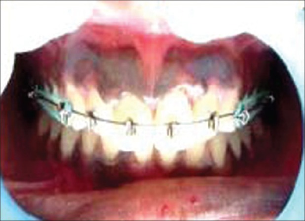
Preoperative view showing unsightly physiologic melanin pigmentation of upper labial gingiva
Figure 2.
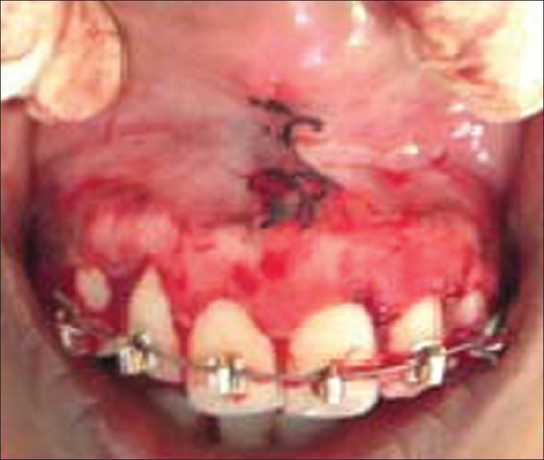
View immediately after depigmentation, No. 15 blade being used to remove the pigment layer and frenectomy done
Figure 3.
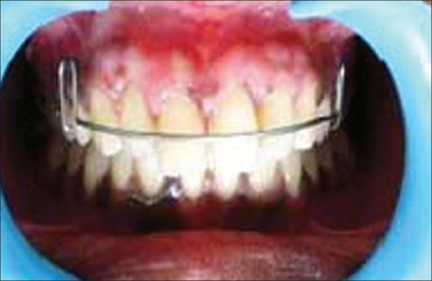
3 months postoperative view. Depigmented gingiva that is pink and healthy
Case 2
A young female patient aged 23 years visited the Department of Periodontics, JKK Nattraja Dental College and Hospital with the chief complaint of blackish gums which esthetically interfered with her smile. The DOPI score was 3 and revealed very high smile line [Figure 4]. Considering the patient's concern, depigmentation procedure was planned using electrocautery. A loop electrode was used for depigmentation of gingiva [Figure 5]. It was used in a light brushing strokes and the tip was kept in motion all the time to avoid excessive heat buildup and destruction of the tissues. Finally, perio-pack was placed over the wound area and oral hygiene instructions were given. The pack was removed after 1 week and the area debrided. Pigmentation was absent in the newly formed epithelium, with the gingiva appearing pale pink in color after a period of 3 months [Figure 6].
Figure 4.
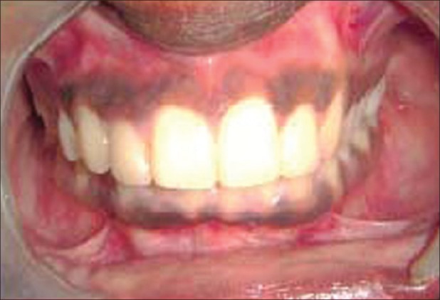
Preoperative view showing highly pigmented upper labial gingival
Figure 5.
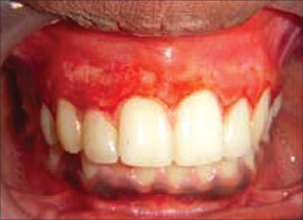
Immediate postoperative view, using electrocautery
Figure 6.
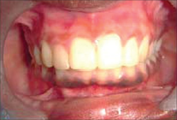
3 months postoperative view
Case 3
A 25-year-old female patient complaining of heavily pigmented gums visited Department of Periodontics, JKK Nattraja Dental College and Hospital, with a request for esthetic treatment of black gums. On examination, her DOPI score was 3 and the patient had a very high smile line [Figure 7]. The patient and the staffs were protected from laser by wearing manufacturer's spectacles. After adequate anesthesia was given, diode laser in contact mode was used. The ablation was operated using a hand piece with fiber optic filament, 320 μm in diameter set at 0.8 W. The procedure was performed in a contact mode in cervico-apical direction on all pigmented areas [Figure 8]. No pain or bleeding complications were observed during and after the procedure. One day postoperatively, it revealed no pain or discomfort [Figure 9]. The ablated wound healed completely in 1 week. Three months after the ablation, the gingiva was generalized pink in color and healthy in appearance [Figure 10].
Figure 7.
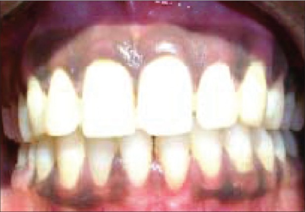
Preoperative picture of 25-year-old female complaining of black-colored gums
Figure 8.
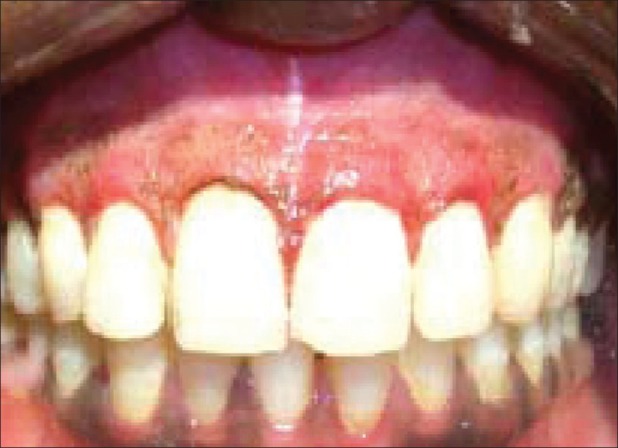
Immediate postoperative picture, diode laser used
Figure 9.
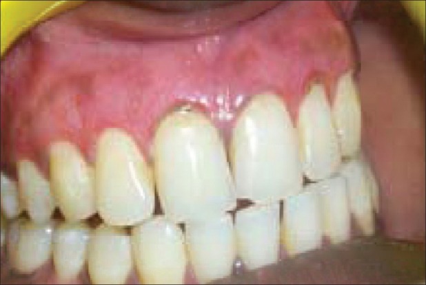
One day postoperative view
Figure 10.
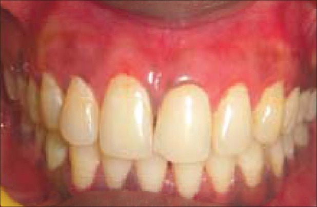
3 months postoperative picture showing healthy gingiva with no increase in the extent of repigmented areas
Discussion
Pigmented gingival tissue many a times forces the patients to seek cosmetic treatment.[4] Several treatment modalities have been suggested and presented in the literature, ranging from a simple scalpel method to sophisticated lasers.[7] According to Cicek (2003),[2] melanin pigmentation is caused by melanin deposition by active melanocytes located in the basal layer of oral epithelium.[8]
In the present case series, three techniques were selected for depigmentation, which included scalpel, electrosurgery, and laser. Following surgical procedure, patients were recalled at 1 and 3 months to evaluate recurrence of pigments. All the techniques produced successful results with good patient satisfaction.
The depigmentation procedure by scalpel technique is simple, easy to perform, noninvasive, and above all, cost effective. According to Almas and Sadiq (2002),[3] the scalpel wound heals faster than that in other techniques. In our study also, the healing by scalpel was faster than that by cautery and laser. But scalpel surgery causes unpleasant bleeding during and after the operation. It is also necessary to cover the exposed lamina propria with periodontal dressing for 7–10 days.[9]
The superior efficiency of electrosurgery than scalpel seen in the present case series could be explained based on Oringer's (1975)[10] “exploding cell theory.” According to the theory, it is predicted that the electrical energy leads to molecular disintegration of melanin cells present in basal and suprabasal cell layers of operated and surrounding sites. Thus, electrosurgery has a strong influence in retarding migration of melanin cells from the locally situated cells, which were detected clinically to be removed. Cicek (2003)[2] reported that there is no bleeding and there is minimal patient discomfort while using electrocautery. But electrosurgery also has its own limitations in that its repeated and prolonged use induces heat accumulation and undesired tissue destruction.[10]
Another effective treatment modality employed in the present case series is the depigmentation by diode laser. Here, radiation energy is transformed into ablation energy, resulting in cellular rupture and vaporization with minimal heating of the surrounding tissue.[11] According to Atsawasuwan and Greethong (1999),[12] laser beam produces bloodless field for surgery, causes minimum damage to the periosteum and underlying bone, and the treated gingiva and mucosa do not need any dressing. This has the advantages of easy handling, short treatment time, hemostasis, and decontamination and sterilization effects. But this approach needs expensive and sophisticated equipment, which makes the treatment very expensive.[12] Laser beam even destroys the epithelial cells including those at the basal layer, and hence reduces repigmentation.[13] Thus, repigmentation was minimal and patient compliance was much better in this case series while using lasers with than other techniques.
Pigment recurrence has been documented to occur following the surgical procedure, within 24 days to 8-year long period. A study by Perlmutter et al. (1986)[14] showed that gingival surgical procedures performed solely for cosmetic reasons offer no permanent results. Repigmentation refers to the clinical reappearance of melanin pigment following a period of clinical depigmentation. The mechanism suggested for the spontaneous repigmentation is that the melanocytes from the normal skin proliferate and migrate into the depigmented areas.[14]
Conclusion
The growing esthetic concern requires the removal of unsightly pigmented gingival areas to create a pleasant and confident smile, which altogether may alter the personality of an individual. This could be easily attained by using any of the above three methods used in this case series. But the repigmentation seems to be less using laser and cautery. The methods used here produced desired results, and above all, the patients were satisfied with the outcome, which is the ultimate goal of any therapy that is carried out.
Footnotes
Source of Support: Nil
Conflict of Interest: None declared.
References
- 1.Prasad D, Sunil S, Mishra R, Sheshadri Treatment of gingival pigmentation: A case series. Indian J Dent Res. 2005;16:171–6. doi: 10.4103/0970-9290.29901. [DOI] [PubMed] [Google Scholar]
- 2.Cicek Y, Ertas U. The normal and pathological pigmentation of oral mucous membrane: A review. J Contemp Dent Pract. 2003;4:76–86. [PubMed] [Google Scholar]
- 3.Almas K, Sadiq W. Surgical Treatment of Melanin-Pigmented Gingiva: An Esthetic Approach. Indian J Dent Res. 2002;13:70–3. [PubMed] [Google Scholar]
- 4.Deepak P, Sunil S, Mishra R, Sheshadri Treatment of gingival pigmentation: A case series. Indian J Dent Res. 2005;16:171–6. doi: 10.4103/0970-9290.29901. [DOI] [PubMed] [Google Scholar]
- 5.Dummet CO, Barens G. Oromucosal pigmentation: An updated literary review. J Periodontol. 1971;42:726–36. doi: 10.1902/jop.1971.42.11.726. [DOI] [PubMed] [Google Scholar]
- 6.Liebart ME, Deruelle CF, Santini A, Dillier FL, Corti VM. Smile line and periodontium visibility. Perio. 2004;1:17–25. [Google Scholar]
- 7.Prasad SS, Neeraj A, Reddy NR. Gingival depigmentation: A case report. People's. J Science Research. 2010;3:27–9. [Google Scholar]
- 8.Kathariya R, Pradeep AR. Split mouth de-epithelization techniques for gingival depigmentation: A case series and review of literature. J Indian Soc Periodontol. 2011;15:161–8. doi: 10.4103/0972-124X.84387. [DOI] [PMC free article] [PubMed] [Google Scholar]
- 9.Pal TK, Kapoor KK, Parel CC, Mukharjee K. Gingival melanin pigmentation: A study on its removal for esthetics. J Indian Soc Periodontol. 1994;3:52–4. [Google Scholar]
- 10.Oringer MJ, editor. Electrosurgery in Dentistry. 2nd ed. Philadelphia: W.B. Saunders Co; 1975. [Google Scholar]
- 11.Ishikawa I, Aoki A, Takasaki AA. Potential application of Erbium: YAG laser in periodontics. J Periodontol Res. 2004;39:275–85. doi: 10.1111/j.1600-0765.2004.00738.x. [DOI] [PubMed] [Google Scholar]
- 12.Atsawasuwan P, Greethong K, Nimmanon V. Treatment of gingival hyperpigmentation for esthetic purposes by Nd: YAG laser: Report of 4 cases. J Periodontol. 2000;71:315–21. doi: 10.1902/jop.2000.71.2.315. [DOI] [PubMed] [Google Scholar]
- 13.Tal H, Oegiesser D, Tal M. Gingival depigmentation by erbium: YAG laser: Clinical observations and patient responses. J Periodontol. 2003;74:1660–7. doi: 10.1902/jop.2003.74.11.1660. [DOI] [PubMed] [Google Scholar]
- 14.Perlmutter S, Tal H. Repigmentation of the gingiva following surgical injury. J Periodontol. 1986;57:48–50. doi: 10.1902/jop.1986.57.1.48. [DOI] [PubMed] [Google Scholar]


