Abstract
A double left anterior descending (LAD) coronary artery emerging from the left and right coronary arteries is classified among rare coronary anomalies. We herein report a 73-year-old man presenting with acute coronary syndrome (posterolateral myocardial infarction). He was admitted with typical chest pain, and due to his progressive ischemic changes on electrocardiography (ECG) and elevated cardiac enzyme, he was candidated for cardiac catheterization. The coronary angiography revealed an anomalous LAD from the right sinus of Valsalva. The unusual coronary anatomy was perfectly matched with the distribution of ischemia and its clinical evidence on echocardiography and ECG. The culprit lesion was stented, and the patient was discharged in good physical condition from the hospital.
Keywords: Coronary vessel anomalies, Myocardial infarction, Acute coronary syndrome
Introduction
Coronary anomalies are reported in 1 to 5% of coronary angiographic examinations; they are usually asymptomatic, and are discovered accidentally.1 An anomalous origin of the left anterior descending artery (LAD) from both right and left sinus of Valsalva is considered one of the rarest anomalies.2–7 The following report describes a patient presenting with an acute coronary syndrome, whose coronary angiography revealed an unusual course of the LAD from the right and left coronary arteries.
Case Report
A 73-year-old man was admitted to our emergency department with chest pain. As he stated, he had been suffering from pain without radiation for the past 30 minutes, accompanied by nausea, vomiting, and diaphoresis. He claimed that he had had a previous similar episode the day before his admission, but the pain had resolved spontaneously after about one hour. He had a history of cigarette smoking, hypertension, and respiratory disease, for which he had received no appropriate and regular medical care. He had used Atenolol for the last three months in an irregular manner. No abnormal findings were detected on his physical examination (blood pressure = 140/80 mmHg, heart rate = 60 bpm, and normal breathing and heart sounds). His initial electrocardiogram (ECG) showed a left-axis deviation and left anterior hemiblock with ischemic changes (inverted T wave) on its pericardial leads. He had elevated cardiac troponin, and no other significant abnormalities were detected in his blood tests. He underwent medical management for acute coronary syndrome, and his pain was reduced within his first hour of admission. Echocardiography was performed for the patient and revealed a moderate left ventricular (LV) dysfunction with an LV ejection fraction of 40% and anterior and lateral wall hypokinesia. Mild LV hypertrophy was also detected. The patient was scheduled for coronary angiography on the following day. No chest pain or other symptoms were reported during the first night. The next morning, the ECG showed the evidence of a posterior (lateral) myocardial infarction (MI).
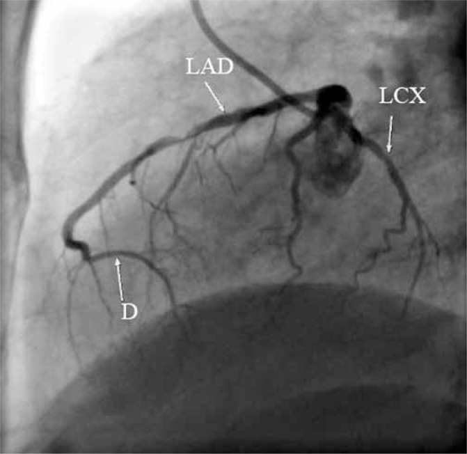
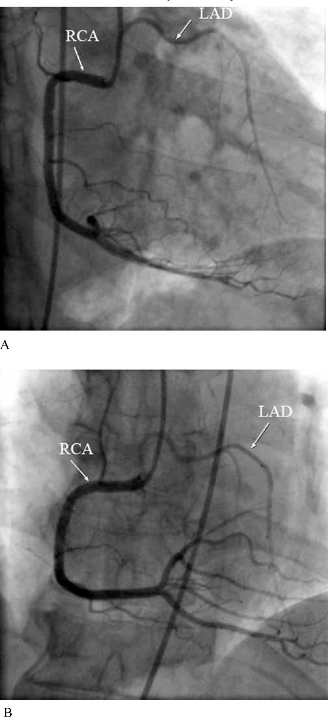
The patient underwent cardiac catheterization via the standard Judkins techniques. The selective angiography of the left coronary artery showed a critical and thrombotic stenosis on the midportion of the LAD as well as a moderate lesion on the midportion of the left circumflex (LCx) artery (Figure 1). Mild stenoses were also detected at the proximal end of the LAD and high obtuse marginatus. The LAD ran through the interventricular groove but at the end of the midportion, it deviated from its usual course and transformed to a diagonal and supplied the anterolateral area of the LV. On the right side, the selective coronary artery injection revealed a dominant right coronary artery (RCA) and also an anomalous coronary artery arising just after the origin the RCA (Figure 2). The accessory vessel ran into the left side and coursed into the mid and distal portions of the anterior interventricular groove and supplied the apex area. There were small septal arteries separating from it. On the left ventriculography, hypokinetic anterolateral and diaphragmatic walls were seen. The culprit lesions on the LAD were stented with a drug-eluting stent (DES), and the patient was discharged from the hospital with an acceptable condition on the fourth day of his hospitalization.
Figure 1.
Selective coronary angiography at the straight lateral view, showing that the LAD terminated suddenly without reaching the apex LAD, Left anterior descending artery; LCX, Left circumflex artery; D, Diagonal artery
Figure 2.
Selective coronary angiography at the left anterior oblique (A) and anterior-posterior (B) views demonstrating the aberrant LAD LAD, Left anterior descending artery; RCA, Right coronary artery
Discussion
Different types of congenital coronary anomalies have been reported, most of them having been discovered incidentally through routine coronary angiography. A double LAD is among the rarest anomalies and its incidence ranges from 0.01 to 0.03% in different studies.5 In their classification, Spindola-Franko described four types of double LAD8: type I–III arises from the left side, and the last type (apparently the rarest) emerges from the right coronary sinus. Here, we present a patient whose coronary anatomy consisted of a double LAD type IV. He was admitted to our department with acute coronary syndrome, and his ECG showed evidence of anterior and posterior (lateral) MI. On selective coronary angiography, the LAD had a thrombotic lesion on its mid course and it turned toward the anterolateral region, which is usually supposed to be supplied by a diagonal. Probably the lesion at the midportion of the LAD temporarily blocked the blood flow in the posterolateral area and caused the MI. On the right side, the RCA was dominant, and an anomalous branch arose from the origin of the RCA and coursed to the left side through the distal interventricular groove. The septal arteries branching from the anomalous vessel and also its left-sided course proved its identity as the LAD. As was mentioned before, the last type of a double LAD has a very low incidence. More often than not, the anomaly is suspected by visualization of an “avascular area” in the distribution of the left coronary artery and the absence of collaterals.3 In the present report, the LAD changed its usual destination and ran into the posterolateral area; and the apex, which is usually supplied by the distal portion of the LAD, was supplied by the anomalous branch: the second LAD.
The anatomical and physiological understandings of coronary artery anomalies have both clinical and therapeutical applications. The relationship between an anomalous coronary artery, aorta, and pulmonary trunk and the possibility of the compression of the aberrant vessel by the two great arteries could be a source of different clinical presentations (i.e., angina, syncope, MI, and sudden cardiac death) and sometimes mandate surgical correction. Apart from the interarterial course, recent studies have shown that an intramural course could be identified in some anomalous coronary artery emerging from the opposite Valsalva sinus.9 The clinical impact of such entity remains unclear. Fortunately, the course of the anomalous branch usually could be detected by conventional angiography. The operator should always pay attention to the avascular region and search for a possible anomalous circulation. However, as Matsumoto et al. reported, sometimes the aberrant vessel cannot be found by coronary artery angiography and multi-detector-row computed tomography may be helpful.10 Beside the clinical consequences of anomalous vessels, knowing the anatomical variation is crucial at the time of surgery. Grave surgical complications may occur by cutting an aberrant vessel.3
In conclusion, we presented a rare anatomical variation of the LAD, known as a double LAD type IV. The LAD arose aberrantly from the right sinus of Valsalva and supplied the area abandoned by the main LAD. Operators should always be aware of an anomalous circulation and seek to define its anatomical and clinical roles.
Acknowledgments
The authors wish to acknowledge Dr. Melody Farrashi for English editing.
References
- 1.Angelini P, Velasco JA, Flamm S. Coronary anomalies: incidence, pathophysiology, and clinical relevance. Circulation. 2002;105:2449–2454. doi: 10.1161/01.cir.0000016175.49835.57. [DOI] [PubMed] [Google Scholar]
- 2.Turhan H, Atak R, Erbay AR, Senen K, Yetkin E. Double left anterior descending coronary artery arising from the left and right coronary arteries: a rare congenital coronary artery anomaly. Heart Vessels. 2004;19:196–198. doi: 10.1007/s00380-003-0747-3. [DOI] [PubMed] [Google Scholar]
- 3.Kosar F. An unusual case of double anterior descending artery originating from the left and right coronary arteries. Heart Vessels. 2006;21:385–387. doi: 10.1007/s00380-006-0916-2. [DOI] [PubMed] [Google Scholar]
- 4.Voudris V, Salachas A, Saounotsou M, Sionis D, Ifantis G, Margaris N, Koroxenidis G. Double left anterior descending artery originating from the left and right coronary artery: a rare coronary artery anomaly. Cathet Cardiovasc Diagn. 1993;30:45–47. doi: 10.1002/ccd.1810300112. [DOI] [PubMed] [Google Scholar]
- 5.Kunimoto S, Sato Y, Kunimasa T, Kasamaki Y, Takayama T, Matsumoto N, Kasama S, Yoda S, Saito S, Hirayama A. Double left anterior descending artery arising from the left and right coronary arteries: depiction at multidetector-row computed tomography. Int J Cardiol. 2009;20:e54–56. doi: 10.1016/j.ijcard.2007.08.019. 132. [DOI] [PubMed] [Google Scholar]
- 6.Tuncer C, Batyraliev T, Yilmaz R, Gokce M, Eryonucu B, Koroglu S. Origin and distribution anomalies of the left anterior descending artery in 70,850 adult patients: multicenter data collection. Catheter Cardiovasc Interv. 2006;68:574–585. doi: 10.1002/ccd.20858. [DOI] [PubMed] [Google Scholar]
- 7.Yilmaz A, Yalta K, Turgut OO, Yilmaz MB, Karadas F, Erdem A, Ozyol A, Tandogan I. The left anterior descending artery arising from the right sinus of Valsalva: a case report. Adv Ther. 2007;24:178–181. doi: 10.1007/BF02850006. [DOI] [PubMed] [Google Scholar]
- 8.Spindola-Franko H, Grose R, Solomon M. Dual left anterior descending coronary artery: angiographic description of important anatomic variants and surgical implications. Am Heart J. 1983;105:445–455. doi: 10.1016/0002-8703(83)90363-0. [DOI] [PubMed] [Google Scholar]
- 9.Angelini P, Walmsley RP, Libreros A, Ott DA. Symptomatic anomalous origination of left coronary artery from the opposite sinus of valsalva: clinical presentations, diagnosis, and surgical repair. Tex Heart Inst J. 2006;33:171–179. [PMC free article] [PubMed] [Google Scholar]
- 10.Matsumoto M, Yokoyama K, Yahagi T, Kikushima K, Watanabe K, Tani S, Anazawa T, Kawamata H, Nagao K, Hirayama A. Double left anterior descending artery arising from right and left sinus of Va lsalva in patient with acute coronary syndrome. Int J Cardiol. 2011;149:e40–42. doi: 10.1016/j.ijcard.2009.03.049. [DOI] [PubMed] [Google Scholar]


