Abstract
Water motion probed by intrinsic tryptophan shows the significant slowdown around protein surfaces but it is unknown how the ultrafast internal conversion of two nearly degenerate states of Trp (1La and 1Lb) affects the initial hydration in proteins. Here, we used a mini-protein with ten different tryptophan locations one at a time through site-directed mutagenesis and extensively characterized the conversion dynamics of the two states. We observed all the conversion time scales in 40-80 fs by measurement of their anisotropy dynamics. This result is significant and shows no noticeable effect on the initial observed hydration dynamics and unambiguously validates the slowdown of hydration layer dynamics as shown here again in two mutant proteins.
Tryptophan (Trp or W) has been developed as a powerful optical probe to study protein hydration dynamics1-5 with site-directed mutagenesis.3-5 The recent series of characterizations of hydration dynamics on various proteins showed the slowdown of the hydrating water motions near protein surfaces.6-11 The obvious evidence is that at the blue side of the emission such as at 305 or 310 nm, the femtosecond-resolved fluorescence transients significantly slow down compared with that at the same wavelength in bulk water. It has been suspected that the complexity of excited states (1La and 1Lb) may smear the initial ultrafast decay dynamics at the blue-side emission in proteins. The 1La (S2) state in polar environments lies below the 1Lb (S1) state due to its larger static dipole moment.12 Ultrafast internal conversion through conical intersection (CI) was proposed from the higher 1Lb to lower 1La state13-15 and observed to occur in ~40 fs in bulk water.16,17 The internal conversion from 1La to 1Lb in gas phase (or in vacuum) has also been observed in 20-100 fs.18 Typically, when we use 290 nm to excite tryptophan in proteins, both states are excited simultaneously.19 Thus, one critical question is what time scales of the CI dynamics of 1Lb to 1La in proteins are and related to this, how this dynamics affects the initial protein hydration.
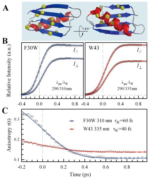
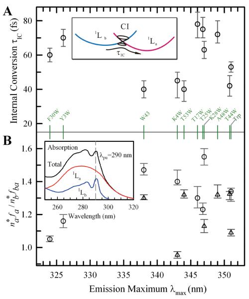
To resolve this critical issue, we scanned the GB1 protein (the B1 immunoglobin-binding domain of protein G) by placing Trp to different positions one at a time with site-directed mutagenesis (Fig. 1A). GB1 is a small domain protein with 56 amino-acid residues containing only one single tryptophan residue (W43).20 Nine mutant proteins were made from double mutation by first replacing W43 to F43. Since the CI dynamics is ultrafast and the absorption of 1Lb and 1La is overlapped, we examined the fluorescence anisotropy dynamics after initial excitation to understand the CI dynamics and extract their time scales. Due to the nearly perpendicular transition dipoles of the two states,12a we should observe the ultrafast change of anisotropy. Upon 290-nm fs-pulse excitation, we actually prepared a coherent superposition of nearly degenerate 1La and 1Lb states. The evolution of anisotropy with time for such coherent states has been well studied by Wynne and Hochstrasser, especially for symmetric molecules.21 With the ultrafast conversion by CI for a coherent superposition state, the Jonas group has recently carried out a series of theoretical and experimental studies to understand the molecular mechanism.22 However, the actual coupling between the dephasing and CI processes for tryptophan is unknown, we proposed two possible models to simulate our experimental results: One is called the sequential model, i.e., the coherent state decays to 1La and 1Lb states with the dephasing time T2 and then molecules in 1Lb are converted into 1La state by CI with the time τIC. The other model is a parallel one and both the dephasing and CI processes occur at the same time. The detailed kinetics for the two models are given in the Supporting Information. Surprisingly, both models give the similar CI times (τIC) for each mutant while the sequential model shows the dephasing times (T2) in 30-70 fs and the parallel model around 300 fs. However, the sequential model gives a better fit (see Fig. S1 in the SI). Figure 1B shows the typical fluorescence transients either at 310 or 335 nm with the two different polarization detections (parallel and perpendicular) for the mutant of F30W and wide-type W43. The solid lines are the fitting results using the sequential model (S9 and S10 equations in the SI) with the two state dipole ratio (μb/μa) of 0.455.23 Figure 1C is the resulting anisotropy dynamics with the solid fitting lines (S11 equation in the SI). We obtained the internal conversion time (by CI) of 60 fs for F30W and 40 fs for W43 and a similar dephasing time of 50 fs for both proteins. Due to the limited temporal resolution of 360-400 fs determined by the water Raman signal at 320 nm, the initial anisotropy value is not very high (not 0.6-0.7 as expected for a coherent superposition of two nearly degenerate states) and drops to 0.2-0.35. In Fig. 1C, the anisotropy promptly decays to a constant value on such ultrafast time window and this value is directly related to , i.e., proportional to (S13 equation in the SI). and are the initially excited populations in 1La and 1Lb states, respectively, which are directly proportional to their extinction coefficients. The constants fa and fba (fa / fba = β1) are relative emission coefficients, at a given wavelength, of the initially excited 1La state and the 1La state that is transferred from the 1Lb state through CI, respectively. By fitting both the transients and anisotropy dynamics, we obtained the CI dynamics of tryptophan in the ten proteins and the related initial absorption coefficients of 1La and 1Lb states at 290 nm. Figure 2A shows the obtained CI time scales of the wide type and nine mutants and Figure 2B basically gives the ratio of initial excited 1La and 1Lb populations (if fa = fba), i.e., the relative absorption coefficients of the two states at 290 nm. Significantly, all CI dynamics (insert in Fig. 2A) occur in 40-80 fs in all the mutant proteins and are independent on the emission maximum, i.e., local environment total polarity. The CI dynamics can vary by a factor of 2 but are all less than 100 fs, within several vibrational periods, similar to the values observed in gas phase18 and bulk water.16 The theoretical calculations showed single/double-bond rearrangements and out-of-plane molecular distortions responsible for the CI process14 and thus these structural changes seem not to be affected by the local physical constraints due to the small amplitude motions during CI. The CI dynamics could be mainly affected by the relative energy levels of 1La and 1Lb states at t=0 which are determined by the local electrostatics of initial configurations upon excitation. Thus, due to no obvious trend of τIC with emission maxima (Fig. 2A), the initial energy-level ordering of 1La states determined by the ground-state equilibrium configurations in the ten proteins at t=0 can be different from the final energy-level ordering after environment relaxation (solvation) which determines the emission maxima of the proteins, as shown in the inset of Figure 3A, reflecting the different stabilizations of the excited state by the equilibrated ground-state (t=0) and excited-state (t=∞) configurations. The dephasing times (T2) of all ten proteins are also similar in 30-70 fs.
Figure 1.
(A) Structure of the native state of GB1 with 9 designed tryptophan mutants and the wild type (W43) labeled by yellow balls. (B) Femtosecond-resolved parallel (I∥) and perpendicular (I⊥) fluorescence transients of mutant F30W gated at 310 nm (left, blue) and wild-type W43 gated at 335 nm (right, red). (C) Corresponding anisotropies of the two mutants in (B). The symbols (squares and triangles) are the experimental data and the solid lines are the best simulations from our model.
Figure 2.
(A) Derived internal conversion time scales (τIC) of all the nine mutants and the wild type of GB1 as well as Trp in bulk water with respect to the emission maxima. The inset shows a sketch of conical intersection (CI) between 1La and 1Lb states of Trp. (B) Distributions of the fitting parameter for 10 GB1 proteins and free Trp in water. Circles and triangles represent the results from 310 and 335-nm measurements, respectively. The inset shows the total absorption spectrum of Trp with deconvolution of relative contributions of 1La and 1Lb states from ref. 19. All mutants are shown in the middle with green ticks corresponding to the data points.
Figure 3.
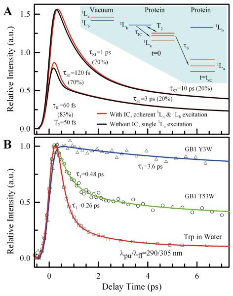
(A) Comparison of simulated transients with and without IC in short time range. With IC, the dephasing dynamics is also included. The solvation parameters used in the simulation are shown beside the transients. The inset shows the schematic, relative energy levels of 1La and 1Lb states in proteins before (t=0) and after (t=tsc) solvation, as well as in vacuum. At t=0, three dynamics, dephasing (T2), CI (τIC) and solvation (τS) start to occur. (B) Normalized femtosecond-resolved fluorescence transients of two GB1 mutants Y3W and T53W as well as Trp in bulk water gated at the blue-side emission (305 nm), showing drastically different initial decay behaviors and reflecting the slowdown of hydration layer dynamics.
Fig. 2B shows all initial ratios of the excited 1La and 1Lb populations larger than 1.0, close to the reported value of 1.2 in ref. 19 at 290-nm excitation (insert in Fig. 2B), and the clear difference of the anisotropy plateaus at 310 and 335 nm. For each protein, the plateau constant at 310 nm is always larger than that at 335 nm, indicating that the ratio of fa/fba at the shorter wavelength is always larger than that at the longer wavelength. Note that fa/fba indicates the difference of emission coefficients of the initial excited 1La state and the transferred 1La state at the same emission wavelength (310 or 335 nm), reflecting that the emission at the same wavelength could be from the different vibronic 1La states and that the transferred 1La is not at the same energy level of the initial excited 1La state, consistent with the CI mechanism.13-15
Knowing the CI dynamics of tryptophan in the proteins, we simulated two solvation dynamics, ultrafast and fast, to mimic the fluorescence transients at 305 nm with two different solvation time scales in Fig. 3A and to examine how the CI dynamics affect the solvation dynamics. One assumes the solvation dynamics in 120 fs (70%) and 3 ps (20%) and the other one is 1 ps (70%) and 10 ps (20%). Both simulated transients have the same lifetime components, 500 ps (5%) and 3 ns (5%). Clearly, with the CI and dephasing dynamics, the overall solvation dynamics with the ultrafast solvation component (120 fs) show a minor change with a slightly increase in amplitude. For the fast solvation (1 ps), the simulations show nearly the similar transients with and without the CI dynamics. Thus, the CI dynamics will not smear the ultrafast solvation behavior and could apparently “enhance” such ultrafast relaxation process at least in amplitude. Hence, in studies of any protein hydration/solvation probed by Trp, if we observe the slow fluorescence decay transients at 305 nm, the observed slow dynamics should truly reflect the slowdown of hydrating water motions around the protein. In Fig. 3B, we show the fluorescence transients at 305 nm for two mutant proteins of GB1 (Y3W and T53W) in comparison with the transient in bulk water at the same experimental conditions. For T53W, the fluorescence emission maximum is at 344 nm. The probe is exposed to hydration water at the protein surface and can detect several layers of hydration water.2-4 For Y3W, the emission peak is at 325.8 nm. The probe is nearly buried in the protein and can only measure inner water layer(s) at the water-protein interface.2-4 Clearly, the initial fluorescence decay dynamics at 305 nm slow down to 0.48 and 3.6 ps and thus, at the protein surface the protein hydration dynamics, compared with the free-water dynamics, unambiguously slow down and are not affected by the CI dynamics. Thus, the extensive characterization of the CI dynamics of Trp (1La and 1Lb states) in the proteins here validates the slowdown of hydration layer dynamics24 and reflects the nature of water-protein interfacial interactions confined around nano-scale protein surfaces.
Supplementary Material
ACKNOWLEDGMENTS
We thank Prof. Thomas Magliery (Ohio State University) for generously providing us the GB1 plasmid. Also thanks to Thomas Haver for help with experiment, Yangzhong Qin and Dr. O. Bräm (Prof. M. Chergui’s group) for helpful discussion of anisotropy dynamics. Special thanks to one referee for pointing out ref. 21 about the anisotropy dynamics of a coherent superposition state. This work was supported in part by the National Science Foundation (Grant CHE0748358) and the National Institute of Health (Grant GM095997).
Footnotes
Supporting Information Detailed descriptions of the two models used in data analysis. This material is available free of charge via the Internet at http://pubs.acs.org.
REFERENCES
- (1).Zhong D, Pal SK, Zewail AH. Chem. Phys. Lett. 2011;503:1–11. [Google Scholar]
- (2).Zhong D. Adv. Chem. Phys. 2009;143:83–149. [Google Scholar]
- (3)(a).Zhang LY, Wang LJ, Kao YT, Qiu WH, Yang Y, Okobiah O, Zhong D. Proc. Natl. Acad. Sci. U.S.A. 2007;104:18461–18466. doi: 10.1073/pnas.0707647104. [DOI] [PMC free article] [PubMed] [Google Scholar]; (b) Zhang LY, Yang Y, Kao YT, Wang LJ, Zhong D. J. Am. Chem. Soc. 2009;131:10677–10691. doi: 10.1021/ja902918p. [DOI] [PubMed] [Google Scholar]
- (4).Qiu WH, Kao YT, Zhang LY, Yang Y, Wang LJ, Stites WE, Zhong D, Zewail AH. Proc. Natl. Acad. Sci. U.S.A. 2006;103:13979–13984. doi: 10.1073/pnas.0606235103. [DOI] [PMC free article] [PubMed] [Google Scholar]
- (5)(a).Xu JH, Toptygin D, Graver KJ, Albertini RA, Savtchenko RS, Meadow ND, Roseman S, Callis PR, Brand L, Knutson JR. J. Am. Chem. Soc. 2006;128:1214–1221. doi: 10.1021/ja055746h. [DOI] [PubMed] [Google Scholar]; (b) Xu JH, Chen JJ, Toptygin D, Tcherkasskaya O, Callis PR, King J, Brand L, Knutson JR. J. Am. Chem. Soc. 2009;131:16751–16757. doi: 10.1021/ja904857t. [DOI] [PMC free article] [PubMed] [Google Scholar]
- (6)(a).Peon J, Pal SK, Zewail AH. Proc. Natl. Acad. Sci. U.S.A. 2002;99:10964–10969. doi: 10.1073/pnas.162366099. [DOI] [PMC free article] [PubMed] [Google Scholar]; (b) Pal SK, Peon J, Zewail AH. Proc. Natl. Acad. Sci. U.S.A. 2002;99:1763–1768. doi: 10.1073/pnas.042697899. [DOI] [PMC free article] [PubMed] [Google Scholar]; (c) Pal SK, Zewail AH. Chem. Rev. 2004;104:2099–2124. doi: 10.1021/cr020689l. [DOI] [PubMed] [Google Scholar]
- (7)(a).Cohen BE, McAnaney TB, Park ES, Jan YN, Boxer SG, Jan LY. Science. 2002;296:1700–1703. doi: 10.1126/science.1069346. [DOI] [PubMed] [Google Scholar]; (b) Abbyad P, Shi XH, Childs W, McAnaney TB, Cohen BE, Boxer SG. J. Phys. Chem. B. 2007;111:8269–8276. doi: 10.1021/jp0709104. [DOI] [PMC free article] [PubMed] [Google Scholar]; (c) Abbyad P, Childs W, Shi XH, Boxer SG. Proc. Natl. Acad. Sci. U.S.A. 2007;104:20189–20194. doi: 10.1073/pnas.0706185104. [DOI] [PMC free article] [PubMed] [Google Scholar]
- (8)(a).Bagchi B. Chem. Rev. 2005;105:3197–3219. doi: 10.1021/cr020661+. [DOI] [PubMed] [Google Scholar]; (b) Bhattacharyya K, Bagchi B. J. Phys. Chem. A. 2000;104:10603–10613. [Google Scholar]
- (9)(a).Qiu WH, Wang LJ, Lu WY, Boechler A, Sanders DAR, Zhong D. Proc. Natl. Acad. Sci. U.S.A. 2007;104:5366–5371. doi: 10.1073/pnas.0608498104. [DOI] [PMC free article] [PubMed] [Google Scholar]; (b) Qiu WH, Zhang LY, Kao YT, Lu WY, Li TP, Kim J, Sollenberger GM, Wang LJ, Zhong D. J. Phys. Chem. B. 2005;109:16901–16910. doi: 10.1021/jp0511754. [DOI] [PubMed] [Google Scholar]; (c) Qiu WH, Zhang LY, Okobiah O, Yang Y, Wang LJ, Zhong D, Zewail AH. J. Phys. Chem. B. 2006;110:10540–10549. doi: 10.1021/jp055989w. [DOI] [PubMed] [Google Scholar]; (d) Kim J, Lu WY, Qiu WH, Wang LJ, Caffrey M, Zhong D. J. Phys. Chem. B. 2006;110:21994–22000. doi: 10.1021/jp062806c. [DOI] [PubMed] [Google Scholar]
- (10)(a).Chang CW, Guo LJ, Kao YT, Li J, Tan C, Li TP, Saxena C, Liu ZY, Wang LJ, Sancar A, Zhong D. Proc. Natl. Acad. Sci. U.S.A. 2010;107:2914–2919. doi: 10.1073/pnas.1000001107. [DOI] [PMC free article] [PubMed] [Google Scholar]; (b) Chang CW, He TF, Guo LJ, Stevens JA, Li TP, Wang LJ, Zhong D. J. Am. Chem. Soc. 2010;132:12741–12747. doi: 10.1021/ja1050154. [DOI] [PMC free article] [PubMed] [Google Scholar]
- (11)(a).Li TP, Hassanali AA, Kao Y-T, Zhong D, Singer SJ. J. Am. Chem. Soc. 2007;129:3376–3382. doi: 10.1021/ja0685957. [DOI] [PubMed] [Google Scholar]; (b) Toptygin D, Woolf TB, Brand L. J. Phys. Chem. B. 2010;114:11323–11337. doi: 10.1021/jp104425t. [DOI] [PubMed] [Google Scholar]
- (12)(a).Callis PR. Methods Enzymol. 1997;278:113–150. doi: 10.1016/s0076-6879(97)78009-1. [DOI] [PubMed] [Google Scholar]; (b) Vivian JT, Callis PR. Biophy J. 2001;80:2093–2109. doi: 10.1016/S0006-3495(01)76183-8. [DOI] [PMC free article] [PubMed] [Google Scholar]; (c) Callis PR, Liu T. J. Phys. Chem. B. 2004;108:4248–4259. [Google Scholar]
- (13).Sobolewski AL, Domcke W. Chem. Phys. Lett. 1999;315:293–298. [Google Scholar]
- (14).Giussani A, Merchán M, Roca-Sanjuán D, Lindh R. J. Chem. Theory Comput. 2011;7:4088–4096. doi: 10.1021/ct200646r. [DOI] [PubMed] [Google Scholar]
- (15)(a).Brand C, Küpper J, Pratt DW, Leo Meerts W, Krügler D, Tatchen J, Schmitt M. Phys. Chem. Chem. Phys. 2010;12:4968–4979. doi: 10.1039/c001776k. [DOI] [PubMed] [Google Scholar]; (b) Böhm M, Tatchen J, Krügler D, Kleinermanns K, Nix MGD, LeGreve TA, Zwier TS, Schmitt M. J. Phys. Chem. A. 2009;113:2456–2466. doi: 10.1021/jp810502v. [DOI] [PubMed] [Google Scholar]
- (16).Bräm O, Oskouei AA, Tortschanoff A, van Mourik F, Madrid M, Echave J, Cannizzo A, Chergui M. J. Phys. Chem. A. 2010;114:9034–9042. doi: 10.1021/jp101778u. [DOI] [PubMed] [Google Scholar]
- (17).Zhong D, Pal SK, Zhang DQ, Chan SI, Zewail AH. Proc. Natl. Acad. Sci. U.S.A. 2002;99:13–18. doi: 10.1073/pnas.012582399. [DOI] [PMC free article] [PubMed] [Google Scholar]
- (18).Montero R, Conde ÁP, Ovejas V, Castaño F, Longarte A. J. Phys. Chem. A. 2012;116:2698–2703. doi: 10.1021/jp207750y. [DOI] [PubMed] [Google Scholar]
- (19).Valeur B, Weber G. Photochem. Photobiol. 1977;25:441–444. doi: 10.1111/j.1751-1097.1977.tb09168.x. [DOI] [PubMed] [Google Scholar]
- (20).Gronenborn AM, Filpula DR, Essig NZ, Achari A, Whitlow M, Wingfield PT, Clore GM. Science. 1991;253:657–661. doi: 10.1126/science.1871600. [DOI] [PubMed] [Google Scholar]
- (21)(a).Wynne K, Hochstrasser RM. Chem. Phys. 1993;171:179–188. [Google Scholar]; (b) Wynne K, Hochstrasser RM. J. Raman Spectrosc. 1995;26:561–569. [Google Scholar]; (c) Wynne K, LeCours SM, Galli C, Therien MJ, Hochstrasser RM. J. Am. Chem. Soc. 1995;117:3749–3753. [Google Scholar]
- (22).Farrow DA, Qian W, Smith ER, Ferro AA, Jonas DM. J. Chem. Phys. 2008;128(No. 144510) doi: 10.1063/1.2837471. [DOI] [PubMed] [Google Scholar]; (b) Farrow DA, Smith ER, Qian W, Jonas DM. J. Chem. Phys. 2008;129(No. 174509) doi: 10.1063/1.2982160. [DOI] [PubMed] [Google Scholar]; (c) Peters WK, Smith ER, Jonas DM. In Conical Intersections: theory, computation and experiment. 2011:715–745. [Google Scholar]
- (23).Schenkl S, van Mourik F, van der Zwan G, Haacke S, Chergui M. Science. 2005;309:917–920. doi: 10.1126/science.1111482. [DOI] [PubMed] [Google Scholar]
- (24).Sterpone F, Stirnemann G, Laage D. J. Am. Chem. Soc. 2012;134:4116–4119. doi: 10.1021/ja3007897. [DOI] [PubMed] [Google Scholar]
Associated Data
This section collects any data citations, data availability statements, or supplementary materials included in this article.


