Abstract
Background:
The significant increase in the number of human subcutaneous dirofilariasis in recent years, suggests the appearance of a new health problem in the old world with most cases reported from Mediterranean countries. Besides the present case, eleven cases of human subcutaneous dirofilariasis have been detected in Iran, three of which belong to Gilan Province, northern Iran.
Methods:
We present an autochthonous case of subcutaneous Dirofilaria repens infection in a 39-year-old woman from Kouchesfahan district of Gilan Province, manifest as an itching and highly erythmatous subcutaneous tender nodule on her right thigh. The nodule was excised by a dermatologist as a suspected case of cutaneous fascioliasis.
Results:
Microscopic examination of the excised nodule revealed the presence of D. repens.
Conclusion:
Since Gilan Province is the endemic region for human fascioliasis and several cases of cutaneous fascioliasis have been detected in the province during last two decades, we propose the physicians and pathologists to take in to account subcutaneous dirofilariasis as an emergent zoonosis causing dermal and visceral lesions which may sometimes misdiagnose as malignant tumors, and also as differential diagnosis of cutaneous fascioliasis.
Keywords: Human dirofilariasis, Dirofilaria repens, Ectopic fascioliasis, Iran
Introduction
Dirofilariasis, the infection caused by filarial nematodes belonging to the genus Dirofilaria (Nematoda, Filarioidea, Onchocerchidae), is a zoonosis commonly found in dogs and other carnivores worldwide (1–3). Mosquitoes of the genera Culex, Aedes and Anopheles are the vectors and humans are the accidental hosts (3, 4). The most common clinical manifestation of D. repens infection in humans appears as a small, slowly growing, erythmathous subcutaneous nodule (2, 5). Human dirofilariasis due to D. repens commonly distributed in Mediterranean countries and South Asia. The majority of the cases have been reported from European Union (6). The most affected European countries are Italy, France, Greece and Spain (3, 7). In Asia, Sri Lanka is the most affected country (3).
Although until the middle of last century only a few cases of subcutaneous dirofilariasis have been reported, the increasing number of human infections during last few decades suggesting an emergent zoonosis (2, 3). In addition to our report, eleven cases of human dirofilariasis caused by D. repens have been detected in Iran, of which three cases reported from Gilan province, at the littoral of Caspian Sea, northern Iran (8–15). Animal dirofilariasis has also been reported in dogs, foxes, cats and jackals in different regions of the country (8, 16, 17).
Gilan Province is the endemic area for human fascioliasis and occurrence of two large outbreaks has resulted in thousands of human infections mainly in Rasht and Anzali cities (18, 19). During last two decades several cases of cutaneous fascioliasis have also been encountered in the province (unpublished data). Since the clinical picture of cutaneous fascioliasis resemble to that of cutaneous dirofilariasis, dermatologists and pathologists should take into account dirofilariasis as a differential diagnosis.
In addition, there are some reports concerning the visceral infections due to D. repens, especially in the ocular region, upper limbs, lung, mesentery, breast and male genitalia (3, 20). The involvement of these locations has almost always led to diagnosis of malignant tumors requiring drastic surgery and has thus highlighted the importance of the parasite in human pathology (3).
Here we present an autochthonous case of subcutaneous D. repens infection in Gilan Province.
Materials and Methods
A 39 yr old woman who suffered from an itching and erythmatous subcutaneous tender nodule on her right thigh, presented to a private dermatology clinic in Rasht district, capital of Gilan Province. Physical examination confirmed a single, slightly swelling, erythmatous nodule of about 1.5 cm in diameter that was firm and slightly painful when touched.
The nodule was excised under local anesthesia, as a probable case of cutaneous fascioliasis, put in 10% formalin and sent to the laboratory. In laboratory multiple histological sections of the specimen were stained with haematoxylin-eosin (H & E), examined and diagnosed by a parasitologist as D. repens. Several photos of the sections were also taken and sent to some expert researchers abroad to verify our diagnosis.
Results
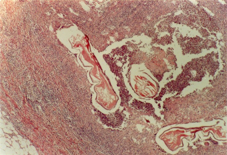
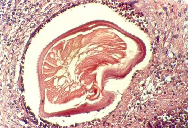
Histopathological findings revealed an eosinophilic abscess surrounding by a foreign body reaction (Fig. 1). An inflammatory cell infiltration was seen around the parasite. In the central area, the sections of the parasite were delimited by patchy granulomatous reactions consisting of epithelioid cells, histiocytes, eosinophils and foreign body giant cells. The external longitudinal cuticular ridges and a well-developed muscle layer were the main diagnostic features seen in transverse sections of the parasite (Fig. 2). Our diagnosis verified by dirofilariasis experts abroad. The patient was a resident of Kouchesfahan of Rasht district with no history of any travel out of the province at least for 2 yr before appearance of the nodule. Her lesion healed in several weeks following the removal of the nodule.
Fig. 1:
Sections of D. repens in an eosinophilic abscess.
Fig. 2:
Section of D. repens with external longitudinal cuticular ridges and a well-developed muscle layer
Discussion
The public health importance of human dirofilariasis has been significantly increased following dramatic rise in the number of cases reported during last few decades. From 1885, in which the first case of human infection due to D. repens reported, until 2000 about 782 cases of human dirofilariasis have been detected in 37 countries in the Old World(2, 3). This dramatic increase in the number of reported cases suggests dirofilariasis as an emergent parasitic zoonosis. The most infected countries are Italy (298 cases), Sri Lanka (132 cases), Russia (Siberia inclusive) (83 cases), France (76 cases), Ukraine (51 cases) and Greece (27 cases) (3).
Although improvement in the diagnostic skills of the pathologists and greater medical awareness might be incriminated in the detection of more human cases in the endemic areas, the real increase in the incidence of human infections might be associated with changes in the climatic conditions (temperature, relative humidity, rainfall and evaporation), expanded vector breading season, the abundance of microfilaraemic reservoir hosts, the increasing frequency of travel to endemic countries and outdoors human activities. The increase in the population of the vectors and the infection of reservoir hosts are directly associated with climatic changes, which in turn influenced in the increase of the incidence of human infections (2, 3, 7).
Besides the present case, eleven cases of human dirofilariasis due to D. repens have been detected in Iran, of which three cases (27.3%) were detected in Gilan Province. The amount of rainfall and relative humidity in Gilan are several times higher than the mean values of the whole country. Presence of a very short dry season, abundance of vegetations, mainly rice fields, and long-lasting water collections which may surround human dwellings, in addition to adequate temperatures, are very suitable for mosquito reproduction and disease establishment in the province. Furthermore, Gilan Province is a coastal touristy area and hundred thousands of Iranian and also many foreign tourists visit this province annually, especially during the transmission seasons, so they will be potentially at risk of the infection during their stay.
Since human infections are usually asymptomatic and the physicians and pathologists in the province are not familiar with dirofilariasis, the number of real cases of human infections might be much higher than the reported cases.
In spite of the detection of several cases of subcutaneous dirofilariasis in Gilan, unfortunately no study on animal reservoirs has been carried out up to date, but studies in adjacent province of Mazandaran, with almost similar climatic conditions, have shown the infection rate of 60.8% and 10% in dogs and jackals respectively (8, 16). To determine the real situation of dirofilariasis in Gilan a comprehensive study on human and animal dirofilariasis is needed.
On the other hand, Gilan Province is the endemic region of human fascioliasis in Iran, where two outbreaks have occurred in 1987 and 1998 and affected about 15000 people mainly in Rasht and Bandar-Anzali districts, from which three cases of human subcutaneous dirofilariasis have been reported (18, 19). Also several cases of cutaneous fascioloasis have been detected in Gilan during last two decades (Unpublished data). Interestingly, the present case of dirofilariasis was clinically suspected as a probable case of cutaneous fascioliasis by an expert dermatologist because of the similarity of clinical manifestations. So, subcutaneous dirofilariasis should be considered as a differential diagnosis of cutaneous fascioliasis in the area.
Summing up, due to non-stop increasing in the number of human dirofilariasis worldwide and the suitable conditions for transmission of the infection in Gilan, we suggest the pathologists who working in the area to be familiar with the microscopic aspects of human dirofilariasis and similarly the clinicians to take in to account the dirofilariasis when encounter any solitary nodule of uncertain nature in subcutaneous tissues and mucus membranes.
Acknowledgments
The authors would like to thank Dr E Maltezos and Dr E Siviridis from Democritus University of Thrace, Alexandroupolis, Greece and Dr WM Tilakaratne from University of Peradeniya, Sri Lanka for their assistance in verifying our diagnosis. We are also graceful of Dr Pampiglione S for his nice cooperation by sending his publications to the authors.
References
- 1.MacDougall LT, Magoon CC, Fritsche TR. Dirofilaria repens manifesting as a breast nodule: diagnostic problems and epidemiologic considerations. Am J Clin Pathol. 1992;97(5):625–30. doi: 10.1093/ajcp/97.5.625. [DOI] [PubMed] [Google Scholar]
- 2.Pampiglione S, Rivasi F, Angeli G, Boldorini R, Incensati RM, Pastormerlo M, Pavesi M, Ramponi A. Dirofilariasis due to Dirofilaria repens in Italy, an emergent zoonosis: report of 60 new cases. Histopathology. 2001;38:344–45. doi: 10.1046/j.1365-2559.2001.01099.x. [DOI] [PubMed] [Google Scholar]
- 3.Pampiglione S, Rivasi F. Human dirofilariasis due to Dirofilaria (Nochtiella) repens: an update of world literature from 1995–2000. Parasitologia. 2000;(3–4):42. 231–54. [PubMed] [Google Scholar]
- 4.Lucy S, Davada K, Subramanian H. Dirofilariosis in dogs and humans in Kerala. Indian J Med Res. 2005;121:691–93. [PubMed] [Google Scholar]
- 5.Tilakaratne WM, Pitakotuwage TN. Intra-Oral Dirofilaria repens infection: report of seven cases. J Oral Pathol Med. 2003;32(8):502–5. doi: 10.1034/j.1600-0714.2003.00183.x. [DOI] [PubMed] [Google Scholar]
- 6.Muro A, Genchi C, Cordero M, Simon F. Human dirofilariasis in the European Union. Parasitol Today. 1999;15:386–89. doi: 10.1016/s0169-4758(99)01496-9. [DOI] [PubMed] [Google Scholar]
- 7.Simon F, Lopez-Besmonte J, Marcos-Atxutegi C, Morchon R, Martin-Pacho JR. What is happening outside North America regarding human dirofilariasis. Vet Parasitol. 2005;133:181–89. doi: 10.1016/j.vetpar.2005.03.033. [DOI] [PubMed] [Google Scholar]
- 8.Azari S. Review of Dirofilariasis in Iran. J Med Fac of Gilan Univ Med Sci Iran. 2007;15(60):102–12. [Google Scholar]
- 9.Athari A, Rohani S. Sari, Iran: 2001. Ocular dirofilariosis: report of a rare case. In abstract book of the Third Iranian National Congress on Parasitology; p. 280. [Google Scholar]
- 10.Athari A. Zoonotic subcutaneous dirofilariasis in Iran. Arch Iran Med. 2003;6:63–65. [Google Scholar]
- 11.Degardin P, Simonart JM. Dirofilariosis a rare usually imported dermatosis. Dermatology. 1996;192(4):398–99. doi: 10.1159/000246430. [DOI] [PubMed] [Google Scholar]
- 12.Rohani S, athari A. Ocular dirofilariasis in Iran. A case report. Med J Islamic Rep Iran. 2003;17:85–86. [Google Scholar]
- 13.Maraghi S, Rahdar M, Akbari H, Radmanesh M, Saberi AA. Human dirofilariasis due to Dirofilaria repens in Ahvaz, Iran: A report of three cases. Pak J Med Sci. 2006;22(2):211–13. [Google Scholar]
- 14.Siavashi MR, Massoud J. Human cutaneous dirofilariasis in Iran: a report of two cases. Iranian J Med Sci. 1995;20:85–6. [Google Scholar]
- 15.Jamshidi A, Jamshidi M. Periocular dirofilariasis. Scientific Journal of the Eye Bank of I.R.Iran. 2006;2(11):257–59. [Google Scholar]
- 16.Sadeghian A. Helminth parasites of stray dogs and jackals in Shahsavar area, Caspian region, Iran. J Parasitol. 1969;55(2):372–74. [PubMed] [Google Scholar]
- 17.Sadjjadi SM, Mehrabani D, Oryan A. Dirofilariasis of stray dogs in Shiraz, Iran. J Vet Parasitol. 2004;18(2):181–82. doi: 10.1016/s0304-4017(99)00151-x. [DOI] [PubMed] [Google Scholar]
- 18.Massoud J. Fascioliasis outbreak and drug test (Triclabendazole) in Caspian Sea Littoral, northern part of Iran. Bulletin de la Societe Franςaise de Parasitologie. 1990;8:438. [Google Scholar]
- 19.Assmar M, Milaninia A, Amirkhani A, Yadegari D, Forghan-Parast K, Nahravanian H, Piazak N, et al. Seroepidemiological investigation of fascioliasis in northern Iran. Med J Islamic R Iran. 1991;5:23–7. [Google Scholar]
- 20.Maltezos ES, Sivridis EL, Giatromanolaki AN, Simopoulos CE. Human subcutaneous Dirofilariasis: A report of three cases manifesting as breast or axillary nodules. Scot Med J. 2002;47(4):86–8. doi: 10.1177/003693300204700404. [DOI] [PubMed] [Google Scholar]


