Abstract
This case report presents a coincidence of trisomy 18 and balanced Robertsonian translocation (13;14). Aneuploidy was suspected based on anomalies detected in ultrasound scan and confirmed with karyotype. In a 31 years-old healthy woman with a history of one miscarriage, second trimester ultrasound scan reported IUGR (<3rd percentile) with normal amniotic fluid, bilateral choroid plexus cysts, suspicious agenesis of corpus callosum and clenched hands. Amniocentesis was performed and karyotype was 46xx,der(13;14) (q10;q10),+18. Maternal karyotype was 45xx,der(13;14)(q10;q10). Pregnancy was continued due to legal limitation for termination after 20 weeks gestation. Delivery was done at 36 weeks gestation. A female newborn was borned and a physical feature was hypotonia, small mouth, prominent occiput, low-set and posteriorly rotated ears, clenched hands with overlapping fingers and rocker bottom feet. Ultrasound scan and echocardiography detected agenesis of corpus callosum and VSD, ASD, PDA and cardiomegaly. These features are typical of trisomy 18. Balanced Robertsonian translocation usually has no phenotypic expression. Genetic counseling and prenatal diagnosis for future pregnancy was recommended.
Keywords: Robertsonian translocation, Trisomy 18, Prenatal diagnosis, Interchromosomal effect
Introduction
Chromosomal abberations include two broad categories: numerical and structural anomalies. Trisomy 18 is the second most common autosomal trisomy, also known as Edward syndrome, is a well-known human aneuploidy with a short life time expectancy. It has an overall frequency of 1 in 3000 up to 1 in 8000 newborns, 3 to 4 times more common in females than in males (1), and is a maternal age related trisomy (2).
Robertsonian translocation (Rob) is centric fusion of two acrocentric chromosomes, and is the most common chromosomal translocation, occurs 1 per 1000 in general population. The most common Rob is between chromosomes 13 and 14 (13q;14q) which constitutes 75% of all Robs. As long as the fused q arms are intact, the Rob carriers are usually phenotypically normal (3). However unbalanced gametes of heterozygous carriers are common and give rise to monosomic or trisomic fetuses. Most monosomies and trisomies are lethal and spontaneously are lost early in pregnancy (4). Observed incidence of normal/balanced offspring is 13.28% (5). Coincidence of Rob(13;14) with trisomy 18 has been reported as denovo event (6) or in offspring of Rob carriers, both are rare conditions. This rather uncommon observation has been interpreted as interchromosomal effect (ICE), which lead to missegregation of the chromosomes not involved in the translocation. The issue is still very controversial (7–8).
Ultrasound findings, maternal serum markers, and parental risk factors for genetic disease are all considered in determining the risk that the fetus is affected. Fetal karyotyping is suggested when structural anomalies or early intrauterine growth retardation,IUGR (weight below the 10th percentile for gestational age) is detected (9).
Case report
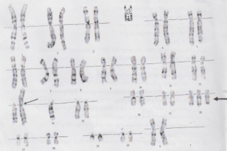
A 31 years-old healthy mother, gravid 2 and 1 miscarriage (blighted ovum), her mother had an intrauterine fetal demise without gross anomaly. The father was 30 years-old and healthy, family history was unremarkable. Second trimester screening quadruple test result was low risk for trisomy 18, 21 and 13. However, ultrasound scan at 22 week’s gestation revealed a single alive fetus with symmetric IUGR <3%, normal amniotic fluid. Fetal anomaly scan demonstrated: bilateral choroid plexus cysts (8–10 millimeters), suspicious agenesis of corpus callosum and clenched hands. She was referred to our unit for karyotyping. An amniocentesis was performed at 22 weeks gestation. Amniocytes were cultured using standard techniques, finding on GTG-banded metaphase spreads showed a karyotype of 46xx, der(13;14)(q10;q10),+18, trisomy 18 and balanced Robertsonian translocation between nonhomologous acrocentric chromosomes 13 and 14. Karyotypes of parents were requested. Father’s was normal and mother’s was described as 45xx,der(13;14)(q10;q10) (Fig. 1).
Fig. 1:
Karyotype: 46xx,der(13;14)(q10;q10),+18
Following genetic counseling and informing of poor prognosis for fetus with Edward syndrome, mother chose to terminate the pregnancy but because of legal limitations after 20 weeks gestation, pregnancy was continued. A female infant was delivered in 36 weeks gestation. She weighed 2000 grams (<3rd percentile), length was 43 centimeters, (<3rd percentile) and head circumference was 31.5 centimeters (10–50th percentile). Physical findings were hypotonia, small mouth, prominent occiput, low-set and posteriorly rotated ears, clenched hands with overlapping fingers and rocker bottom feet. Brain ultrasound scan showed agenesis of corpus callosum and echocardiography reported VSD, ASD, PDA, and cardiomegaly.
She was physically retarded and had problems such as failure to thrive, feeding difficulties, and frequent NICU admissions. She died at 11 months-old.
Discussion
Robs usually have reproductive risks, as our case had a previous miscarriage and a recent pregnancy with aneuploidy A range of 77 to 97% fetuses with trisomy 18 has been detected during second trimester ultrasound (10). Detection rate depends on gestational age, operator’s expertise, and ultrasound machine quality. In our case, aneuploidy was suspected in ultrasound scan, and a rare complex chromosomal aberration of Rob (13;14) and trisomy 18, was diagnosed by karyotype.
The major phenotypic features of the newborn were characteristic of trisomy 18. Balanced Rob usually does not have phenotypic consequences because the lost sections of acrocentric chromosomes do not contain unique genetic sequences. Trisomy 18 cases are usually caused by primary nondisjunction in maternal meiosis II (11). It is postulated that translocations disturb meiotic disjunction. Interchromosomal effect maybe exists, as several reports suggest a higher rate of chromosome abnormalities in Robs (8).
Genetic counseling and consideration of prenatal diagnosis is recommended for families with chromosomal translocations and reproductive failures (7, 8). Although second trimester genetic ultrasound scan is helpful for detection of aneuploidies, first trimester screening has improved detection rate and should be offered to all pregnant women (12), for early detection and safe and legal termination.
Ethical considerations
Ethical issues (Including plagiarism, Informed Consent, misconduct, data fabrication and/or falsification, double publication and/or submission, redundancy, etc) have been completely observed by the authors.
Acknowledgments
The authors declare that there is no conflict of interests.
References
- 1.Snijders RJ, Sebire NJ, Nicolaides KH. Maternal age and gestational age-specific risk for chromosomal defects. Fetal Diagn Ther. 1995 Nov-Dec;10(6):356–67. doi: 10.1159/000264259. [DOI] [PubMed] [Google Scholar]
- 2.Ferguson-Smith MA, Yates JR. Maternal age specific rates for chromosome aberrations and factors influencing them: report of a collaborative european study on 52 965 amniocenteses. Prenat Diagn. 1984 Spring;4:5–44. doi: 10.1002/pd.1970040704. Spec No: [DOI] [PubMed] [Google Scholar]
- 3.Scriven PN, Flinter FA, Braude PR, Ogilvie CM. Robertsonian translocations--reproductive risks and indications for preimplantation genetic diagnosis. Hum Reprod. 2001 Nov;16(11):2267–73. doi: 10.1093/humrep/16.11.2267. [DOI] [PubMed] [Google Scholar]
- 4.Mutton D, Alberman E, Hook EB. Cytogenetic and epidemiological findings in Down syndrome, England and Wales 1989 to 1993. National Down Syndrome Cytogenetic Register and the Association of Clinical Cytogeneticists. J Med Genet. 1996 May;33(5):387–94. doi: 10.1136/jmg.33.5.387. [DOI] [PMC free article] [PubMed] [Google Scholar]
- 5.Zhang YP, Xu JZ, Yin M, Chen MF, Ren DL. [Pregnancy outcomes of 194 couples with balanced translocations] Zhonghua Fu Chan Ke Za Zhi. 2006 Sep;41(9):592–6. (abstract). [PubMed] [Google Scholar]
- 6.Lesniewicz R, Posmyk R, Lesniewicz I, Wolczynski S. Prenatal evaluation of a fetus with trisomy 18 and additional balanced de novo Rob (13;14) Folia Histochem Cytobiol. 2009;47(5):S137–40. doi: 10.2478/v10042-009-0053-8. [DOI] [PubMed] [Google Scholar]
- 7.Douet-Guilbert N, Bris MJ, Amice V, Marchetti C, Delobel B, Amice J, et al. Interchromosomal effect in sperm of males with translocations: report of 6 cases and review of the literature. Int J Androl. 2005 Dec;28(6):372–9. doi: 10.1111/j.1365-2605.2005.00571.x. [DOI] [PubMed] [Google Scholar]
- 8.Munne S, Escudero T, Fischer J, Chen S, Hill J, Stelling JR, et al. Negligible interchromosomal effect in embryos of Robertsonian translocation carriers. Reprod Biomed Online. 2005 Mar;10(3):363–9. doi: 10.1016/s1472-6483(10)61797-x. [DOI] [PubMed] [Google Scholar]
- 9.American college of obstetricians and gynecologists 2000. Intrauterine growth restriction.ACOG practice Bulletin # 12; january.
- 10.DeVore GR. Second trimester ultrasonography may identify 77 to 97% of fetuses with trisomy 18. J Ultrasound Med. 2000 Aug;19(8):565–76. doi: 10.7863/jum.2000.19.8.565. [DOI] [PubMed] [Google Scholar]
- 11.Bugge M, Collins A, Petersen MB, Fisher J, Brandt C, Hertz JM, et al. Non-disjunction of chromosome 18. Hum Mol Genet. 1998 Apr;7(4):661–9. doi: 10.1093/hmg/7.4.661. [DOI] [PubMed] [Google Scholar]
- 12.Wapner R, Thom E, Simpson JL, Pergament E, Silver R, Filkins K, et al. First-trimester screening for trisomies 21 and 18. N Engl J Med. 2003 Oct 9;349(15):1405–13. doi: 10.1056/NEJMoa025273. [DOI] [PubMed] [Google Scholar]


