Abstract
Lipoma is a benign mesenchymal tumor of fat with uncertain pathogenesis. Although the most common mesenchymal neoplasm in trunk and proximal portions of the extremities, it is rarely seen in the oral cavity. Oral lipomas are clinically soft, smooth-surfaced nodular masses that mostly are less than 3 cm in size. Typically the tumor is asymptomatic unless bitten or become noticeable because of their size. The buccal mucosa and buccal vestibule are the most common intraoral sites and account for 50% of all cases. Reported here is a relatively large lipoma of buccal mucosa that was treated surgically under local anesthesia. In an 18-month postsurgical follow up no complication or recurrence has occurred. This case will also be compared to intraoral lipomas reported in Iranian population. As lipomas are usually smaller than 3 cm in diameter, lipoma with the size reported, is of clinical importance. Since the large lipomas are in differential diagnosis with other, even malignant, mesenchymal, or salivary gland tumors. Thus, this case report recommends clinical awareness in diagnosis of large intraoral soft tissue lesions.
Keywords: Buccal mucosa, fatty tumor, lipoma, mouth mucosa
INTRODUCTION
Lipoma is a benign mesenchymal tumor of fat. This tumor is rarely seen in the oral cavity, although by far it represents the most common mesenchymal neoplasm elsewhere such as trunk, shoulders, neck, and axilla. Although the etiology and pathogenesis of lipomas is not well known, lipomas appear to be more common in obese people.[1]
Clinically oral lipomas are seen as yellow (deeper tumors may appear pink) soft, smooth-surfaced nodular masses less than 3 cm in size that might be sessile or pedunculated. Typically the tumor is asymptomatic unless bitten or become noticeable because of their size. Many patients have noticed the lesion months or years before diagnosis.[1]
The most common intraoral site for lipoma is buccal mucosa and buccal vestibule. Lipomas of the oral and maxillofacial region have shown equal predilection for involvement of men and women, although one study has reported a marked male predilection. Lipoma is uncommon in children and affected patients mostly are 40 years of age or older.[1]
Histopathologically most oral lipomas are composed of mature fat cells very similar in appearance to the surrounding normal fat. Individual cells have a clear cytoplasm with a flat nucleus located on the periphery of the cell. Lipoma is usually well circumscribed and may demonstrate a thin fibrous capsule. Lipomas are treated by conservative local excision, and recurrence is rare.[1,2]
CASE REPORT
A 36-year old otherwise healthy male patient was referred to oral surgeon with an asymptomatic, swelling in the right buccal mucosa that he had been aware of for 2 years.
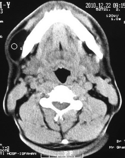
In clinical examination an obvious submucosal swelling with intact overlying mucosa was observed. The clinical appearance was similar to mesenchymal tumors, salivary gland tumors, or even bone tumor with soft tissue invasion. An omnipaque contrast CT scan was taken, in which a well-circumscribed lesion with a density similar to adipose tissue was shown [Figure 1].
Figure 1.

Axial omnipaque contrast CT scan of the lesion
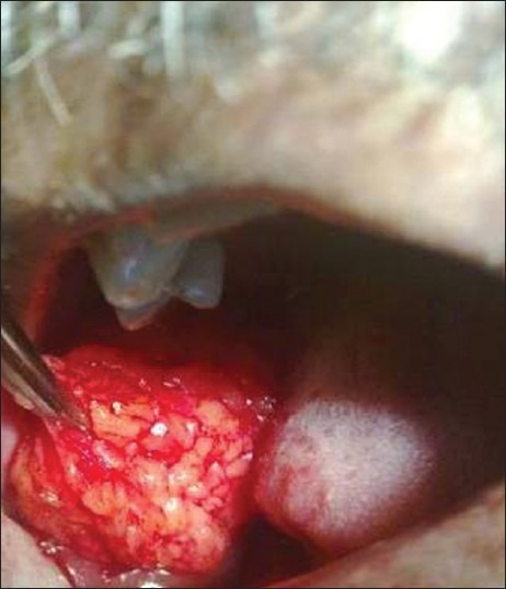
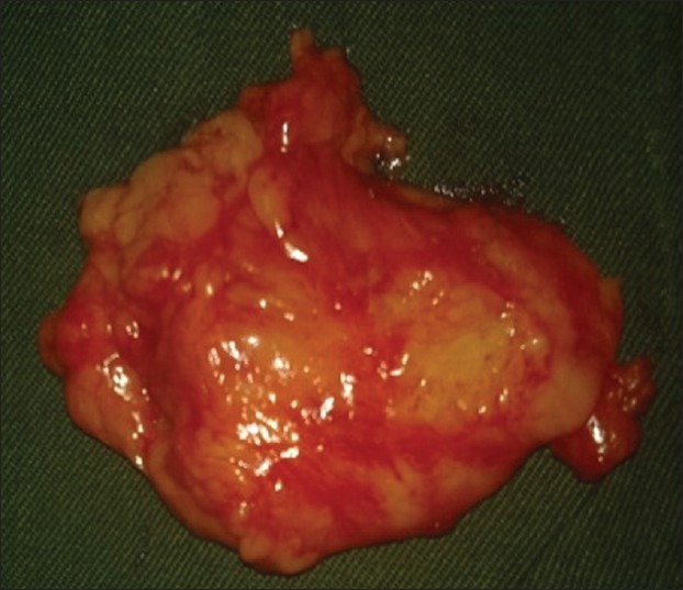
With a provisional diagnosis of oral lipoma under local anesthesia, surgical excision of the lesion was performed. When a deep seated, yellow, well-circumscribed soft tissue mass with a lobulated surface 4 × 3.5 × 1.2 (length × width × thickness) centimeters in size was totally excised and submitted for microscopic examination in 10% buffered formalin [Figures 2 and 3].
Figure 2.

Surgical removal of the lesion via an intraoral approach
Figure 3.

Macroscopic image of the lesion with the lobular surface (4 × 3.5 × 1.2 cm)
In pathology laboratory, the submitted specimen was processed and embedded in paraffin using standard procedures. From the paraffin block a 3 μ section was prepared and stained with Hematoxylin and Eosin stain (H and E) to allow histopathologic examination.
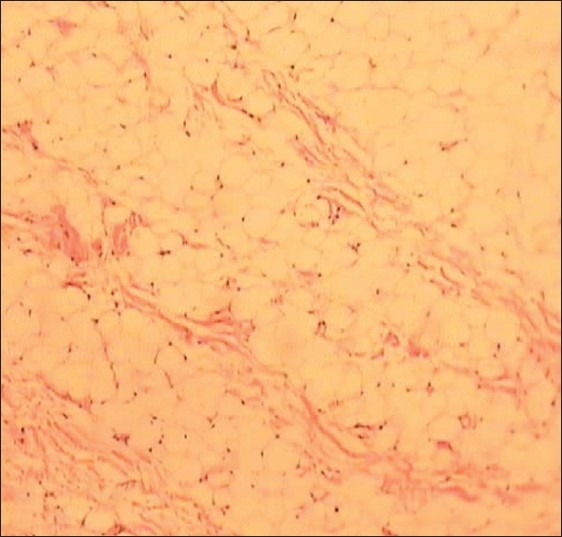
In microscopic examination of H and E stained slides, using light microscope (Olympus, Japan), a well-circumscribed nodular mass was seen. Which showed a distinct lobular pattern composed of mature adipocytes, cells with large clear cytoplasm and a flat dark nucleus on periphery. Thin septa of fibrous tissue was seen between the closely packed normal looking fat cells. At the borders, lesion was surrounded by a thin fibrous capsule. According to histopathologic features a definitive diagnosis of lipoma was made [Figure 4]. In an 18-month postsurgery follow-up no recurrence or complication has occurred.
Figure 4.

Microscopic image of the lesion, closely packed fat cells separated by thin septa of the fibrous tissue (H and E, × 100)
DISCUSSION
Lipoma is a very common benign tumor of adipose tissue, but its presence in oral region is relatively uncommon. Once present, a mucosal oral lipoma may increase to 5 to 6 cm over a period of years, but most cases are less than 3 cm in greatest dimension at diagnosis. Conservative surgical removal is the treatment of choice with occasional recurrences expected.[3]
As mentioned before, it is believed that oral lipomas tend to occur with equal predilection for involvement of men and women. But Furlong[4] et al. mentioned that oral lipoma is seen more frequently in men (as presented here). Although it should be mentioned here that Freitas[5] et al. showed oral lipoma has a predilection of occurrence for women. The place of involvement was the most common place for occurrence of intraoral lipomas.
For size only few large lipomas have been reported in the English literature.[6] As the mean size of oral lipoma in 450 cases was 1.66cm (0.2-10cm) in diameter.[7]
In Iranian population we found only five entities introducing intra oral lipoma. A case of oral lipoma was reported in a 57-year old woman with a size of 1.5 cm in diameter.[8] Saghafi[9] et al. reported a case of osteolipoma in a 68-year old male patient, 1.8 cm in the largest dimension. Tavakoli[10] et al. reported two cases of oral Lipoma one: in a 50-year old male in junction of hard and soft palate with 1.5 cm in diameter and second: a 63-year old male patient with the involvement of lateral border of tongue where the greatest dimension of the lesion reported was 1 cm.
The lesion reported here is to a great extent larger than other intraoral lipomas reported in Iran. This report and the reports of Saghafi, Tavakoli may recommend a male predilection for lipoma of oral cavity in Iran. Despite mentioned case reports patient reported here is under 40 year of age.
CONCLUSION
Although intraoral lipoma is rare, authors recommend the clinical awareness that lipoma can occur in oral cavity, even larger than we usually expect.
Footnotes
Source of Support: Nil
Conflict of Interest: None declared.
REFERENCES
- 1.Nevill B, Damm D, Allen C, Bouquot J. Oral and Maxillofacial Pathology. 3rd ed. Missouri: Saunders; 2009. pp. 523–4. [Google Scholar]
- 2.Regezi J, Sciubba J, Jordan R. Oral Pathology Clinical Pathologic Correlations. 5th ed. Philadelphia: Saunders; 2008. pp. 176–7. [Google Scholar]
- 3.Bouquot J, Nikai H. Lesions of the Oral cavity. In: Gneep D, editor. Diagnostic surgical pathology of the head and neck. Philadelphia: Saunders; 2001. pp. 192–3. [Google Scholar]
- 4.Furlong MA, Fanburg-Smith JC, Childers EL. Lipoma of the oral and maxillofacial region: Site and subclassification of 125 cases. Oral Surg Oral Med Oral Pathol Oral Radiol Endod. 2004;98:441–50. doi: 10.1016/j.tripleo.2004.02.071. [DOI] [PubMed] [Google Scholar]
- 5.Freitas MA, Freitas VS, Lima AA, Pereira FB, dos Santos JN. Intraoral lipomas: A study of 26 cases in a Brazilian population. Quintessence Int. 2009;40:79–85. [PubMed] [Google Scholar]
- 6.Colella G, Biondi P, Caltabiano R, Vecchio GM, Amico P, Magro G. Giant intramuscular lipoma of the tongue: A case report and literature review. Cases J. 2009;7:130–3. doi: 10.4076/1757-1626-2-7906. [DOI] [PMC free article] [PubMed] [Google Scholar]
- 7.Studart-Soares E, Costa F, Sousa FB, Alves A, Osterne R. Oral lipomas in a Brazilian population: A 10-year study and analysis of 450 cases reported in the literature. Med Oral Patol Oral Cir Bucal. 2010;15:691–6. doi: 10.4317/medoral.15.e691. [DOI] [PubMed] [Google Scholar]
- 8.Lavaf S, Azizi A, Sajady F. A case report of oral lipoma. Sci Med J Ahwaz Univ Med Sci. 2009;7:130–3. [Google Scholar]
- 9.Saghafi SH, Rahpeyma A, Salehinejad J, Zare R. Osteolipoma of the oral cavity: A case report and review of the literature. Iran J Otorhinolaryngol. 2007;19:161–4. [Google Scholar]
- 10.Tavakoli A, Razavi M, Khabazian A. Lipoma in Oral Mucosa: Two case reports. Dent Res J (Isfahan) 2010;7:41–3. [PMC free article] [PubMed] [Google Scholar]


