Sir,
Conventional rigid Macintosh laryngoscopy is known to fail in patients with mouth opening of less than 25 mm.[1] Nasal fibreoptic tracheal intubation (FOI) has been suggested as the technique of choice in these patients.[2] Recently, we were faced with a situation of failed FOI in a patient with restricted mouth opening secondary to neglected faciomaxillary trauma. In the absence of lightwand availability, expertise in blind nasotracheal intubation and reluctance to adopt retrograde tracheal intubation as an initial strategy, we successfully intubated this patient using a combination of C-Mac videolaryngoscopy and dynamic alteration in endotracheal tube (ETT) shape using Schroeder's stylet via the right nostril.
A 27-year-old, ASA I female patient with past history of faciomaxillary trauma 6 weeks back was posted for multiple plating of fractured faciomaxillary bones. Mouth opening was restricted to less than 18 mm. The patient was irritable and generally uncooperative for awake airway management.
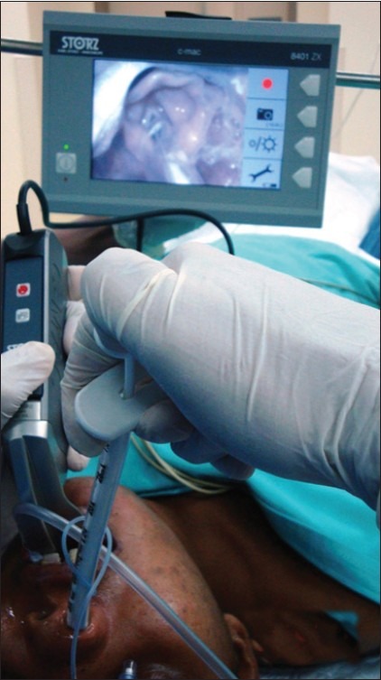
Anaesthesia was induced with gradually increasing concentrations of sevoflurane while maintaining spontaneous breathing for FOI (Plan A). Early in the induction period, the patient developed obstructed breathing. A gently placed 7.0 mm ID nasopharyngeal airway overcame the upper airway obstruction. After reaching an adequate depth of anaesthesia, a flexible fibrescope (Olympus LF-TP) with a pre-mounted 6.5 mm ID armoured ETT was introduced following removal of the nasopharyngeal airway. Possibly, during placement of the nasopharyngeal airway, some degree of bleeding might have occurred that blurred the view of the fibrescope and fibrescopy had to be abandoned. Our next plans (Plan B) were to use the Macintosh-shaped C-Mac videolaryngoscope and guide the nasotracheal tube into the glottis. This laryngoscope has a slim blade profile (14 mm height), which gives more space for its use in patients with limited mouth opening. It failed to visualize the glottis either directly or indirectly on the monitor. It was now suggested to use the D-blade of the C-MAC videolaryngoscope, which has blade angulation of more than 60°. A 50% glottic view was now obtained. Unfortunately, the size 6.5 mm ID polyvinyl chloride ETT passed could not be sighted in the video screen and hence failed to be advanced towards the glottis. This has been observed by others also.[3] Subsequently, a conventional stylet was used to shape the ETT into a gentle C shape as suggested earlier.[4] The styleted ETT via the right nostril could be visualized on the video screen but, despite repeated attempts and optimal external laryngeal manipulation, it could not be negotiated into the glottis. At this stage, it was decided to use a well-lubricated Schroeder's directional stylet (SDS) (SteBar Instrument Corp.) to manipulate the shape of the ETT dynamically as it was directed towards the glottis. This quality of SDS has been previously used to achieve blind nasotracheal intubation of a patient with temporomandibular joint ankylosis.[5] The curvature of the ETT could now be easily changed with the use of SDS, and the tip of the ETT entered between the vocal cords fairly easily [Figure 1] but impacted in the vestibule of the larynx. Withdrawing the SDS and raising the head slightly helped in negotiation of the impacted ETT into the trachea. Correct tracheal intubation was further confirmed by capnography. During this procedure with the C-Mac videolaryngoscope, the patient was administered 2% sevoflurane in 100% oxygen via a 14 gauge catheter attached to the side channel of the C-MAC blade. Oxygen saturation, mean arterial pressure and heart rate remained between 97 and 99%, 68 and 98 mmHg and 88 and102/min, respectively.
Figure 1.

Schroeder's directional stylet in endotracheal tube aiding entry into the glottis using D-blade of the C-Mac videolaryngoscope
In conclusion, a combination of D-blade of the C-Mac videolaryngoscope and Schroeder's directional stylet may provide an alternative strategy for tracheal intubation in patients with restricted mouth opening, with the added advantage of oxygenation during intubation.
REFERENCES
- 1.Aiello G, Metcalf I. Anaesthetic implications of temporomandibular joint disease. Can J Anaesth. 1992;39:610–6. doi: 10.1007/BF03008329. [DOI] [PubMed] [Google Scholar]
- 2.Kulkarni DK, Prasad AD, Rao SM. Experience in fiberoptic nasal intubation for temporomandibular joint ankylosis. Ind J Anaesth. 1999;43:26–9. [Google Scholar]
- 3.Maassen R, Lee R, Hermans B, Marcus M, van Zundert A. A comparison of three videolaryngoscopes; the Macintosh laryngoscope blade reduces, but does not replace, routine stylet use for intubation in morbidly obese patients. Anesth Analg. 2009;109:1560–5. doi: 10.1213/ANE.0b013e3181b7303a. [DOI] [PubMed] [Google Scholar]
- 4.Behringer EC, Kristensen MS. Evidence for benefit vs novelty in new intubation equipment. Anaesthesia. 2011;66(Suppl 2):57–64. doi: 10.1111/j.1365-2044.2011.06935.x. [DOI] [PubMed] [Google Scholar]
- 5.Mahajan R, Shafi F, Sharma A. Use of Shroeder's directional stylet to enhance navigability during nasotracheal intubation. J Anesth. 2010;24:150–1. doi: 10.1007/s00540-009-0838-0. [DOI] [PubMed] [Google Scholar]


