Abstract
Context:
It has been proved that lip prints are analogous to thumb prints. A correlation between thumb prints and sagittal dental malocclusion has already been established. Soft tissue is gaining more importance in judgement of deformity or identity of a patient.
Aim:
To find a correlation between sagittal skeletal jaw relation and lip prints.
Settings and Design:
Descriptive, cross-sectional, comparative, single-blind, hospital-based study.
Materials and Methods:
A total of 90 patients were categorized into skeletal class I, class II, and class III, comprising 30 patients in each group with equal gender distribution. Dolphin imaging (10.5) software was used for analyzing sagittal jaw relation. Lip prints obtained from these 90 patients were analyzed.
Statistical Analyses Used:
Karl Pearson's correlation coefficient, Chi-square test, t-test, Spearman's co-efficient, analysis of variance (ANOVA).
Results:
It was observed that angle ANB (Angle formed between points nasion[N] to Subnasal[A] and nasion[N] to supramental [B]) and beta angle were statistically significant, revealing a strong negative correlation (-0.9060) with different classes of jaw relation. Significant difference was observed between genders in all the three classes. Significant difference was observed in relation to lip print and the quadrants of upper and lower lips. A statistical significance was noted on the right side of both upper and lower arches.
Conclusion:
This study shows that lip prints can be employed for sagittal jaw relation recognition. A further study on various ethnic backgrounds with a larger sample size in individual group is necessary for comparing lip prints and malocclusion.
Keywords: ANB, beta angle, cheiloscopy, lip prints, sagittal jaw relation, WITS appraisal
Introduction
Every human being is distinct and discernible in that they exhibit their own pattern of characteristics.[1] Lip prints are normal lines and fissures in the form of wrinkles and grooves present in the zone of transition of human lip, between the inner labial mucosa and outer skin, the examination of which is referred to as “cheiloscopy.”[2,3]
The biological phenomenon of systems of furrows on the red part of human lips was first described by an anthropologist Fischer in 1902, as quoted by Sivapathasundaram et al.[3] However, until 1930, anthropology merely mentioned the existence of furrows without suggesting a practical use for the phenomenon. In 1961, the first research in Europe was carried out on the subject of lip prints in Hungary. Lip print evaluation gained importance when lip traces were found on a glass door at the scene of murder.[3,4] The search for cheiloscopic correlation to various other factors is yet unanswered.
Earlier studies have indicated that lip prints can be used for personal identification as well as sex determination.[2,3] Mc Donel in 1972 studied identical twins and concluded that both the lip prints showed some similarities as told by Agarwal.[4] It has been proved that lip prints are analogous to thumb prints.[2] A correlation between thumb prints and sagittal dental malocclusion has already been established.[5] It is stated that fingers, palms, lip, alveolus, and palate develop during the same embryonic period.[6] Lip prints are established at a very early period in comparison to sagittal jaw relation and dental relation.[6,7] Establishing a correlation between sagittal jaw relation and lip prints would benefit the clinician by predicting the type of malocclusion and can also provide additional information on individual personal identity.[7] No previous studies reported on the correlation between lip prints and sagittal jaw relation. The aim of our study was to find out any correlation that exists between cheiloscopy and sagittal skeletal jaw relations. The objective was to compare the gender variation of lip prints and of sagittal skeletal jaw relation as well as to relate the different lip print patterns with those of sagittal skeletal jaw relations.
Materials and Methods
A descriptive, cross-sectional, single-blind, hospital-based study was conducted in the Department of Orthodontics and Dentofacial Orthopedics. Ethical clearance was obtained from the ethical committee of the Sumandeep Vidyapeeth university. Informed consent was obtained from each subject prior to the study. Patients having any developmental anomaly or any pathology on lips and jaws, those who were unable to open their mouth, and those who did not give informed consent were excluded from the study. A pilot study was conducted on 15 patients (5 in each group) to know the feasibility and acceptability of the study. Based on secondary literature available, a sample size of 90 subjects was found to be appropriate. A convenience sample of 90 patients, in the age group of 18–25 years, from the outpatient Department of Orthodontics was included in our study.
Digital lateral cephalogram
The digital cephalograms were recorded with Kodak 8000C machine utilized for taking both lateral cephalogram and orthopantomographs. For taking the cephalograms, 6 kV, 12 mA current and an exposure time of 0.8 sec was used. Images were recorded on a CCD and were processed with the help of Kodak dry view 8150. All 90 digital cephalograms were analyzed using Dolphin imaging (10.5) software [Figure 1].
Figure 1.
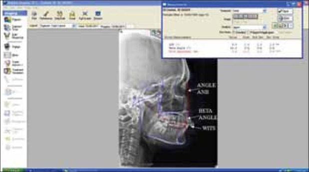
Lateral cephalometric analysis done by Dolphin imaging 10.5
Establishment of sagittal jaw position
Determining skeletal jaw relation is dependent on factors like position of maxilla and mandible with reference to cranial base [Figure 2]. Cephalometric analyses regularly used are Downs’, Steiner's, McNamara's, etc. to determine the sagittal jaw position and relation of an individual.[8–15] If all the parameters of Downs’, Steiner's and McNamara's analyses are in range, then the individual is categorized into skeletal class I jaw relation, having normal anteroposterior relationship of maxilla and mandible with respect to cranial base. But if the parameters vary from the ranges mentioned in the table, the sagittal jaw position is considered to be affected and class II or class III sagittal skeletal jaw relation develops [Table 1].[8–15]
Figure 2.
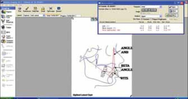
Representing various planes used for determining cephalometric sagittal jaw relation
Table 1.
Various cephalometric parameters for sagittal jaw position
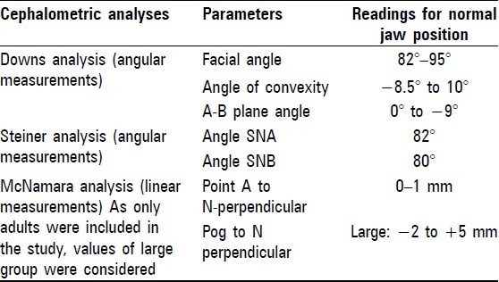
Establishment of sagittal jaw relation
Patient's initial sagittal jaw position was concluded based on Downs’,[8,9] Steiner's,[10,11] and McNamara's analyses.[12,13] Individuals were further divided into skeletal class I, class II, and class III groups with the help of angle ANB (Angle formed between points nasion[N] to Subnasal[A] and nasion[N] to supramental [B]),[10,11] WITS (Millimetric reading taken on occlusal plane- perpendicular of point A and B to occlusal plane) appraisal,[14,15] and beta angle.[16] Digital cephalograms of the first come 90 subjects (equal number of males and females) was taken and 30 subjects (15 males and 15 females) were assigned to each group (i.e., class I, class II, and class III). Dolphin imaging (10.5) software was employed for a second time to achieve specific details like angle ANB,[10,11] WITS appraisal,[14,15] and beta angle.[16] Keeping the norms into consideration, the cephalograms were categorized into class I, class II, and class III when at least two norms coincided, according to Jacobson and Baik, as mentioned in Table 2.[10,14,16] All cephalometric analyses were performed by one individual to prevent any inter-observer bias.
Table 2.
Various cephalometric parameters for sagittal jaw relation

Lip print recording
A dark-colored lipstick was applied uniformly with one stroke on upper and lower lips. Patient was asked to rub upper and lower lip. After 2 minutes, lip impression was made on a transparent self-adhesive tape having a width of 48 mm. This lip impression was immediately pasted on a white bond paper as proposed by Sivapathasundaram et al.[3] Magnifying glass lens was used for the analysis of lip prints and the field of observation was restricted to 10 mm on each side of the quadrant.[2,3] All lip print analyses [Figures 3 and 4] were done by another observer who was blinded in relation to clinical examination and cephalometric analysis of the patient. Tsuchihashi's classification of lip print [Figure 5] was used to analyze the lip prints.[1]
Figure 3.
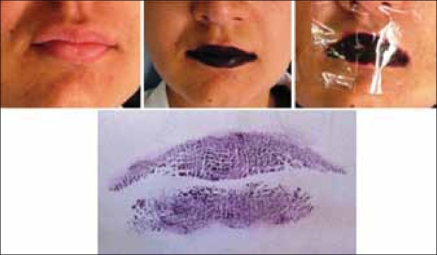
Lip print recording
Figure 4.
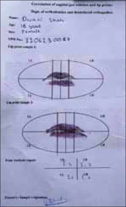
Analysis of lip prints
Figure 5.
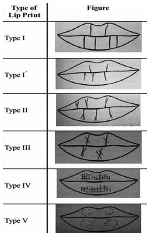
Suzuki and Tsuchihashi's classification of lip print [(courtesy Dr. Sivapathasundharam B, Dr. Ajayprakash P, and Dr. Sivakumar G, Lip prints (cheiloscopy) Indian J Dent Res, 10:234-37, 2001]
Tsuchihashi's classification for lip print identification
Courtesy Dr. Sivapathasundharam B, Dr. Ajayprakash P, and Dr. Sivakumar G[3]
Type 1: Clear-cut grooves running vertically across the lips
Type 1’: Straight grooves which disappear half way instead of covering the entire lip
Type 2: Fork grooves in their course
Type 3: Intersecting grooves
Type 4: Reticulate grooves
Type 5: Undetermined
Statistical analyses
SPSS 16.0 version software was used for statistical analysis. A confidence interval of 95% and a significance level of 5% were set. Comparison of three skeletal groups, class I, class II, and class III, with respect to angle ANB, WITS appraisal, and beta angle was made by one-way analysis of variance (ANOVA) and Tukey's multiple post hoc procedures. Correlation among all the parameters in class I, class II, and class III groups was determined by Karl Pearson's correlation coefficient method. Chi-square test was utilized to analyze significance of lip prints in terms of gender, quadrant, and among all the three sagittal groups. Spearman's correlation was used to compare lip prints and sagittal jaw relation.
Results
There were 30 subjects each in the three groups, accounting to a total of 90 subjects. The mean age of the study subjects was 19.5 ± 2.97 years. There was a significant difference in age when the three groups (class I 19.37 ± 3.22 years, class II 18.53 ± 2.75 years, class III 20.70 ± 2.94 years) were compared (P=0.0001, S).
The mean value of angle ANB(in mm) was 1.93 ± 0.45, 7.27 ± 1.11, and -3.7 ± 1.73 for skeletal class I, class II, and class III, respectively [Graph 1]. Mean value of WITS appraisal(in mm) was 0.33 ± 0.96, 4.44 ± 1.9, and -2.50 ± 0.86 for skeletal class I, class II, and class III, respectively, as observed in Graph 2. Beta angle (in mm) had a mean value of 29.23 ± 2.33, 23.93 ± 1.96, and 38.67 ± 2.44 for skeletal class I, class II, and class III, respectively [Graph 3]. Karl Pearson's correlation co-efficient indicated statistically significant difference between skeletal class I, class II, and class III with respect to angle ANB, WITS appraisal, and beta angle [Table 3]. When gender was taken as a variable and compared for angle ANB (P=0.54, NS), WITS appraisal (P=0.35, NS), and beta angle (P=0.76, NS), there was no significant difference.
Graph 1.
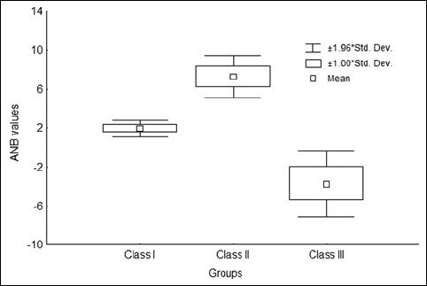
Comparison of three groups with respect to ANB values. F-value = 613.0771, P-value = 0.0000, S
Graph 2.
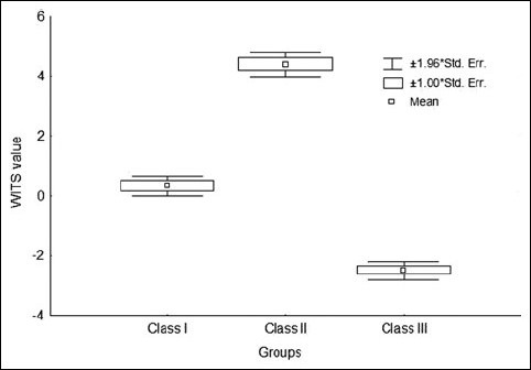
Comparison of three groups with respect to wits values. F-value = 351.3208, P-value = 0.0000, S
Graph 3.
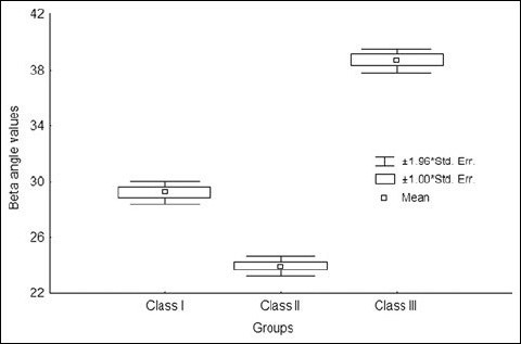
Comparison of three groups with respect to Beta angle values. F-value = 328.9314, P-value = 0.0000, S
Table 3.
Correlation among all the parameters in total samples of all three groups by Karl Pearson's correlation coefficient method

Lip prints were examined in relation to upper and lower lips which were further subdivided into right and left quadrants. Among all the four quadrants, it was observed that a combination of 1,3; 1’,3; and 2,3 types of lip prints were predominant in skeletal class I group of individuals. 1,4 and 3,4 types of lip print combinations were predominant among skeletal class III group of patients. 1,2 type of lip print combination was observed to be more predominant among skeletal class II individuals as observed in Tables 4 and 5. A significant difference was observed among skeletal class I, class II, and class III groups. Right quadrant in both upper and lower lips revealed statistical significance.
Table 4.
Distribution of study subjects according to groups and types of lip prints in upper lip
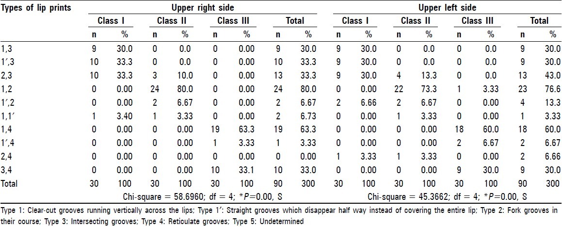
Table 5.
Distribution of study subjects according to groups and types of lip prints in lower lip
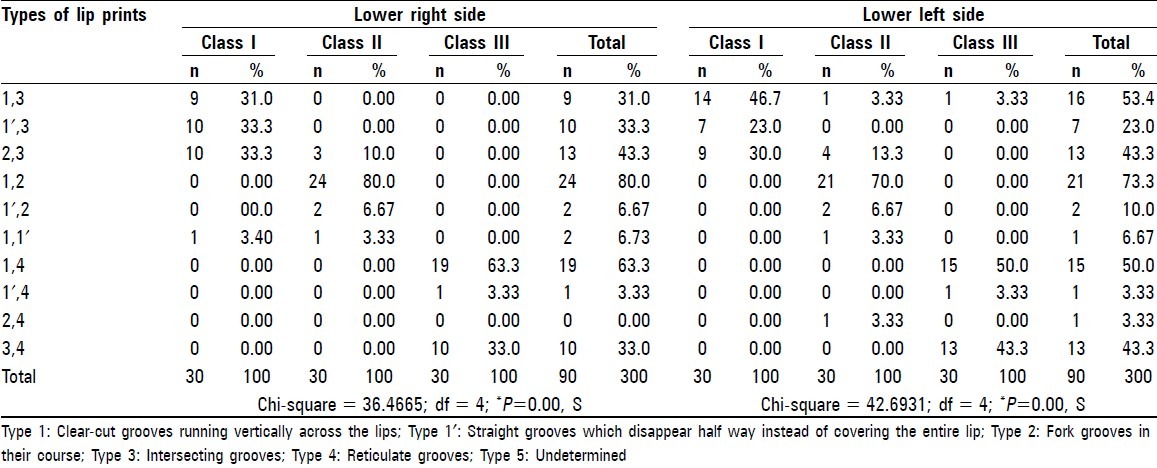
There was an association of combination of lip print patterns with respect to angle ANB, WITS appraisal, and beta angle as shown in Table 6, whereas individual lip prints did not show any significance. There was significant difference when angle ANB was compared with lip print patterns on upper and lower lips. However, the upper right lip print combinations (1–4) were not significant (P=0.0939). WITS appraisal had a statistically strong association with that of lip print patterns. Beta angle showed no significant association with the lower right and left lip print combinations (1–4) but had a significant relation on the same side with 1’,1 combination of lip prints as shown in Table 6.
Table 6.
Correlation of ANB, WITS, and beta angles, with lip print pattern by Spearman's rank correlation

Regarding the gender difference, females had more of 1’ (75.66%), 1 (58.98%), 3 (55.77%), and 4 (73.11%) types of lip print patterns when compared to males. Lip print pattern 2 was more common among males (61.3%).
Discussion
Thumb prints, lip prints, and dental examination are routinely used for forensic examination.[1,11] It has been proved that lip prints are analogous to thumb prints and is confirmed that specific lip prints are common among individuals of specific gender.[2,3] Profile photograph indicates probable sagittal skeletal relation and can be used for personal identification.[7,12] Probability of trauma and sublimation of records occurs more on soft tissue.[13] Hence, dental records are gaining more importance nowadays,[13] and an attempt was made to correlate sagittal skeletal jaw relation with the type of lip prints. As there is lack of literature for this relation, valid comparisons could not be made with other studies. Among all the four quadrants of the lip, it was observed that 1,3; 1’,3; and 2,3 types of lip print combinations were predominant in skeletal class I group of individuals. 1,4 and 3,4 types of lip print combinations were predominant among skeletal class III group of patients. 1,2 type of lip print combination was observed to be more predominant among skeletal class II group of patients. Statistically significant difference was noted between angle ANB and beta angle, revealing a strong negative correlation (-0.9060).
Sagittal jaw relation
The angular and linear measurements in various analyses that have been proposed for anteroposterior measurement could be inaccurate because they depend on various factors. Hence, an accurate assessment of anteroposterior jaw relationship is critical.[14,15] In the present study, the mean values of angle ANB were 1.93 ± 0.45, 7.27 ± 1.11, and –3.7 ± 1.73 for skeletal class I, class II, and class III, respectively. This observation is in concurrence with earlier literature.[8] We observed a smaller value with respect to angle ANB in skeletal class I samples in comparison to the observation made by Patel et al.[17] Mean values of WITS appraisal were –0.33 ± 0.96, 4.44 ± 1.9, and –2.50 ± 0.86 for skeletal class I, class II, and class III, respectively, and are similar to those reported in an earlier study.[7] Beta angle showed mean values of 29.23 ± 2.33, 23.93 ± 1.96, and 38.67 ± 2.44 for skeletal class I, class II, and class III, respectively, as observed in Graph 3. This observation is in correlation with earlier literature.[13] In the present study, variation of measurement was observed in skeletal class II and class III groups, indicating severe malocclusion. However, we could not find any studies in this context among the Indian population. A further study on what could be the probable cause of this severity of malocclusion among the Indian population can be conducted.
Lip prints
Type 1 and 1’ lip prints were more common among females. Type 2 lip prints were more common among males, which correlates with earlier studies.[14–16,18] However, we could not correlate lip prints with the quadrants, as mentioned previously.[16,18] A study conducted among Saudi subjects showed different results, with horizontal pattern of grooving reported to be more common among females. However, we could not compare our results with those of that study as it used a classification of nine types of grooves. In this study, lip print pattern revealed no association with sagittal skeletal jaw relation. Since a maximum of four types of lip prints was observed among all the three groups, combination of two types of lip prints was analyzed. The lip print pattern analysis criteria used in our study were different from those of previous studies. This might be due to lack of standard universal pattern for lip print analysis.[19,20]
Based on lip prints, skeletal class I and class III could be identified more expediently in comparison to skeletal class II. Skeletal class II had a combination of 1,2 type of lip prints present predominantly among all the four quadrants. Type 1 and type 2 lip prints signify the gender of an individual. Hence, identifying skeletal class II sagittal jaw relation would be difficult in comparison to other groups. We could not find any studies to correlate in this context.
In conclusion, the present study has shown that lip prints can be employed for sagittal jaw relation recognition. Similar studies with a larger sample size with different ethnic groups are necessary for comparing lip prints and malocclusion. A further extensive study is necessary to be carried out in relation to lip prints and skeletal class II.
Acknowledgments
We would like to thank the participants for their cooperation throughout the study procedure. I thank my Head of the Department, my colleagues for guidance, and the post graduate students for the support extended to me during the study. I also thank Dr. Aswini Y. B., Reader, Department of Public Health Dentistry, and Dr. Varun Sardana, Senior Lecturer, Department of Pedodontics and Preventive Dentistry, Teerthanker Mahaveer Dental College, Moradabad, for extending their special help to me in editing and reviewing the manuscript.
Footnotes
Source of Support: Nil
Conflict of Interest: None declared
References
- 1.Tsuchihashi Studies on personal identification by means of lip prints. Forensic sci. 1974;3:233–48. doi: 10.1016/0300-9432(74)90034-x. [DOI] [PubMed] [Google Scholar]
- 2.Sharma P, Saxena S, Rathod V. Cheiloscopy; the study of lip prints in sex identification. J Forensic Dent Sci. 2009;1:24–7. [Google Scholar]
- 3.Sivapathasundharam B, Prakash PA, Sivakumar G. Lip prints (cheiloscopy) Indian J Dent Res. 2001;12:234–7. [PubMed] [Google Scholar]
- 4.Agarwal A. The importance of lip prints (forensic files) [Last Accessed from 2008, Oct 24]. Available from: http://lifeloom.com//IIAgarwal.html .
- 5.Tikare S, Rajesh G, Prasad KW, Thippeswamy V, Javali SB. Dermatoglyphics–a marker for malocclusion? Int Dent J. 2010;60:300–4. [PubMed] [Google Scholar]
- 6.Kanematsu N, Yoshida Y, Kishi N, Kawata K, Kaku M, Maeda K, et al. Study on abnormalities in the appearance of finger and palm prints in children with cleft lip, alveolus, and palate. J Maxillofac Surg. 1986;14:74–82. doi: 10.1016/s0301-0503(86)80265-x. [DOI] [PubMed] [Google Scholar]
- 7.Profitt WR, Fields HW, Sarver DM. 4th ed. Missouri: Mosby imprint, Elsevier; 2007. Contemporary Orthodontics; p. 195. [Google Scholar]
- 8.Jacobson A, Jacobson RL. 2nd ed. Illinois: Quintessence Publishing; 2006. Radiographic Cephalometry: From Basics to 3-d Imaging; p. 72. [Google Scholar]
- 9.Jacobson A, Jacobson RL. 2nd ed. Illinois: Quintessence Publishing; 2006. Radiographic Cephalometry: From Basics to 3-d Imaging; p. 99. [Google Scholar]
- 10.Jacobson A, Jacobson RL. 2nd ed. Illinois: Quintessence Publishing; 2006. Radiographic Cephalometry: From Basics to 3-d Imaging; pp. 112–22. [Google Scholar]
- 11.Cavard S, Alvarez JC, De Mazancourt P, Tilotta F, Brousseau P, de la Grandmaison GL, et al. Forensic and police identification of “X” bodies. A 6-years French experience. Forensic Sci Int. 2011;204:139–43. doi: 10.1016/j.forsciint.2010.05.022. [DOI] [PubMed] [Google Scholar]
- 12.Lynnerup N, Bojesen S, Kuhlman MB. Matching profiles of masked perpetrators: A pilot study. Med Sci Law. 2010;50:200–4. doi: 10.1258/msl.2010.010013. [DOI] [PubMed] [Google Scholar]
- 13.Valenzuela A, Martin-de las Heras S, Marques T, Exposito N, Bohoyo JM. The application of dental methods of identification to human burn victims in a mass disaster. Int J Legal Med. 2000;113:236–9. doi: 10.1007/s004149900099. [DOI] [PubMed] [Google Scholar]
- 14.Baik CY, Ververidou M. A new approach of assessing sagittal discrepancies: the Beta angle. Am J Orthod Dentofacial Orthop. 2004;126:100–5. doi: 10.1016/j.ajodo.2003.08.026. [DOI] [PubMed] [Google Scholar]
- 15.Gul-e-Erum , Fida M. A comparison of cephalometric analyses for assessing sagittal jaw relationship. J Coll Physicians Surg Pak. 2008;18:679–83. [PubMed] [Google Scholar]
- 16.Vahanwala S, Parekh BK. Study of lip prints as an aid to forensic methodology. JIDA. 2000;71:268–71. [Google Scholar]
- 17.Patel HM, Joshi MR. A study of the differences between the dento-facial patterns associated with Class II division 1 malocclusion and normal occlusion. J Indian Orthod Soc. 1977;9:1–10. [PubMed] [Google Scholar]
- 18.Vahanwala S, Nayak CD, Pagare SS. Study of Lip – Prints as Aid for sex determination. Medico legal update. 2005;5:93–8. [Google Scholar]
- 19.El Domiaty MA, Al-gaidi SA, Elayat AA, Safwat MD, Galal SA. Morphological patterns of lip prints in Saudi Arabia at Almadinah Almonawarah province. Forensic Sci Int. 2010;200:179. doi: 10.1016/j.forsciint.2010.03.042. [DOI] [PubMed] [Google Scholar]
- 20.More C, Patil R, Asrani M, Gondivkar S, Patel H. Cheiloscopy – A Review. Indian J Forensic Med Toxicol. 2009;3:17–20. [Google Scholar]


