Abstract
A former 34-week-old female infant with Down syndrome underwent surgical correction of a congenital heart defect at 5 months of age. Her postoperative course was complicated by severe pulmonary hypertension and junctional ectopic tachycardia. Following treatment with amiodarone infusion, she developed laboratory indices of acute liver injury. At their peak, liver transaminase levels were 19 to 35 times greater than the upper limit of normal. Transaminitis was accompanied by coagulopathy, hyperammonemia, and high serum lactate and lipid levels. Hepatic laboratory abnormalities began to resolve within 48 hr of stopping amiodarone infusion. Heart rate control was achieved concurrently with discovery of laboratory test result abnormalities, and no further antiarrhythmic therapy was required. The intravenous formulation of amiodarone contains the diluent polysorbate 80, which may have hepatotoxic effects. Specifically, animal studies suggest that polysorbate 80 may destabilize cell membranes and predispose to fatty change within liver architecture. Polysorbate was implicated in infant fatalities from E-ferol use in the 1980s. This case illustrates a possible adverse event by the Naranjo probability scale. Given the extent of clinically apparent hepatic injury, this patient was not rechallenged with amiodarone during the remainder of her hospitalization. With amiodarone now used as first-line pharmacologic therapy for critical tachyarrhythmia in this population, the number of children exposed to this drug should be expected to increase. Laboratory indices of liver function should be evaluated at initiation of amiodarone therapy, as well as frequently throughout duration of therapy. Consideration should be given to polysorbate-free formulation of intravenous amiodarone for use in the cohort with congenital cardiac disease.
INDEX TERMS: amiodarone, congenital heart disease, hepatotoxicity, junctional ectopic tachycardia, polysorbate 80
INTRODUCTION
Junctional ectopic tachycardia (JET) is a narrow-complex tachyarrhythmia encountered in the acute postoperative setting. Incidence of JET is highly dependent on the cardiac lesion and specific surgical repair, but patients at highest risk appear to be those with Tetralogy of Fallot (TOF), ventricular septal defects, and atrioventricular (AV) septal defects.1 Local trauma or surgical irritation near the AV node leads to enhanced automaticity of the cardiac conduction bundle, manifesting as acceleration in heart rate with loss of AV synchrony.1 JET often develops during the time of peak postoperative cardiac edema, which further compromises ventricular filling and diastolic function. Due to the recalcitrant nature of JET, amiodarone is often used concurrently with other treatments.
Although generally considered a class III agent, amiodarone has a unique antiarrhythmic profile derived from all four Vaughn Williams classes. It has shown particular promise in postoperative congenital cardiac patients2 and is now first-line pharmacologic therapy for critical tachyarrhythmia.1 Acute hepatotoxicity following intravenous (IV) amiodarone administration has been reported in several collections of adult case studies.3–5 In 2005, Rätz Bravo et al6 reviewed 25 such adult cases reported between 1986 and 2002. Most cases occurred in patients aged 50 to 70 years with atrial tachyarrhythmia. Liver enzyme abnormalities occurred within 3 days of starting parenteral amiodarone in approximately 88% of cases.6 Establishing a causal relationship in these cases remains difficult, as most patients are critically ill with multiorgan dysfunction. However, 3 (of the 25) cases commented on a return of hepatotoxicity after rechallenge with intravenous amiodarone.6
In contrast to adult reports, amiodarone hepatotoxicity literature in the congenital cardiac population is scarce. Given this population's increased risk of rhythm disturbance and subsequent exposure to amiodarone, we present the case of a 5-month-old female with suspected amiodarone-induced hepatotoxicity after continuous infusion for the treatment of postoperative JET. This case emphasizes the need to monitor laboratory indices of hepatocellular function early and often during therapy and proposes consideration of the alternate formulation of amiodarone in this vulnerable population.
CASE REPORT
A former 34-week-old African-American female with Down syndrome presented for operative repair of TOF with AV septal defect at 5 chronologic months of age. This child experienced minimal congestive heart failure symptoms as an outpatient, required no preoperative diuretics, and weighed 2.6 kg at admission. Concern for pulmonary hypertension prompted the surgeon to leave a residual atrial septal defect for shunting and initiate preemptive inhaled nitric oxide therapy. Surgical repair required 4.5 hr of cardiopulmonary bypass and 3.5 hr of aortic cross-clamp time. At operative conclusion, she presented with complete heart block necessitating AV sequential pacing to separate from bypass. She was transitioned to the intensive care unit sedated, intubated, and pacemaker-dependent. Vasoactive infusions upon transfer consisted of milrinone (1 mcg/kg/min) and epinephrine (0.1 mcg/kg/min). Her rhythm evolved into an accelerated junctional rhythm, with heart rate into the 160s, and few distinct “p” waves, visualized by electrocardiography. However, with supportive critical care, her blood pressure and markers of end-organ perfusion remained adequate throughout the first postoperative night. Her condition changed the following afternoon with JET acceleration into the 190s, accompanied by hemodynamic compromise.
At this juncture, several therapies were undertaken simultaneously to effect heart rate control. Initial attempts were made at overdrive atrial pacing to restore AV synchrony, but these attempts were unsuccessful. Due to the proarrhythmic effect of catecholamines in the setting of JET,1 epinephrine infusion was reduced to minimal levels (0.03 mcg/kg/min). Replacement of electrolytes, including calcium, potassium, and magnesium, was optimized through IV therapy. In anticipation of a noncompliant right ventricle associated with her TOF repair, intravascular volume status was challenged with several intermittent fluid boluses until no further improvement in blood pressure was observed. She was adequately sedated, neuromuscularly blocked, and moderately cooled (36.5°C). Despite these measures, her JET persisted. Amiodarone therapy was then initiated.
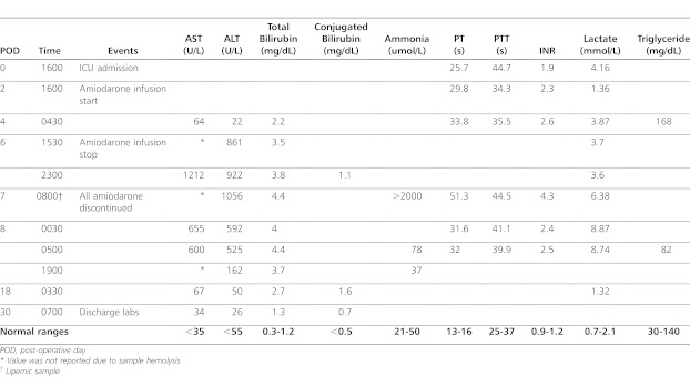
On the evening of postoperative day (POD) 2, she received two amiodarone IV bolus doses of 5 mg/kg each prior to beginning a 10 mg/kg/day continuous infusion. She remained on this infusion for 96 consecutive hours (POD 2–6). By the evening of POD 6, her junctional rhythm had slowed to the 120s, permitting AV sequential pacing and discontinuation of the continuous infusion. Coincident with heart rate control, laboratory values suspicious for liver injury were first observed. However, given the severity of her illness, enteral amiodarone therapy was pursued, pending reevaluation of hepatic function the following morning. Unfortunately, rapidly progressive laboratory signs of liver injury prompted complete discontinuation of the drug at that time. Specific laboratory indices of both hepatocellular injury and synthetic dysfunction are outlined in Table 1. At their peak, her liver transaminase levels exceeded 35 times the upper limit of normal for aspartate aminotransferase (AST) and 19 times the upper limit of normal for alanine aminotransferase (ALT). In addition, she showed evidence of hyperammonemia, mild coagulopathy, and lactate dysmetabolism. This patient received a total of 148 mg of amiodarone (135 mg of IV formulation). Serum amiodarone levels were not drawn.
Table 1.
Patient's biochemical profile during admission
Consultants from toxicology, clinical pharmacy, cardiology, and gastroenterology commented on this patient's evolving hepatic injury, and all consultants cited amiodarone as the likely causative agent. To support this conclusion, several potential mechanisms of causation were explored. Some consultants questioned whether a history of prematurity could predispose to drug-induced hepatic insult. While a valid concern in severe prematurity, this patient's prematurity state was mild (6 weeks). Hepatic enzymatic and metabolic processes should be adequate at 3.5 months corrected gestational age. Knowing amiodarone can transiently depress blood pressure during administration, several consultants questioned whether hypoperfusion or a low cardiac output state contributed to her liver dysfunction. However, concurrent with amiodarone administration, she received blood pressure support via an epinephrine infusion. As a result, she did not experience clinically significant hypotension during this time. While invasive cardiac output monitoring was not used, her hemodynamic findings were deemed adequate, as other end organs appeared well perfused. For example, with special regard to her renal system, she maintained age-appropriate urine output and creatinine levels. In regard to coadministered medication effects, she was exposed to a variety of hepatically metabolized analgesics (acetaminophen, morphine, fentanyl, and methadone) in the days prior to and during amiodarone administration. However, these agents were administered by appropriate weight-based dosing strategies. Metabolism of analgesic agents was clinically assessed via pain scoring systems with established thresholds prompting need for additional doses. Total IV nutrition, which can produce elevated transaminases and conjugated hyperbilirubinemia after chronic use, did not begin until the evening of POD 6. Thus, parenteral nutrition did not overlap with administration of parenteral amiodarone. Finally, as recalcitrant as this patient's JET was to treatment, her pulmonary hypertension was similarly problematic. In fact, most considered right-heart failure stemming from intractable pulmonary hypertension a contributing factor to her hepatic dysfunction, yet transthoracic echocardiography on the morning of amiodarone discontinuation (POD 7) suggested only moderate pulmonary hypertension, with half-systemic right ventricular pressures. Global systolic function of the right ventricle was interpreted as normal. Liver ultrasonography revealed biliary and hepatic vessels with normal waveforms, velocities, and appropriate directionality.
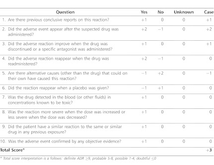
Hepatic laboratory value abnormalities began to show resolution within 48 hr of stopping IV amiodarone therapy and were fully normalized prior to discharge home (POD 30). With concurrent heart rate control, no further antiarrhythmic therapy was required during the remainder of her hospitalization. Given the extent of clinically apparent hepatic injury, this patient was not rechallenged with amiodarone for cause. As shown in Table 2, this case constituted a possible adverse event by the Naranjo adverse drug reaction probability scale.7 A voluntary US Food and Drug Administration (FDA) MedWatch report was filed after patient discharge annotating the suspected link between IV amiodarone administration and development of hepatotoxicity in this particular patient. The Institutional Review Board waived need for review.
Table 2.
Naranjo Adverse Drug Reaction Score
DISCUSSION
Amiodarone hydrochloride is metabolized via CYP3A4 to the active metabolite N-desethylamiodarone. Metabolite serum concentrations rival the parent compound in chronic therapy, and animal studies suggest 70% of the antiarrhythmic effect of amiodarone may be attributed to this active metabolite.8 The volume of distribution is large, with adipose tissue as a major site of collection.9 When therapy is begun, the drug redistributes into peripheral tissues 80 times faster than it is eliminated from the body, making continuous IV infusion the most efficient means of maintaining stable serum concentrations.10 Due to extensive drug storage, elimination half-life of the parent compound and the metabolite are variable. Antiarrhythmic effects may persist up to 6 months following discontinuation of the drug.9
Hepatotoxicity can range from mild transaminitis to fulminant liver failure. There is lack of consensus regarding the degree of transaminitis that constitutes a clinically significant effect.10 The FDA finds clinical significance when transaminases exceed 3 times the upper limit of normal.11 With AST 35 times and ALT 19 times the upper limit of normal, our patient satisfied this definition. When her hepatic function was first evaluated 2 days after initiation of amiodarone, only mild elevation in AST was noted. On subsequent recheck 48 hours later, both AST and ALT levels were markedly abnormal. Perhaps of greater importance were the abnormal markers of hepatic synthetic and metabolic function, including coagulopathy, hyperammonemia, and high serum lactate and lipid levels. These laboratory findings are consistent with those found in previously published reports of amiodarone-induced hepatotoxicity in adults. In the study by Rätz Bravo6 in particular, AST and ALT elevations above 10 times the upper limit of normal were observed in 22 of the 25 case reports. Hyperbilirubinemia was observed in half of those cases.
Rhodes et al12 were the first to suggest polysorbate 80, a solubilizing agent, as the hepatotoxic component in the IV formulation of amiodarone. The injectable product has a ratio of 2 mg polysorbate 80 for every 1 mg of amiodarone.13 Our patient thus received 270 mg of polysorbate 80 (103 mg/kg) in addition to 135 mg of IV amiodarone. No human toxicity values are published, but the 50% lethal dose (LD50) for rats and mice given IV polysorbate 80 is 1790 mg/kg and 4500 mg/kg, respectively.14 Animal models suggest that parenteral polysorbate 80 is rapidly degraded by serum esterases.15 Furthermore, human studies of polysorbate 80 elimination in adult cancer patients cite a short terminal half-life of 0.607 ± 0.245 hr and total plasma clearance of 7.7 ± 2.9 L/hr.16 This short-terminal half-life may explain the rapid resolution in hepatic injury after discontinuation of parenteral amiodarone. With antiarrhythmic effects persisting for months due to redistribution of amiodarone into peripheral tissues, rapidity of hepatic insult resolution attests more to discontinuation of polysorbate than to discontinuation of amiodarone. While data for pathogenic mechanisms in humans are mainly speculative, polysorbate may destabilize cell membranes and predispose to steatosis. This process has been observed in cultured rat hepatocytes within hours of exposure.17 Polysorbate 80 may also directly inhibit hepatic CYP3A4, resulting in elevated serum levels of the parent compound or other coadministered medications.18 The hepatotoxic potential of polysorbate gained notoriety after the E-ferol tragedy in the 1980s. E-ferol was an IV formulation of vitamin E marketed in December 1983 as antioxidant therapy for premature infants. The formulation contained a mixture of polysorbate 80 (9%) and polysorbate 20 (1%) as solubilizing agents. After only 4 months of use, 38 infant deaths were reported in 11 states.19 While hepatic histology results from infants receiving E-ferol suggested a more cytotoxic than steatotic process, few investigations supported vitamin E content as the responsible culprit, thus leaving the mixture of polysorbate as suspect.19
If one is concerned about polysorbate in the IV formulation, one may question whether administering initial doses in the oral formulation, which does not contain polysorbate, would be a safer but equally effective alternative. Several authors have noted that oral amiodarone therapy is well tolerated in patients even following acute hepatitis with parenteral therapy.3,20 However, onset of action following oral administration is estimated to be 2 to 3 days,21 which is not an acceptable time course for a patient with hemodynamic compromise in the critical care environment. Furthermore, given the potential for multiorgan dysfunction in patients with critical illness, poor gastrointestinal function may lead to unreliable absorption.
While the vehicle in the IV amiodarone formulation is the likely cause of liver toxicity in this patient, hepatic injury from progressive cardiac dysfunction deserves special consideration. Also known as cardiac hepatopathy, elevated filling pressures from a failing right heart result in venous stasis within the hepatobiliary system. A constellation of clinical and chemical abnormalities, including hepatomegaly, ascites, transaminitis, and decrement in synthetic function, may be observed. Liver histology obtained by biopsy may reveal fibrotic changes in hepatic architecture, as well as dilated vasculature, inflammation, or hemorrhage.22 Liver biopsy was not pursued in this patient, but transthoracic echocardiography and abdominal ultrasonography were used as noninvasive surrogates. Neither study supported right-heart dysfunction as the probable explanation in this case.
CONCLUSIONS
With appreciation for growing use of amiodarone for critical tachyarrhythmia, the polysorbate-free formulation merits consideration for priority use in the congenital cardiac disease population. This cohort incurs risk of postoperative complications to include arrhythmia, cardiopulmonary arrest, and organ dysfunction stemming from sequelae of cardiopulmonary bypass. While the FDA approved a polysorbate-free formulation of amiodarone (Nexterone, Baxter Healthcare Corporation, Deerfield, IL) in late 2008,23 patents will preclude a generic formulation for several years. Higher cost of the brand name product may prove fiscally prohibitive for widespread use in many institutions. However, use of a polysorbate-free formulation in a congenital cardiac patient who develops progressive hepatic dysfunction may afford the clinician diagnostic clarity. Clinicians caring for these patients are encouraged to evaluate laboratory indices of hepatocellular and synthetic liver function both at amiodarone initiation and at frequent intervals throughout therapy.
ACKNOWLEDGMENT
The University of Virginia Institutional Review Board waived review of this report.
ABBREVIATIONS
- ALT
alanine aminotransferase
- AST
aspartate aminotransferase
- AV
atrioventricular
- FDA
Food and Drug Administration
- INR
international normalized ratio
- IV
intravenous
- JET
junctional ectopic tachycardia
- LD50
50 percent lethal dose
- POD
postoperative day
- PT
prothrombin time
- PTT
partial thromboplastin time
- TOF
tetrology of fallot
Footnotes
DISCLOSURE The authors declare no conflicts or financial interest in any product or service mentioned in the manuscript, including grants, equipment, medications, employment, gifts, and honoraria.
REFERENCES
- 1.Haas NA, Plumpton K, Justo R, et al. Postoperative junctional ectopic tachycardia (JET) Z Kardiol. 2004;93(5):371–380. doi: 10.1007/s00392-004-0067-3. [DOI] [PubMed] [Google Scholar]
- 2.Perry JC, Fenrich AL, Hulse JE, et al. Pediatric use of intravenous amiodarone: efficacy and safety in critically ill patients from a multicenter protocol. J Am Coll Cardiol. 1996;27(5):1246–1250. doi: 10.1016/0735-1097(95)00591-9. [DOI] [PubMed] [Google Scholar]
- 3.Pye M, Northcote RJ, Cobbe SM. Acute hepatitis after parental amiodarone administration. Br Heart J. 1988;59(6):690–691. doi: 10.1136/hrt.59.6.690. [DOI] [PMC free article] [PubMed] [Google Scholar]
- 4.Gregory SA, Webster JB. Acute hepatitis induced by parenteral amiodarone. Am J Med. 2002;113(3):254–255. doi: 10.1016/s0002-9343(02)01149-x. [DOI] [PubMed] [Google Scholar]
- 5.Rizzioli E, Incasa E, Gamberini S, et al. Acute toxic hepatitis after amiodarone intravenous loading. Am J Emerg Med. 2007;25(9):1082.e1–1082.e4. doi: 10.1016/j.ajem.2007.02.045. [DOI] [PubMed] [Google Scholar]
- 6.Rätz Bravo AE, Drewe J, Schlienger RG. et al. Hepatotoxicity during rapid intravenous loading with amiodarone: description of three cases and review of the literature. Crit Care Med. 2005;33(1):128–134. doi: 10.1097/01.ccm.0000151048.72393.44. [DOI] [PubMed] [Google Scholar]
- 7.Naranjo CA, Busto U, Sellers EM, et al. A method for estimating the probability of adverse drug reactions. Clin Pharmacol Ther. 1981;30(2):239–245. doi: 10.1038/clpt.1981.154. [DOI] [PubMed] [Google Scholar]
- 8.Nattel S, Davies M, Quantz M. The antiarrhythmic efficacy of amiodarone and desethylamiodarone, alone and in combination, in dogs with acute myocardial infarction. Circulation. 1988;77(1):200–208. doi: 10.1161/01.cir.77.1.200. [DOI] [PubMed] [Google Scholar]
- 9.Podrid PJ. Amiodarone: reevaluation of an old drug. Ann Intern Med. 1995;122(9):689–700. doi: 10.7326/0003-4819-122-9-199505010-00008. [DOI] [PubMed] [Google Scholar]
- 10.Babatin M, Lee SS, Pollak PT. Amiodarone hepatotoxicity. Curr Vasc Pharmacol. 2008;6(3):228–236. doi: 10.2174/157016108784912019. [DOI] [PubMed] [Google Scholar]
- 11.US Food and Drug Administration. Guidance for Industry, Drug-Induced Liver Injury: Premarketing Clinical Evaluation: 2009. Silver Spring, MD: US Department of Health and Human Services Food and Drug Administration ; 75 Federal Register 14602–14603; 2009. [Google Scholar]
- 12.Rhodes A, Eastwood JB, Smith SA. Early acute hepatitis with parenteral amiodarone: a toxic effect of the vehicle? Gut. 1993;34(4):565–566. doi: 10.1136/gut.34.4.565. [DOI] [PMC free article] [PubMed] [Google Scholar]
- 13.Philadelphia, PA: Wyeth Laboratories; 2001. Cordarone intravenous [package insert] [Google Scholar]
- 14.US Dept of Health and Human Services, National Toxicity Program. Testing Status of Agents at NTP. 2012 CAS Registry Number: 9005-65-6 Toxicity Effects. http://ntp.niehs.nih.gov/index.cfm?objectid=E8841408-BDB5-82F8-FC7F7D3E0F941C7E. Accessed July 17. [Google Scholar]
- 15.van Tellingen O, Beijnen JH, Verweij J, et al. Rapid esterase-sensitive breakdown of polysorbate 80 and its impact on the plasma pharmacokinetics of docetaxel and metabolites in mice. Clin Cancer Res. 1999;5(10):2918–2924. [PubMed] [Google Scholar]
- 16.ten Tije AJ, Loos WJ, Verweij J, et al. Disposition of polyoxyethylated excipients in humans: implications for drug safety and formulation approaches. Clin Pharmacol Ther. 2003;74(5):509–510. doi: 10.1016/j.clpt.2003.08.004. [DOI] [PubMed] [Google Scholar]
- 17.Yamaki T, Tsu-ura Y, Watanabe K, et al. Acute and reversible fatty metamorphosis of cultured rat hepatocytes. Pathol Int. 1997;47(2–3):103–111. doi: 10.1111/j.1440-1827.1997.tb03728.x. [DOI] [PubMed] [Google Scholar]
- 18.Mountfield RJ, Senepin S, Schleimer M, et al. Potential inhibitory effects of formulation ingredients on intestinal chromosome P450. Int J Pharm. 2000;211(1–2):89–92. doi: 10.1016/s0378-5173(00)00586-x. [DOI] [PubMed] [Google Scholar]
- 19.Balistreri WF, Farrell MK, Bove KE. Lessons from the E-ferol tragedy. Pediatrics. 1986;78(3):503–506. [PubMed] [Google Scholar]
- 20.James PR, Hardman SMC. Acute hepatitis complicating parenteral amiodarone does not preclude subsequent oral therapy. Heart. 1997;77(6):583–584. doi: 10.1136/hrt.77.6.583. [DOI] [PMC free article] [PubMed] [Google Scholar]
- 21.Philadelphia, PA: Wyeth Laboratories; 2004. Cordarone tablets [package insert] [Google Scholar]
- 22.Myers RP, Cerini R, Sayegh R, et al. Cardiac hepatopathy: clinical hemodynamic, and histologic characteristics and correlations. Hepatology. 2003;37(2):393–400. doi: 10.1053/jhep.2003.50062. [DOI] [PubMed] [Google Scholar]
- 23.US Food and Drug Administration. Silver Spring, MD: US Department of Health and Human Services, Food and Drug Administration; 2012. Drugs@FDA: FDA Approved Drug Products. http://www.accessdata.fda.gov/scripts/cder/drugsatfda/index.cfm?fuseaction=Search.Set_Current_Drug&ApplNo=022325&DrugName=NEXTERONE&ActiveIngred=AMIODARONE%20HYDROCHLORIDE&SponsorApplicant=BAXTER%20HLTHCARE&ProductMktStatus=1&goto=Search.DrugDetails. Accessed July 22. [Google Scholar]


