

Abstract
Background
As our understanding of hip pathology evolves, the focus is shifting toward earlier identification of hip pathology. Therefore, it is vitally important to elucidate intra-articular versus extra-articular pathology of hip pain in every step of the patient encounter: history, physical examination, and imaging.
Questions/Purposes
The objective was to address the following research questions: (1) Can an algorithmic approach to physical examination of a painful non-arthritic hip provide a more accurate diagnosis and improved treatment plan? (2) Does an anatomical layered concept of clinical diagnosis improve diagnostic accuracy? (3) What are the diagnostic tools necessary for the accurate application of a four-layer (osteochondral, inert, contractile, and neuromechanical) diagnosis?
Methods
An unrestricted computerized search of MEDLINE was conducted. Different terms were used in various combinations.
Results
An algorithmic approach to physical examination of a painful nonarthritic hip, including history, physical examination (specific tests), and advanced imaging allow for better interpretation of debilitating intra- and extra-articular disorders and their effect on core performance. Additionally, it improves our understanding as to how underlying abnormal joint mechanics may predispose the hip joint and the associated hemipelvis to asymmetric loads. These abnormal joint kinematics (layer I) can lead to cartilage and labral injury (layer II), as well as resultant injury to the musculotendinous (layer III) and neural structures (layer IV) about the hip joint and the hemipelvis. The layer concept is a systematic means of determining which structures about the hip are the source of hip pathology and how to best implement treatment.
Conclusions
A clear understanding of the differential diagnosis of hip pain through a detailed and systematic physical examination, diagnostic imaging assessment, and the interpretation of how mechanical factors can result in such a wide range of compensatory injury patterns about the hip can facilitate the diagnosis and treatment recommendations.
Keywords: hip pain, mechanical hip pain, intra-articular hip pathology, extra-articular hip pathology, physical examination of the hip joint
Introduction
The young patient presenting with a painful non-arthritic hip often presents a diagnostic dilemma. Hip pain in young adults often is characterized by nonspecific symptoms, normal imaging studies, and vague findings from the history and physical examination [39, 79]. Therefore, identifying the source and mechanism of the pain to determine proper treatment can be difficult. As our understanding of hip pathology evolves, and arthroscopic and other hip preserving operative techniques continue to improve, the focus is shifting toward earlier identification of hip pathology.
This shift has been facilitated by the improvement of the understanding of the functional anatomy around the hip joint. Advancements in magnetic resonance imaging (MRI) have broadened the differential diagnosis of pain around the hip joint and improved the treatment of these problems. The distinction between the various intra- and extra-articular pain causes of hip pain is important for treating these patients [109]. Intra-articular causes of hip pain, which are usually addressed arthroscopically, are labral tears, loose bodies, femoroacetabular impingement (FAI), synovitis, tears of the ligamentum teres, and chondral injury. Extra-articular causes that can be managed either surgically (endoscopically or open) or nonoperatively include extra-articular bony impingement (trochanteric-pelvic impingement, ischio-femoral impingement, and subspine impingement), iliopsoas tendonitis, internal or external snapping hip, abductor tears and greater trochanteric bursitis, femoral neck stress fracture, myotendinous injuries (adductor, proximal hamstring, and rectus femoris), piriformis syndrome, deep gluteal syndrome, sacroiliac joint pain, athletic pubalgia, “sports hernia,” “Gilmore’s groin,” and osteitis pubis (Table 1) [1, 4, 7, 8, 11, 13, 14, 16, 17, 19, 20, 23, 24, 27, 30, 32–35, 37, 40, 42, 45, 47–49, 52, 55–62, 69, 70, 73–76, 78, 81–83, 86–88, 92, 95, 97, 99, 102–105, 108, 109, 111–113, 115–119].
Table 1.
Differential diagnosis of pain around the hip joint
| Intra-articular causes | Extra-articular causes |
|---|---|
| Labral tears | Extra-articular bony impingement |
| Chondral injury | Trochanteric-pelvic impingement |
| Ligamentum teres tears | Ischio-femoral impingement |
| Femoroacetabular impingement (cam, pincer, or combined) | Subspine impingement |
| Synovitis | Capsular problems |
| Loose bodies—tumors (SOC, PVNS, OCD, DJD, and AVN) | Capsular laxity or atraumatic instability |
| Adhesive capsulitis | |
| Snapping hip | |
| Internal (iliopsoas over iliopectineal eminence, FH, or LT) | |
| External (posterior border of ITB or anterior GM tendon over GT) | |
| Snapping bottom (proximal hamstring over ischial tuberosity) | |
| Lateral hip pain | |
| Recalcitrant trochanteric bursitis | |
| Gluteus medius and minimus tears | |
| Piriformis syndrome/deep gluteal syndrome | |
| Pubic pain | |
| Osteitis pubis | |
| Athletic pubalgia/sports hernia/Gilmore’s groin | |
| Sacroiliac joint pain | |
| Myotendinous injuries about the hip and pelvis | |
| Proximal adductor | |
| Rectus femoris | |
| Proximal hamstring | |
| Avulsion injuries (ASIS, iliac crest, AIIS, pubis, ischial tuberosity, GT, and LT) | |
| Stress fracture | |
| Nerve compression syndromes |
SOC synovial osteochondromatosis, PVNS pigmented villonodular synovitis, OCD osteochondritis dissecans, DJD degenerative joint disease, AVN avascular necrosis, FH femoral head, LT lesser trochanter, ITB iliotibial band, FM gluteus maximus, GT greater trochanter, ASIS anterior superior iliac spine, AIIS anterior inferior iliac spine
Of all the major joints, the hip remains the most difficult to evaluate for most orthopedic clinicians. Especially in the setting of subtle bony abnormalities, such as FAI, a clinician’s ability to differentiate pain generators in the hip has been ambiguous. Deciphering the etiology of the pathology versus the pain generator is essential in prescribing the proper treatment. The layer concept developed by the senior author (BTK) [26] is a systematic means of determining which structures about the hip are the source of the pathology, which are the pain generators and how to then best implement treatment. Consequently, an organized, structured, and reproducible physical examination, together with an understanding of the osseous, capsular, ligamentous, musculotendinous, and neuromechanical contribution to the underlying pathology, will guide the examiner to accurate treatment recommendations or further diagnostic studies.
The objectives of this project were to address the following research questions: (1) How can an algorithmic approach to physical examination of a painful nonarthritic hip provide a more accurate diagnosis and improved treatment alternatives in the field of Hip preservation surgery? (2) Does a layer concept of clinical diagnosis improve accuracy of diagnosis? (3) Which are the diagnostic tools that can allow for an accurate four-layer (osteochondral, inert, contractile, and neuromechanical) diagnosis?
Search Strategy and Criteria
An unrestricted computerized search of MEDLINE was conducted. The basic initial search included the terms “hip pain” and “physical examination of the hip,” which yielded 706 articles. The following terms were used also in various combinations: “groin pain,” “athletic groin injury,” “intra-articular hip pathology,” “femoroacetabular impingement,” “labral tears,” “extra-articular hip pathology,” “snapping hip,” “greater trochanteric pain syndrome,” “clinical history of hip pain,” “capsular laxity,” “neuromuscular control,” “hip arthroscopy,” and “differential diagnosis of hip pain”. An additional search of the reference lists of the retrieved articles in any language was performed. Although abstracts of English-, French-, and German-language publications were read, only English language works were selected for a final review. Data from abstracts and correspondence were included as long as the data were not subsequently duplicated in published articles. After careful review, 119 articles were included in our study.
Results
Systematic Approach for the Assessment of Hip Pain
Careful assessment of the patient history, clinical examination, and focused diagnostic evaluation is crucial to obtain accurate diagnosis, guide management decisions, and optimize treatment outcomes.
History
The first step in evaluating the hip is to obtain a thorough history from the patient. The presence or absence of trauma, past medical and surgical history, mechanism of injury, as well as type, duration, and severity of symptoms should be determined [85]. Exacerbating (sitting, standing, walking, or sports related) and alleviating factors should be identified. Data on nonsurgical treatments, including activity modifications, oral medications, physical therapy (traditional, active release therapy, and others), intra- or peri-articular injections, and assistive devices should be recorded. Intra- versus extra-articular disorders should be delineated. Typically, intra-articular pathology presents as groin pain that may radiate to the knee. Patients with intra-articular hip pathology often report the “C-Sign” [68]. Pain around the greater trochanter associated with snapping can be snapping hip syndrome. Pain located in the lower abdomen and/or at the adductor tubercle can indicate athletic pubalgia. Pain located in the thigh, buttocks, or radiating below the knee is likely to originate from the lumbar spine or buttock or proximal thigh musculature [25]. Back pain, weakness or numbness, and exacerbation with coughing or sneezing may indicate thoracolumbar pathology [68].
Physical Examination
An appropriate physical examination should begin with documentation of vital signs including patient temperature. In any febrile patient with hip pain, septic hip arthritis and other clinical entities that may produce fever and pain radiating to the hip should be ruled out [25]. Attention should be paid to the position in which the patient keeps the hip while at rest. Patients with synovitis or a hip effusion will often keep the hip in a flexed, abducted, and externally rotated position, as this position places the hip capsule at its largest potential volume. A systematic and reproducible physical examination of the hip is described below in five parts: the standing, seated, supine, lateral, and prone examinations.
Standing Assessment
This part of the evaluation should include evaluation of general body habitus, specifically gait and alignment, and single leg stance. The clinician should observe for abnormal gait patterns such as the antalgic gait, the abductor-deficient gait (also known as the Trendelenburg gait), pelvic wink, excessive internal or external rotation, short leg limp, and abnormal foot progression. An antalgic gait is an indication of hip, pelvis, or low back pain [10, 93]. Common key points of evaluation should include stride length, stance phase, foot rotation (internal/external progression angle), and the pelvic rotation in the X- and Y-axes [72, 93, 100]. An antalgic gait will have a shortened stance phase to limit the duration of weight bearing on the affected side [25].
A Trendelenburg gait is characterized by abductor weakness. Clinically, the gluteus medius and minimus are not strong enough to keep the pelvis level, and consequently, the pelvis will drop on the contralateral side during the stance phase of gait. As this weakness progresses, a compensatory shift of weight toward the affected side may occur.
Special attention should be given to a limp and the foot progression. A limp with an excessive external foot progression could be a sign of trauma or effusion, femoral retroversion or FAI. A limp with an excessive internal foot progression could indicate acetabular retroversion or increased femoral anteversion. Attention should also be given to any clicking or snapping the physician or patient hears. This audible sign could indicate psoas contracture (coxa sultans interna), tightness of the iliotibial band (ITB) (coxa sultans externa), or intra-articular pathology.
An equally important aspect in examining general body habitus is alignment. The clinician should compare the patient’s shoulder heights with the heights of the iliac crests to further any leg length discrepancy (LLD) issues. Anterior superior iliac spine (ASIS), iliac crest, and posterior superior iliac spine should be easily palpated in order to assess pelvic alignment. A tilted pelvis may indicate either LLD or an underlying scoliosis. A true LLD is present when the bony structures are of different proportions. This may occur due to tibial or femoral growth plate injury, significant angular hip deformity, or congenital hypoplasia. Leg length is determined the distance measured between the ASIS and the distal aspect of the ipsilateral medial malleolus [38, 101]. A functional LLD is present when the leg lengths are equal in the presence of pelvic obliquity. This is assessed clinically by measuring the distance from the umbilicus to bilateral medial malleoli. Scoliosis, muscle spasms, contractures of the hip joint, or deformities of the pelvis have been implicated as a frequent cause of functional LLD [63, 91]. Evaluation of the spine will facilitate the overall assessment, which should be initially evaluated with forward bending and recording the range of motion (ROM). Inspection of the spine from behind will allow the detection of types of scoliosis. Lateral inspection of the lumbar spine is valuable for detecting kinetic or postural abnormalities such as paravertebral muscle spasm or excessive lordosis.
A single leg stance phase stance is similar to Trendelenburg test and is helpful in identifying a patient with weakened abductor muscles. It should be performed on both legs for comparison, and the nonaffected leg should be examined first. This assessment evaluates the proper mechanics of the hip abductor musculature and neural loop of proprioception. It is performed by having the patient standing with the feet shoulder width apart and then lifting the unaffected leg forward to 45° of hip flexion and 45° of knee flexion and holding this position for 6 s. A positive test is a pelvic shift or a decrease of more than 2 cm [65].
Seated Assessment
The seated examination consists of the neurocirculatory evaluation and the rotational ROM. The neurocirculatory evaluation is composed of the motor function, perceived sensation, and circulation appraisal. The motor portion includes assessing muscles supplied by the femoral, obturator, superior gluteal, and sciatic nerves. The sensory assessment includes evaluation of the sensory nerves originating from the L2 through S1 levels; both sides should be compared to evaluate uniformity. Pain originating from neuralgia occurs on the anterior and lateral thigh and should be ruled out [10, 96]. Neurologic function can be further assessed by the deep tendon reflexes. A straight leg raise is a valuable tool in detecting radicular neurological symptoms [107]. The vascular assessment includes evaluating the pulses of the dorsalis pedis, posterior tibial arteries, and popliteal. The skin and lymphatics are also quickly evaluated for scarring, swelling, or side-to-side asymmetry.
In the seated position, the pelvis is better stabilized with a fixed angle of 90° at the hip joint, allowing for a more accurate assessment of hip rotation. Differences in the degree of internal (IR) and external rotation (ER) may exist in extension and flexion. There should be at least 10° of IR for normal hip function. Diminished IR suggests intra-articular pathology [93, 111]. Patients with FAI or rotational constraint from increased or decreased femoral and/or acetabular anteversion can present with significant side-to-side measurement differences [64].
Supine Assessment
Except for further distinguishing intra from extra-articular pathology, the first step of supine assessment completes the hip ROM, concentrating on flexion, extension, adduction, and abduction. The internal rotation block test can be utilized in order to measure IR, taking care to stabilize the pelvis and bring the hip in 90° of flexion with neutral abduction angle. At this position, the hip is internally rotated till it is mechanically stopped; ROM is dictated by a firm endpoint or by patient’s pain.
For hip flexion and extension, it is important to distinguish motion from the hip joint itself from compensatory motion in the pelvis and lumbar spine [90]. Flexion is recorded by having both knees flexed and brought toward the patient’s chest, flattening the lumbar spine and keeping the knees flexed to oppose any hamstring tightness. Normal flexion is 110°. In order to evaluate hip extension, both hips are first maximally flexed at the same time. The side being tested for extension is then lowered to the table, while the contralateral side is held tightly flexed. Neutral extension is considered when the posterior aspect of the extending thigh makes contact with the examination table. If the thigh cannot reach the table, this is a sign of flexion contracture and represents a positive Thompson test [93]. Abduction and adduction are measured with the hip extended.
Palpation for localized tenderness is an important aspect of the supine examination. The abdominal examination should include inspection and palpation for fascial hernias; isometric contraction of the rectus abdominus and obliques can facilitate their detection. The region of the ilioinguinal ligament should be evaluated and the presence or absence of a Tinel’s sign at this level indicative of femoral nerve pathology should be recorded [93]. Tenderness and swelling at the iliac crest following direct trauma are caused by hematoma formation and is commonly known as a “hip pointer.” Apophyseal avulsion fractures/injury of the sartorius and rectus femoris off the ASIS and AIIS, respectively, are common in adolescent athletes. Clinically, heterotopic bone formation and chronic healed AIIS avulsions can lead to AIIS/subspine impingement (Fig. 1). Compression of the lateral femoral cutaneous nerve under the inguinal ligament (meralgia parasthetica) may produce dysesthesias over the proximal anterolateral thigh. Tenderness at the pubic symphysis or ramus may occur as the result of recurrent stress created by powerful adductors and rectus abdominus/conjoined tendon. The resisted sit-up test is helpful in diagnosing a sports hernia. Tenderness to palpation of the origin and proximal tendon of the adductor longus and pain at this site (tendonitis) are provoked by resisted adduction of the hip and resisted sit-ups with the knees flexed at 90°. Tenderness just superior to the greater trochanter is indicative of gluteus medius tendonitis. Tenderness over the greater trochanter is seen with trochanteric bursitis, whereas tenderness posterior to the greater trochanter is suggestive of piriformis tendonitis or deep gluteal syndrome. Hamstring avulsion injuries are associated with acute tenderness at the ischial tuberosity. Ischiogluteal bursitis, or weaver’s bottom, is frequently found in seated athletes such as bikers, rowers, and equestrian athletes [22].
Fig. 1.

Chronic avulsion injury of the direct head of rectus femoris resulting in heterotopic bone formation and secondarily to AIIS/ subspine impingement
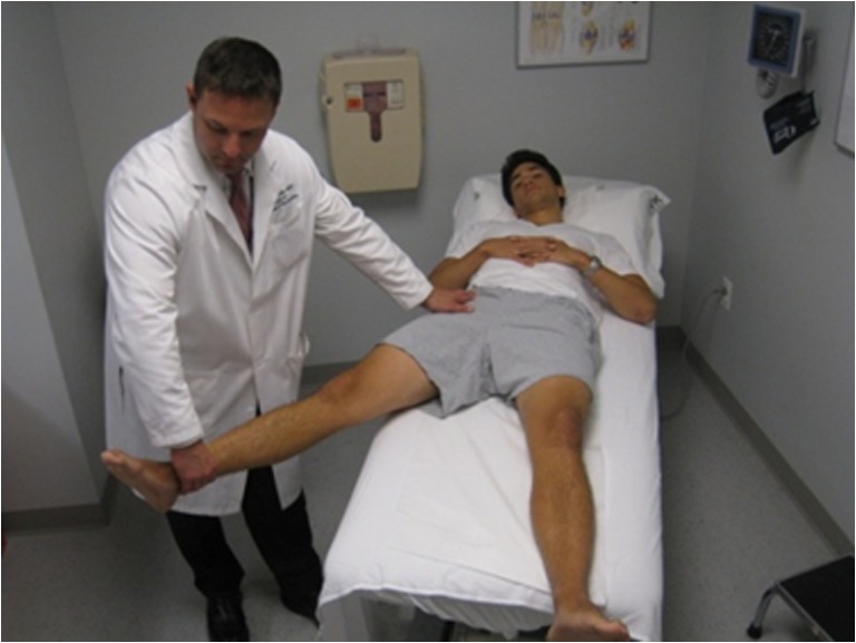
There are specific provocative maneuvers that can enhance the physical examination. The flexion/adduction/internal rotation (FADDIR) test is traditionally performed in the supine position with passive movement of the thigh into 90° of flexion, adduction, and IR (Fig. 2) [50]. Usually, there is anterior or anteromedial pain (positive test) due to impingement of the anterior and anterolateral part of the femoral neck against the superior and anterior acetabular rim.
Fig. 2.
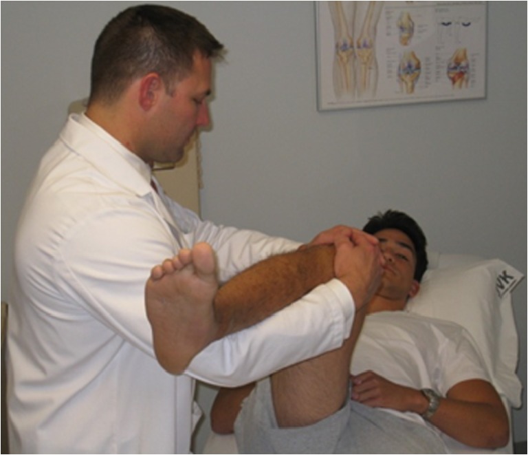
FADDIR test. With the patient in the supine position, the examiner passively brings the hip into 90° of flexion, adduction, and internal rotation. This test can also be performed in the lateral position
The subspine impingement test is performed in the supine position with passive movement of the thigh into maximum flexion, with neutral adduction, and IR. Reproduction of anterior pain indicates impingement of the distal (anterolateral) and medial part of the femoral neck against the AIIS.
The superolateral impingement test is performed with passive movement of the thigh into flexion and ER (Fig. 3). Recreation of anterolateral pain indicates impingement of the superior and superolateral part of the head–neck junction against the superior or acetabular rim.
Fig. 3.
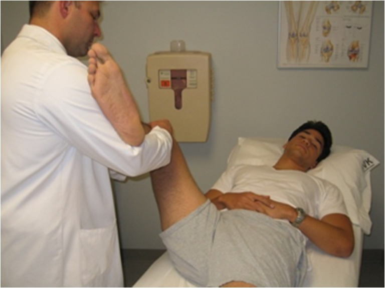
Superolateral impingement test. With the patient in the supine position, the examiner passively brings the hip into flexion, and external rotation
The dynamic external rotatory impingement test (DEXRIT) and the dynamic internal rotatory impingement test (DIRI) are similar to the traditional McCarthy’s test [15, 71]. Both are performed with the contralateral leg maximally flexed to eliminate lumbar lordosis and the affected hip brought to flexion 90°. In the DEXRIT, the hip is passively ranged through a wide arc of abduction and ER. In the DIRI, the hip is passively ranged through a wide arc of adduction and IR. For both maneuvers, the reproduction of patient’s pain in a specific position will correlate with site of bony impingement in a clockwise fashion.
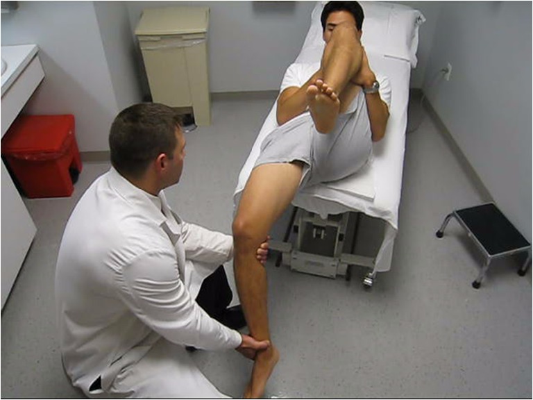
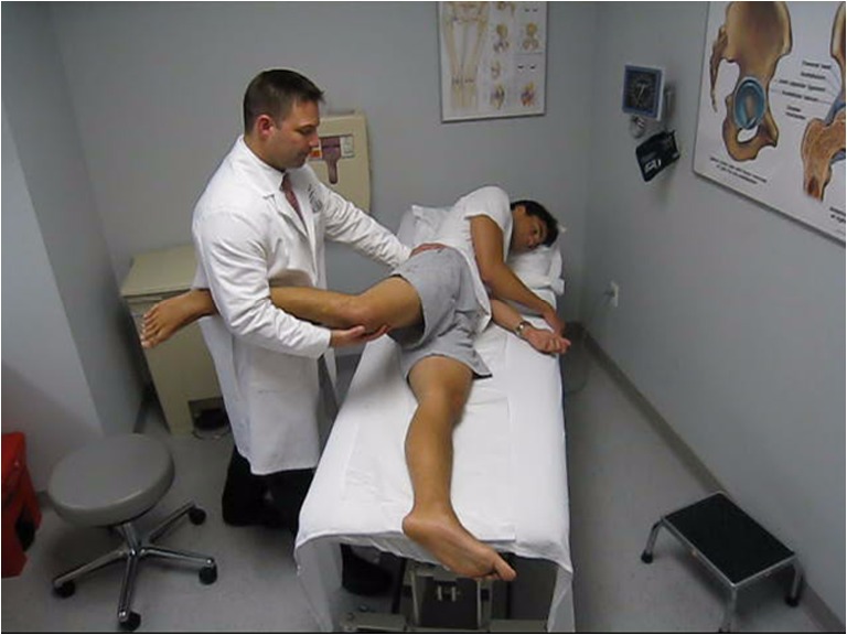
The Patrick flexion abduction external rotation (FABER) test facilitates the differentiation of hip pain in the abducted position. It is performed by laying the ankle of the affected leg across the thigh of the nonaffected leg proximal to the knee joint, creating a Fig. 4 position. This position displaces the anterosuperior part of the femoral head–neck junction to the 12 o’clock position of the acetabular rim. By applying downward pressure onto the knee of the affected leg lateral pain is indicative of superolateral and lateral FAI; groin pain reflects iliopsoas pathology, or psoas impingement against the femoral head [93], or anterior capsule irritation; posterolateral pain is indicative of ischio-trochanteric impingement, especially in cases with increased femoral anteversion (Fig. 4); posterior pain indicates SI joint pathology. Gaenslen’s test places stress on the sacroiliac (SI) joint and could facilitate the differential diagnosis of posterior hip pain [114].
Fig. 4.
FABER-Trochanteric pain test. With the patient in the supine position, the examiner passively brings the hip into flexion, abduction, and external rotation
The posterior rim impingement test is performed with the patient positioned at the edge of the examination table so that the legs hang freely at the hip, and the patient draws up both legs toward the chest, thus eliminating lumbar lordosis. The affected leg is then extended off the table combined with abduction and ER bringing the hip into full extension (Fig. 5), thus allowing assessment of the congruence of the posterolateral part of the femoral neck against the posterior acetabular rim. A positive test is noted when posterior pain is recreated at this position; if anterior pain is recreated, patient may be diagnosed with hip instability [48, 86, 104].
Fig. 5.
Posterior rim Impingement. The patient is positioned at the edge of the examination table so that the legs hang freely at the hip, and the patient draws up both legs toward the chest, thus eliminating lumbar lordosis. The examiner passively brings the affected leg extended off the table, allowing for full extension of the hip, abduction, and external rotation
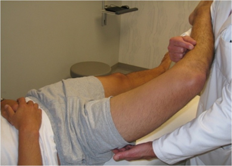
To detect lateral rim impingement the affected leg is abducted (neutral rotation) off the table (Fig. 6). Recreation of lateral pain indicates impingement of the superolateral part of the femoral neck against the superoposterior acetabular rim.
Fig. 6.
Lateral rim impingement. With the patient in the supine position, the examiner passively brings the hip into abduction with neutral rotation
The ischiofemoral impingement sign is attributed to narrowing of the ischiofemoral (distance between lateral cortex of ischial tuberosity and medial cortex of lesser trochanter) and quadratus femoris spaces (space between superolateral surface of hamstring tendons and posteromedial surface of iliopsoas tendon or lesser trochanter). The quadratus femoris muscle may be compressed directly between the lesser trochanter and ischium. Clinically, the symptoms of impingement—pain in the groin and/or buttock which may radiate distally [5, 80, 84]—can be reproduced by a combination of hip extension, adduction, and ER [44]. The insertion of psoas into the lesser trochanter and the origin of the hamstrings may also be affected [84, 110]. Differential diagnosis includes sciatica, chronic hamstring injury, snapping psoas, quadratus femoris tear, and adductor tendonitis [106].
The straight leg raise against resistance test, also known as the Stinchfield test [93], is performed with an active straight leg raise to 45° followed by a direct downward force just proximal to the knee by the examiner. The test is considered positive with reproduction of anterior pain or weakness. This test evaluates hip flexor/psoas strength and indicates intra-articular pathology as the psoas applies pressure on the labrum in active resistance [90].
The Foveal distraction test is performed with axial traction on the abducted 30° leg. This maneuver reduces intra-articular pressure; relief of pain indicates an intra-articular cause.
Lateral Assessment
The lateral assessment (patient lying on the unaffected hip) is very useful in the differential diagnosis of lateral hip pain and can further confirm the presence of intra-articular pathology. Palpation for tenderness focuses especially on the gluteus maximus origin, SI joint, sciatic nerve, piriformis, tensor fascial lata (TFL), ITB, greater trochanteric bursae, and ischial tuberosity [10, 64, 89, 93, 100]. Special attention should be given to the greater trochanter, since it is the site of attachment for five muscles, the gluteus medius and gluteus minimus tendons laterally and the piriformis, obturator externus, and obturator internus more medially. It consists of four distinct facets: the superoposterior, lateral, anterior, and posterior [28]. The posterior facet is the only facet that does not have any distinct tendon attachment but is the primary location of the largest bursae of the peritrochanteric space; consequently, is the likely source of primary pain in patients with true isolated trochanteric bursitis without associated abductor tendon tear [46, 53, 98].
Passive adduction tests are historically similar to Ober’s test performed with the patient positioned on the unaffected hip with the shoulders perpendicular to the table [93]. The examiner assesses full passive hip adduction reproducing the following three tests: (1) the TFL contracture test is performed with the hip and knee in extension, thus placing tension on the TFL when the hip is adducted; (2) the gluteus medius contracture test is performed with 0° of hip extension and 45–90° of knee flexion, thus releasing the ITB and placing tension on the gluteus medius with hip adduction; and (3) the gluteus maximus contracture test is performed with the shoulders rotated back into contact with the table with hip flexion and knee extension, thus placing tension on the gluteus maximus with hip adduction. If the patient cannot perform passive adduction past the midline of the body, all three tests are considered positive, indicative of contracture of each musculature respectively.
The FADDIR and the lateral FABER tests can be performed in the lateral position to confirm intra-articular and peritrochanteric pathology (Fig. 7). Regarding the lateral FABER test, the examiner passively brings the affected hip through a wide arc from flexion to extension in continuous abduction. The lateral position is used to test the normal dynamic pelvic inclination, since the supine position with the contralateral leg flexed eliminates lumbar lordosis. Pelvic inclination may influence physical examination findings, and both positions are valuable in assessment.
Fig. 7.
Lateral FABER test. With the patient in the lateral position, the examiner passively brings the affected hip through a wide arc from flexion to extension in continuous abduction
Prone Assessment
The prone position is optimal for identifying the precise location of pain related to the SI joint region and assessing femoral anteversion. The latter (historically similar to Craig’s test) is performed by flexing the knee to 90°, and using the leg as a lever, the hip is internally rotated until the lateral aspect of the greater trochanter is felt to be most prominent [93]. Femoral anteversion (normally between 8° and 15°) or retroversion is measured by the angle between the tibia and an imaginary vertical line [93]. If there is a significant difference in IR in the extended and seated flexed position, the examiner should differentiate between osseous and ligamentous causes [66]. The Ely and Phelps tests are helpful in diagnosing contractures of the rectus femoris and gracilis muscle, respectively, and both are performed with the patient in the prone position [90].
Due to the higher prevalence of sport-related activities, it is critical to rule out trauma-related cause of hip pain or femoral neck stress fractures. This assessment is performed through the heel strike test, hop test, and the log roll test [10, 65, 90]. If either of these tests is positive, further radiographic evaluation is warranted [10, 90].
Layered Approach to Mechanical Hip Pain
In order to assess the painful hip in a systematic and comprehensive way, the senior author (BTK) has developed an algorithm to approach and understand these often complex compensatory problems. It is described below as the “layered approach” to understand the underlying etiologic factors contributing to pain around the hip joint and associated hemipelvis. During the diagnostic process, it may be helpful to categorize the hip as structurally normal, structurally overcovered, or structurally undercovered. A structurally normal hip will have values that fall within a normal range for center edge angle, hip valgus, and hip version values. A structurally undercovered hip will diagnostically present with anteversion, hip valgus, or dysplastic characteristics. Comparatively, overcoverage will diagnostically present as cam lesion at head neck junction, rim lesion, often associated with acetabular retroversion, acetabular profunda, or acetabular protrusio [6]. Recognizing and attempting to comprehend these osseous, inert, contractile, and neuromechanical relationships and differences, as they relate to normal osseous structure, osseous overcoverage and osseous undercoverage, are what led to the development of the layered concept (Table 2).
Table 2.
Layered approach to clinical assessment of the hip joint (adapted from Draovitch et al. [26])
| Layer | Name | Structures | Purpose | Pathology |
|---|---|---|---|---|
| Layer I | Osteochondral layer | Femur | Joint congruence and normal osteo-/arthro-kinematics | Dynamic impingement |
| Pelvis | Cam, Rim, Femoral retroversion, femoral varus, acetabular overcoverage (focal or global), trochanteric impingement, subspine impingement | |||
| Acetabulum | Static overload | |||
| Acetabular undercoverage, femoral anteversion, Femoral valgus, acetabular version | ||||
| Dynamic Instability | ||||
| Layer 2 | Inert layer | Labrum | Static stability of the hip joint | Labral injury |
| Joint capsule | Ligamentum teres tear | |||
| Ligamentous complex | Ligament tears | |||
| Ligamentum teres | Capsular instability, adhesive capsulitis | |||
| Layer 3 | Contractile layer | Peri-articular musculature | Dynamic stability of the hip, pelvis and trunk | Anterior |
| Anterior structures | Rectus femoris tendonopathy, Psoas, subspine | |||
| Medial structures | Medial | |||
| Posterior structures | Adductor strain, Rectus abdominis strain, osteitis pubis | |||
| Lateral structures | Posterior | |||
| Lumbosacral and pelvic floor | Proximal hamstring, deep gluteal syndrome | |||
| Lateral | ||||
| Abductor tears, ITB syndrome, bursitis | ||||
| Layer 4 | Neuromechanical layer | Neural | Properly sequenced kinetic linking and appropriately balance neuromuscular control presence or absence of neuromechanical shortcomings | Neural |
| Femoral, lateral | Pain syndromes | |||
| Femoral | Neuromuscular dysfunction | |||
| Cutaneous, sciatic, | Spinal referral patterns | |||
| Ilioinguinal, | Nerve entrapments | |||
| Genitofemoral, | Mechanical | |||
| Pudendal, and | Foot structure and mechanics | |||
| Iliohypogastric nerves | Scoliosis | |||
| Regional mechanoreceptors | Pelvic posture over femur | |||
| Thoraco-lumbar mechanichs | Osteitis pubis, Pubic symphysis pathology | |||
| Lower extremity mechanics | Sacro-iliac dysfunction | |||
Layer I (Table 2) is the osteochondral layer, which aims to provide joint congruence and normal osteoarticular kinematics in the normal hip. The structures within this layer are the pelvis, acetabulum, and femur. Abnormalities within this layer can be classified into three distinct groups: (1) static overload, (2) dynamic impingement, and (3) dynamic instability. Anatomical variations resulting in static overload include lateral or anterior acetabular undercoverage/dysplasia, femoral anteversion, and femoral valgus. These structural mechanics lead to eccentric load, abnormal, and increased stress and asymmetric loads between the femoral head and acetabular socket in the axially loaded position (i.e., standing). During hip motion, dynamic factors may contribute to hip pain as abnormal stress and contact between the femoral head and acetabular rim occur. Different structural variations within layer 1 that may contribute to such dynamic impingement include FAI (cam and focal or global pincer impingement), femoral retroversion, and femoral varus (Table 2). When the functional range of motion required to compete in sports or for daily activities is greater than the amount of physiologic motion allowed by the anatomical structures of the hip, a compensatory increase in motion may be provided through layer 1. Specifically, increased motion and consequential stresses through the pubic symphysis, SI joint, and lumbar spine may initiate. When functional range of motion requirements are larger than normal motion limits, forceful anterior contact occurring at the end range of IR may lead to dynamic instability in the form of subtle posterior hip subluxation, which occurs as the femoral head levers out of the hip socket [54]. Various radiographic indices calculated on plain X-rays [standing AP pelvis, elongated-neck lateral view (Dunn lateral radiograph), and false profile], such as Tonnis OA grade, coronal CE angle, Tonnis angle, and variables derived from the three-dimensional CT scan (alpha-angle, beta-angle, McKibbon indices, acetabular version, coronal and sagittal CE angle, neck-shaft angle, and femoral version), which better delineates the bone anatomy, can facilitate the mechanical diagnosis, and specifically whether there is a structurally normal, undercovered, or overcovered hip (Figs. 1, 8, and 9) Computer navigation surgical planning software can be used to confirm and model osseous impingements [12, 36]. These resultant mechanical stresses lead to reactive hip pain related to insufficient congruency or impingement between the head and socket, leading to asymmetric wear of the chondral surfaces of the acetabulum and femoral head with or without associated instability of the hip. Thus, layer I has a direct effect on the inert layer of the hip, or layer II.
Fig. 8.
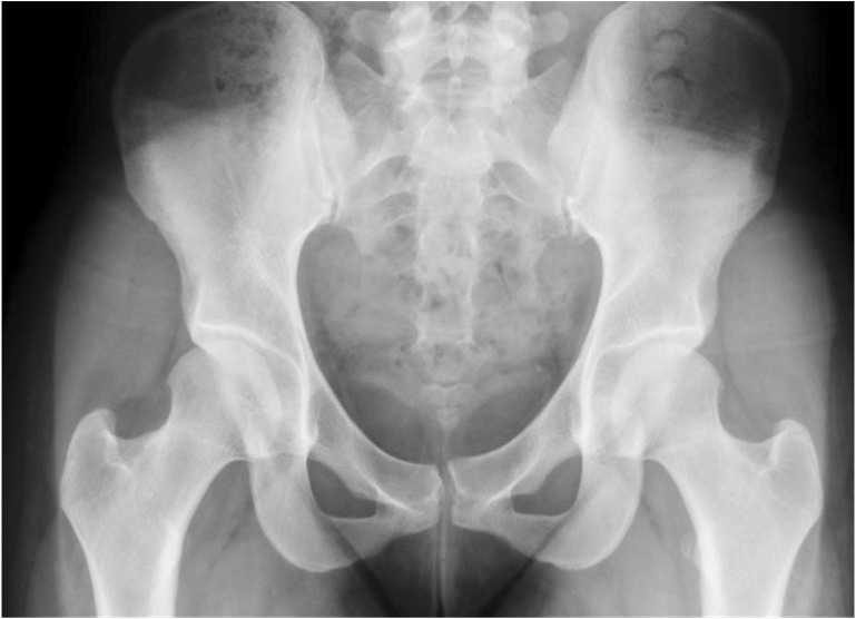
Anteroposterior view of the pelvis demonstrating cross-over sign of the right hip
Fig. 9.

Dunn lateral view of the right hip demonstrating the calculation of the alpha-angle indicative of significant cam deformity (alpha angle > 50º)
Layer II (Table 2) includes the labrum, joint capsule, ligamentous complex, and ligamentum teres. These structures contribute to static stability of the hip joint. When abnormal mechanical stresses are applied to the hip joint secondary to underlying abnormalities within layer 1, pathologies such as labral injury, ligamentum teres tear, capsular irritation and consequent instability or adhesive capsulitis, and various ligament tears can result. MRI can help evaluate the chondral, labral, and capsular damage; specifically, delayed gadolinium-enhanced MRI of cartilage studies can be used to assess the cartilage health [9]. A significant relationship and interaction exists between layers I and II. Venting the capsule and creating a labral tear decreased the forces to distract the head of the femur by 3 mm by 43% and 60%, respectively. Loss of maintaining a suction seal would decrease intra-articular hydrostatic pressure and allow the translation of hip center may be as much as 2–5 mm, thus further stressing inert (layer II) and contractile tissue (layer III) [31]. In addition, a loose hip may lead to abnormal sites of bony impingement. Range of motion requirements of the joint, which are specific-related to the activities, combined with the underlying structural mechanics of layer 1 can predict the type of injury to layer II structures. Intra-articular injections are very useful and reliable to differentiate between intra- and extra-articular hip pathology and specifically to confirm pathology of the inert layer [18].
Layer III (Table 2) is the contractile layer of the hip and hemipelvis. It consists of all musculature around the hemipelvis including the lumbosacral musculature and pelvic floor; it is responsible for the muscular balance and dynamic stability of the hip, pelvis, and trunk. Abnormal mechanics within layers I and II can lead to increased stresses of the lower spine, SI joint, pubic symphysis, and ischium, and secondary increases in the strains of the muscles attached to these pelvic structures. The mechanism may be acute, traumatic, overuse tendinosis, or developmental avulsions [41]. These compensatory injuries can result in a variety of peri-articular muscular structures and can be subcategorized based upon their location (origin or insertion) and relative to the hip joint (anterior, medial, posterior, and lateral). Anterior enthesopathy describes hip flexor strains, psoas impingement, and subspine impingement. Medial enthesopathy encompasses adductor and rectus tendinopathies, which have traditionally been described as athletic pubalgia or “sports hernia.” Posterior enthesopathies include mainly proximal hamstring strains but can also include injuries to the short external rotators including the piriformis and may involve a constellation of pain patterns described as “deep gluteal syndrome,” which involves posterior soft-tissue injury and irritation or compression of the sciatic nerve. Lateral enthesopathies involve the peritrochanteric space and injuries to the gluteus medius and minimus tendons. Casartelli et al. [21] have reported that patients with FAI presented with decreased maximal voluntary contraction levels for the hip adduction (28%), flexion (26%), external rotation (18%), and abduction (11%) when compared with the control group, demonstrating the contractile dysfunction occurring as a result of structural pathology and pain. The TFL has also demonstrated decreased activation during hip flexion in patients diagnosed with FAI. Similarly, an increased cross-sectional area of iliocapsularis muscle in dysplastic hips has been also reported [3], representative of dynamic stabilization to combat loss of inert tissue integrity. MRI and specific injections have been diagnostically sensitive to layer III contractile tissue direct involvement and indirect enthesiopathies. Specific patterns of pathology in layer I can be associated with specific compensatory injury patterns within layer III. Moreover, it has been demonstrated that loss of hip motion will adversely affect motion throughout the entire kinetic chain; specifically, in closed kinetic chain activities, limited hip motion may predispose to noncontact injury [29, 94].
Layer IV (Table 2) is the neurokinetic layer, including the thoracolumbosacral plexus, lumbopelvic tissue, and lower extremity structures. This layer serves as the neuromuscular link and thus functional control of the entire segment as it functions within its environment. Locally at the site of the hip, this layer refers to the neuro-vascular structures, mechanoreceptors, and nociceptors. On a global level, this layer refers to posture and the position of the pelvis over the femur. This may be affected by the result of lumbar pathology on the hip resulting in sacral torsion, rotation of the innominate, or myotomal changes, or changes in foot and ankle mechanics and the response of the lower extremity up to the hip. It also involves looking at functional movement patterns and examining how motor learning affects dynamic movement of the pelvis over the femur or the femur under the pelvis. Compensatory injuries within this layer include nerve compression and pain syndromes, neuromuscular dysfunction, and spine referral patterns. Common peripheral nerve disorders about the hip include lateral femoral cutaneous neuropathy (meralgia paresthetica), femoral neuropathy, sciatic neuropathy (piriformis syndrome), obturator neuropathy, superior and inferior gluteal neuropathies, pudendal neuropathy, and ilioinguinal, iliohypogastric, and genitofemoral neuropathies [2, 43, 51, 67, 69, 77].
Summary
In conclusion, it is vitally important to elucidate intra- versus extra-articular pathology of hip pain in every step of the patient encounter: history, clinical examination, imaging, and mechanical diagnosis. It is critical to follow a systematic approach to physical examination of the hip. It is of paramount importance to comprehend that a loaded pelvis usually rotates over a fixed femur, thus creating anterior and medial forces with instant rotary moments. If these forces are combined with dynamic impingement, static overload to the joint, or instability, it can be explained how structural abnormalities in layer I can result in various damage patterns in layers II, III, and IV, depending on patient’s individual anatomy and required functional range of motion of the hip joint. Therefore, following a functional movement exam and a spine screening (layer IV), the clinical exam of the hip should begin from layer I and move out toward layer III. A series of specific tests may be used to in examining the layers. Overall, the location and quality of the pain should correspond to the mechanical diagnosis and primary and secondary injury patterns. If so, then correcting the mechanical problems and primary and secondary injuries should optimize the outcome.
Disclosures
Each author certifies that he or she has no commercial associations (e.g., consultancies, stock ownership, equity interest, patent/licensing arrangements, etc.) that might pose a conflict of interest in connection with the submitted article.
References
- 1.Allen WC, Cope R. Coxa saltans: the snapping hip revisited. J Am Acad Orthop Surg. 1995;5:303–308. doi: 10.5435/00124635-199509000-00006. [DOI] [PubMed] [Google Scholar]
- 2.Antolak SJ, Jr, Hough DM, Pawlina W, Spinner RJ. Anatomical basis of chronic pelvic pain syndrome: the ischial spine and pudendal nerve entrapment. Med Hypotheses. 2002;3:349–353. doi: 10.1016/s0306-9877(02)00218-9. [DOI] [PubMed] [Google Scholar]
- 3.Babst D, Steppacher SD, Ganz R, Siebenrock KA, Tannast M. The iliocapsularis muscle: an important stabilizer in the dysplastic hip. Clin Orthop Relat Res. 2011;6:1728–1734. doi: 10.1007/s11999-010-1705-x. [DOI] [PMC free article] [PubMed] [Google Scholar]
- 4.Baker CL, Jr, Massie RV, Hurt WG, Savory CG. Arthroscopic bursectomy for recalcitrant trochanteric bursitis. Arthroscopy. 2007;8:827–832. doi: 10.1016/j.arthro.2007.02.015. [DOI] [PubMed] [Google Scholar]
- 5.Bano A, Karantanas A, Pasku D, Datseris G, Tzanakakis G, Katonis P. Persistent sciatica induced by quadratus femoris muscle tear and treated by surgical decompression: A case report. J Med Case Reports. 2010;4:236. [DOI] [PMC free article] [PubMed]
- 6.Bedi A, Dolan M, Leunig M, Kelly BT. Static and dynamic mechanical causes of hip pain. Arthroscopy. 2011;2:235–251. doi: 10.1016/j.arthro.2010.07.022. [DOI] [PubMed] [Google Scholar]
- 7.Bellabarba C, Sheinkop MB, Kuo KN. Idiopathic hip instability. an unrecognized cause of coxa saltans in the adult. Clin Orthop Relat Res. 1998;355:261–271. [PubMed] [Google Scholar]
- 8.Bird PA, Oakley SP, Shnier R, Kirkham BW. Prospective evaluation of magnetic resonance imaging and physical examination findings in patients with greater trochanteric pain syndrome. Arthritis Rheum. 2001;9:2138–2145. doi: 10.1002/1529-0131(200109)44:9<2138::AID-ART367>3.0.CO;2-M. [DOI] [PubMed] [Google Scholar]
- 9.Bittersohl B, Hosalkar HS, Apprich S, Werlen SA, Siebenrock KA, Mamisch TC. Comparison of pre-operative dGEMRIC imaging with intra-operative findings in femoroacetabular impingement: preliminary findings. Skeletal Radiol. 2011;5:553–561. doi: 10.1007/s00256-010-1038-6. [DOI] [PubMed] [Google Scholar]
- 10.Braly BA, Beall DP, Martin HD. Clinical examination of the athletic hip. Clin Sports Med. 2006;2:199–210. doi: 10.1016/j.csm.2005.12.001. [DOI] [PubMed] [Google Scholar]
- 11.Brown RA, Mascia A, Kinnear DG, Lacroix V, Feldman L, Mulder DS. An 18-year review of sports groin injuries in the elite hockey player: clinical presentation, new diagnostic imaging, treatment, and results. Clin J Sport Med. 2008;3:221–226. doi: 10.1097/JSM.0b013e318172831a. [DOI] [PubMed] [Google Scholar]
- 12.Brunner A, Horisberger M, Herzog R. Evaluation of a computed tomography-based navigation system prototype for hip arthroscopy in the treatment of femoroacetabular cam impingement. Arthroscopy. 2009;4:382–391. doi: 10.1016/j.arthro.2008.11.012. [DOI] [PubMed] [Google Scholar]
- 13.Bunker TD, Esler CN, Leach WJ. Rotator-cuff tear of the hip. J Bone Joint Surg Br. 1997;4:618–620. doi: 10.1302/0301-620x.79b4.7033. [DOI] [PubMed] [Google Scholar]
- 14.Burnett RS, Della Rocca GJ, Prather H, Curry M, Maloney WJ, Clohisy JC. Clinical presentation of patients with tears of the acetabular labrum. J Bone Joint Surg Am. 2006;7:1448–1457. doi: 10.2106/JBJS.D.02806. [DOI] [PubMed] [Google Scholar]
- 15.Busconi BD OB. Differential diagnosis of the painful hip. In: McCarthy JC, ed. Early hip disorders. New York: Springer; 2003:7–16.
- 16.Byrd JW. Evaluation and management of the snapping iliopsoas tendon. Instr Course Lect. 2006;55:347–355. [PubMed]
- 17.Byrd JW. Hip arthroscopy: surgical indications. Arthroscopy. 2006;12:1260–1262. doi: 10.1016/j.arthro.2006.08.021. [DOI] [PubMed] [Google Scholar]
- 18.Byrd JW, Jones KS. Diagnostic accuracy of clinical assessment, magnetic resonance imaging, magnetic resonance arthrography, and intra-articular injection in hip arthroscopy patients. Am J Sports Med. 2004;7:1668–1674. doi: 10.1177/0363546504266480. [DOI] [PubMed] [Google Scholar]
- 19.Byrd JW, Jones KS. Traumatic rupture of the ligamentum teres as a source of hip pain. Arthroscopy. 2004;4:385–391. doi: 10.1016/j.arthro.2004.01.025. [DOI] [PubMed] [Google Scholar]
- 20.Byrd JW, Jones KS. Adhesive capsulitis of the hip. Arthroscopy. 2006;1:89–94. doi: 10.1016/j.arthro.2005.10.009. [DOI] [PubMed] [Google Scholar]
- 21.Casartelli NC, Maffiuletti NA, Item-Glatthorn JF, Staehli S, Bizzini M, Impellizzeri FM, Leunig M. Hip muscle weakness in patients with symptomatic femoroacetabular impingement. Osteoarthritis Cartilage. 2011;7:816–821. doi: 10.1016/j.joca.2011.04.001. [DOI] [PubMed] [Google Scholar]
- 22.Cho KH, Lee SM, Lee YH, Suh KJ, Kim SM, Shin MJ, Jang HW. Non-infectious ischiogluteal bursitis: MRI findings. Korean J Radiol. 2004;4:280–286. doi: 10.3348/kjr.2004.5.4.280. [DOI] [PMC free article] [PubMed] [Google Scholar]
- 23.Crawford K, Philippon MJ, Sekiya JK, Rodkey WG, Steadman JR. Microfracture of the hip in athletes. Clin Sports Med. 2006;2:327–335. [DOI] [PubMed]
- 24.Cvitanic O, Henzie G, Skezas N, Lyons J, Minter J. MRI diagnosis of tears of the hip abductor tendons (gluteus medius and gluteus minimus) AJR Am J Roentgenol. 2004;1:137–143. doi: 10.2214/ajr.182.1.1820137. [DOI] [PubMed] [Google Scholar]
- 25.DeAngelis NA, Busconi BD. Assessment and differential diagnosis of the painful hip. Clin Orthop Relat Res. 2003;406:11–18. doi: 10.1097/01.blo.0000043039.84315.be. [DOI] [PubMed] [Google Scholar]
- 26.Draovitch P, Edelstein J, Kelly BT. The layer concept: utilization in determining the pain generators, pathology and how structure determines treatment. Curr Rev Musculoskelet Med. 2012;1:1–8. doi: 10.1007/s12178-011-9105-8. [DOI] [PMC free article] [PubMed] [Google Scholar]
- 27.Dreyfuss P, Dreyer SJ, Cole A, Mayo K. Sacroiliac joint pain. J Am Acad Orthop Surg. 2004;4:255–265. doi: 10.5435/00124635-200407000-00006. [DOI] [PubMed] [Google Scholar]
- 28.Dwek J, Pfirrmann C, Stanley A, Pathria M, Chung CB. MR imaging of the hip abductors: normal anatomy and commonly encountered pathology at the greater trochanter. Magn Reson Imaging Clin N Am. 2005;4:691–704. doi: 10.1016/j.mric.2005.08.004. [DOI] [PubMed] [Google Scholar]
- 29.Ellera Gomes JL, Palma HM, Becker R. Radiographic findings in restrained hip joints associated with ACL rupture. Knee Surg Sports Traumatol Arthrosc. 2010;11:1562–1567. doi: 10.1007/s00167-010-1175-4. [DOI] [PubMed] [Google Scholar]
- 30.Farber AJ, Wilckens JH. Sports hernia: diagnosis and therapeutic approach. J Am Acad Orthop Surg. 2007;8:507–514. doi: 10.5435/00124635-200708000-00007. [DOI] [PubMed] [Google Scholar]
- 31.Ferguson SJ, Bryant JT, Ganz R, Ito K. An in vitro investigation of the acetabular labral seal in hip joint mechanics. J Biomech. 2003;2:171–178. doi: 10.1016/s0021-9290(02)00365-2. [DOI] [PubMed] [Google Scholar]
- 32.Flanum ME, Keene JS, Blankenbaker DG, Desmet AA. Arthroscopic treatment of the painful “internal” snapping hip: results of a new endoscopic technique and imaging protocol. Am J Sports Med. 2007;5:770–779. doi: 10.1177/0363546506298580. [DOI] [PubMed] [Google Scholar]
- 33.Fredericson M, Moore W, Guillet M, Beaulieu C. High hamstring tendinopathy in runners: meeting the challenges of diagnosis, treatment, and rehabilitation. Phys Sportsmed. 2005;5:32–43. doi: 10.3810/psm.2005.05.89. [DOI] [PubMed] [Google Scholar]
- 34.Ganz R, Parvizi J, Beck M, Leunig M, Notzli H, Siebenrock KA. Femoroacetabular impingement: a cause for osteoarthritis of the hip. Clin Orthop Relat Res. 2003;417:112–120. doi: 10.1097/01.blo.0000096804.78689.c2. [DOI] [PubMed] [Google Scholar]
- 35.Griffiths HJ, Utz R, Burke J, Bonfiglio T. Adhesive capsulitis of the hip and ankle. AJR Am J Roentgenol. 1985;1:101–105. doi: 10.2214/ajr.144.1.101. [DOI] [PubMed] [Google Scholar]
- 36.Grissom L, Harcke HT, Thacker M. Imaging in the surgical management of developmental dislocation of the hip. Clin Orthop Relat Res. 2008;4:791–801. doi: 10.1007/s11999-008-0161-3. [DOI] [PMC free article] [PubMed] [Google Scholar]
- 37.Haene RA, Bradley M, Villar RN. Hip dysplasia and the torn acetabular labrum: an inexact relationship. J Bone Joint Surg Br. 2007;10:1289–1292. doi: 10.1302/0301-620X.89B10.17319. [DOI] [PubMed] [Google Scholar]
- 38.Harvey WF, Yang M, Cooke TD, Segal NA, Lane N, Lewis CE, Felson DT. Association of leg-length inequality with knee osteoarthritis: a cohort study. Ann Intern Med. 2010;5:287–295. doi: 10.1059/0003-4819-152-5-201003020-00006. [DOI] [PMC free article] [PubMed] [Google Scholar]
- 39.Hickman JM, Peters CL. Hip pain in the young adult: diagnosis and treatment of disorders of the acetabular labrum and acetabular dysplasia. Am J Orthop (Belle Mead NJ). 2001;6:459–467. [PubMed] [Google Scholar]
- 40.Howell GE, Biggs RE, Bourne RB. Prevalence of abductor mechanism tears of the hips in patients with osteoarthritis. J Arthroplasty. 2001;1:121–123. doi: 10.1054/arth.2001.19158. [DOI] [PubMed] [Google Scholar]
- 41.Ilizaliturri VM, Jr, Camacho-Galindo J, Evia Ramirez AN, Gonzalez Ibarra YL, McMillan S, Busconi BD. Soft tissue pathology around the hip. Clin Sports Med. 2011;2:391–415. doi: 10.1016/j.csm.2010.12.009. [DOI] [PubMed] [Google Scholar]
- 42.Ilizaliturri VM, Jr, Villalobos FE, Jr, Chaidez PA, Valero FS, Aguilera JM. Internal snapping hip syndrome: treatment by endoscopic release of the iliopsoas tendon. Arthroscopy. 2005;11:1375–1380. doi: 10.1016/j.arthro.2005.08.021. [DOI] [PubMed] [Google Scholar]
- 43.Irshad K, Feldman LS, Lavoie C, Lacroix VJ, Mulder DS, Brown RA. Operative management of “hockey groin syndrome”: 12 years of experience in national hockey league players. Surgery. 2001;4:759–764. doi: 10.1067/msy.2001.118093. [DOI] [PubMed] [Google Scholar]
- 44.Johnson KA. Impingement of the lesser trochanter on the ischial ramus after total hip arthroplasty. report of three cases. J Bone Joint Surg Am. 1977;2:268–269. [PubMed] [Google Scholar]
- 45.Kagan A., 2nd Rotator cuff tears of the hip. Clin Orthop Relat Res. 1999;368:135–140. [PubMed] [Google Scholar]
- 46.Kassarjian A, Yoon LS, Belzile E, Connolly SA, Millis MB, Palmer WE. Triad of MR arthrographic findings in patients with cam-type femoroacetabular impingement. Radiology. 2005;2:588–592. doi: 10.1148/radiol.2362041987. [DOI] [PubMed] [Google Scholar]
- 47.Kelly BT, Weiland DE, Schenker ML, Philippon MJ. Arthroscopic labral repair in the hip: surgical technique and review of the literature. Arthroscopy. 2005;12:1496–1504. doi: 10.1016/j.arthro.2005.08.013. [DOI] [PubMed] [Google Scholar]
- 48.Kelly BT, Williams RJ, 3rd, Philippon MJ. Hip arthroscopy: current indications, treatment options, and management issues. Am J Sports Med. 2003;6:1020–1037. doi: 10.1177/03635465030310060701. [DOI] [PubMed] [Google Scholar]
- 49.Khanduja V, Villar RN. The arthroscopic management of femoroacetabular impingement. Knee Surg Sports Traumatol Arthrosc. 2007;8:1035–1040. doi: 10.1007/s00167-007-0319-7. [DOI] [PubMed] [Google Scholar]
- 50.Klaue K, Durnin CW, Ganz R. The acetabular rim syndrome. A clinical presentation of dysplasia of the hip. J Bone Joint Surg Br. 1991;3:423–429. doi: 10.1302/0301-620X.73B3.1670443. [DOI] [PubMed] [Google Scholar]
- 51.Knockaert DC, D’Heygere FG, Bobbaers HJ. Ilioinguinal nerve entrapment: a little-known cause of iliac fossa pain. Postgrad Med J. 1989;767:632–635. doi: 10.1136/pgmj.65.767.632. [DOI] [PMC free article] [PubMed] [Google Scholar]
- 52.Kong A, Vliet A, Zadow S. MRI and US of gluteal tendinopathy in greater trochanteric pain syndrome. Eur Radiol. 2007;7:1772–1783. doi: 10.1007/s00330-006-0485-x. [DOI] [PubMed] [Google Scholar]
- 53.Kramer J, Recht MP. MR arthrography of the lower extremity. Radiol Clin North Am. 2002;5:1121–1132. doi: 10.1016/s0033-8389(02)00057-x. [DOI] [PubMed] [Google Scholar]
- 54.Krych AJ, Larson CM, Byrd TW, Warren RF, Kelly BT. Low energy posterior wall fractures in athletes: Evidence of cam induced posterior hip subluxation. Presented at 2011 NFL Combine Physicians Society Meeting.
- 55.Kusma M, Jung J, Dienst M, Goedde S, Kohn D, Seil R. Arthroscopic treatment of an avulsion fracture of the ligamentum teres of the hip in an 18-year-old horse rider. Arthroscopy. 2004;20:64–66. [DOI] [PubMed]
- 56.Larson CM, Kelly BT, Stone RM. Making a case for anterior inferior iliac spine/subspine hip impingement: three representative case reports and proposed concept. Arthroscopy. 2011;12:1732–1737. doi: 10.1016/j.arthro.2011.10.004. [DOI] [PubMed] [Google Scholar]
- 57.Larson CM, Pierce BR, Giveans MR. Treatment of athletes with symptomatic intra-articular hip pathology and athletic pubalgia/sports hernia: a case series. Arthroscopy. 2011;6:768–775. doi: 10.1016/j.arthro.2011.01.018. [DOI] [PubMed] [Google Scholar]
- 58.Lavigne M, Parvizi J, Beck M, Siebenrock KA, Ganz R, Leunig M. Anterior femoroacetabular impingement: part I. techniques of joint preserving surgery. Clin Orthop Relat Res. 2004;418:61–66. [PubMed] [Google Scholar]
- 59.Lempainen L, Sarimo J, Mattila K, Vaittinen S, Orava S. Proximal hamstring tendinopathy: results of surgical management and histopathologic findings. Am J Sports Med. 2009;4:727–734. doi: 10.1177/0363546508330129. [DOI] [PubMed] [Google Scholar]
- 60.Lequesne M, Djian P, Vuillemin V, Mathieu P. Prospective study of refractory greater trochanter pain syndrome. MRI findings of gluteal tendon tears seen at surgery. Clinical and MRI results of tendon repair. Joint Bone Spine. 2008;4:458–464. doi: 10.1016/j.jbspin.2007.12.004. [DOI] [PubMed] [Google Scholar]
- 61.Lequesne M, Mathieu P, Vuillemin-Bodaghi V, Bard H, Djian P. Gluteal tendinopathy in refractory greater trochanter pain syndrome: diagnostic value of two clinical tests. Arthritis Rheum. 2008;2:241–246. doi: 10.1002/art.23354. [DOI] [PubMed] [Google Scholar]
- 62.Little TL, Mansoor J. Low back pain associated with internal snapping hip syndrome in a competitive cyclist. Br J Sports Med. 2008;4:308–309. doi: 10.1136/bjsm.2007.039560. [DOI] [PubMed] [Google Scholar]
- 63.Longjohn D, Dorr LD. Soft tissue balance of the hip. J Arthroplasty. 1998;1:97–100. doi: 10.1016/s0883-5403(98)90082-1. [DOI] [PubMed] [Google Scholar]
- 64.Margo K, Drezner J, Motzkin D. Evaluation and management of hip pain: an algorithmic approach. J Fam Pract. 2003;8:607–617. [PubMed] [Google Scholar]
- 65.Martin HD, Kelly BT, Leunig M, Philippon MJ, Clohisy JC, Martin RL, Sekiya JK, Pietrobon R, Mohtadi NG, Sampson TG, Safran MR. The pattern and technique in the clinical evaluation of the adult hip: the common physical examination tests of hip specialists. Arthroscopy. 2010;2:161–172. doi: 10.1016/j.arthro.2009.07.015. [DOI] [PubMed] [Google Scholar]
- 66.Martin HD, Savage A, Braly BA, Palmer IJ, Beall DP, Kelly B. The function of the hip capsular ligaments: a quantitative report. Arthroscopy. 2008;2:188–195. doi: 10.1016/j.arthro.2007.08.024. [DOI] [PubMed] [Google Scholar]
- 67.Martin HD, Shears SA, Johnson JC, Smathers AM, Palmer IJ. The endoscopic treatment of sciatic nerve entrapment/deep gluteal syndrome. Arthroscopy. 2011;2:172–181. doi: 10.1016/j.arthro.2010.07.008. [DOI] [PubMed] [Google Scholar]
- 68.Martin HD, Shears SA, Palmer IJ. Evaluation of the hip. Sports Med Arthrosc. 2010;2:63–75. doi: 10.1097/JSA.0b013e3181dc578a. [DOI] [PubMed] [Google Scholar]
- 69.Martinoli C, Miguel-Perez M, Padua L, Gandolfo N, Zicca A, Tagliafico A. Imaging of neuropathies about the hip. Eur J Radiol. 2011. doi:10.1016/j.ejrad.2011.04.034. [DOI] [PubMed]
- 70.Mattila VM, Niva M, Kiuru M, Pihlajamaki H. Risk factors for bone stress injuries: a follow-up study of 102,515 person-years. Med Sci Sports Exerc. 2007;7:1061–1066. doi: 10.1249/01.mss.0b013e318053721d. [DOI] [PubMed] [Google Scholar]
- 71.McCarthy JC, Busconi BD, Owens BD. Assessment of the painful hip. In: IMcCarthy JC ed. Early hip disorders. New York: Springer; 2003:3–6.
- 72.McCarthy J, Noble P, Aluisio FV, Schuck M, Wright J, Lee JA. Anatomy, pathologic features, and treatment of acetabular labral tears. Clin Orthop Relat Res. 2003;406:38–47. doi: 10.1097/01.blo.0000043042.84315.17. [DOI] [PubMed] [Google Scholar]
- 73.Meyers WC, Foley DP, Garrett WE, Lohnes JH, Mandlebaum BR. Management of severe lower abdominal or inguinal pain in high-performance athletes. PAIN (performing athletes with abdominal or inguinal neuromuscular pain study group) Am J Sports Med. 2000;1:2–8. doi: 10.1177/03635465000280011501. [DOI] [PubMed] [Google Scholar]
- 74.Meyers WC, McKechnie A, Philippon MJ, Horner MA, Zoga AC, Devon ON. Experience with “sports hernia” spanning two decades. Ann Surg. 2008;4:656–665. doi: 10.1097/SLA.0b013e318187a770. [DOI] [PubMed] [Google Scholar]
- 75.Moeller JL. Pelvic and hip apophyseal avulsion injuries in young athletes. Curr Sports Med Rep. 2003;2:110–115. doi: 10.1249/00149619-200304000-00011. [DOI] [PubMed] [Google Scholar]
- 76.Mullis BH, Dahners LE. Hip arthroscopy to remove loose bodies after traumatic dislocation. J Orthop Trauma. 2006;1:22–26. doi: 10.1097/01.bot.0000188038.66582.ed. [DOI] [PubMed] [Google Scholar]
- 77.Murata Y, Ogata S, Ikeda Y, Yamagata M. An unusual cause of sciatic pain as a result of the dynamic motion of the obturator internus muscle. Spine J. 2009;6:e16–e18. doi: 10.1016/j.spinee.2009.01.004. [DOI] [PubMed] [Google Scholar]
- 78.Niva MH, Kiuru MJ, Haataja R, Pihlajamaki HK. Fatigue injuries of the femur. J Bone Joint Surg Br. 2005;10:1385–1390. doi: 10.1302/0301-620X.87B10.16666. [DOI] [PubMed] [Google Scholar]
- 79.Northmore-Ball MD. Young adults with arthritic hips. BMJ. 1997;7103:265–266. doi: 10.1136/bmj.315.7103.265. [DOI] [PMC free article] [PubMed] [Google Scholar]
- 80.O’Brien SD, Bui-Mansfield LT. MRI of quadratus femoris muscle tear: another cause of hip pain. AJR Am J Roentgenol. 2007;5:1185–1189. doi: 10.2214/AJR.07.2408. [DOI] [PubMed] [Google Scholar]
- 81.Orava S, Ala-Ketola L. Avulsion fractures in athletes. Br J Sports Med. 1977;2:65–71. doi: 10.1136/bjsm.11.2.65. [DOI] [PMC free article] [PubMed] [Google Scholar]
- 82.Paajanen H, Hermunen H, Karonen J. Pubic magnetic resonance imaging findings in surgically and conservatively treated athletes with osteitis pubis compared to asymptomatic athletes during heavy training. Am J Sports Med. 2008;1:117–121. doi: 10.1177/0363546507305454. [DOI] [PubMed] [Google Scholar]
- 83.Parvizi J, Leunig M, Ganz R. Femoroacetabular impingement. J Am Acad Orthop Surg. 2007;9:561–570. doi: 10.5435/00124635-200709000-00006. [DOI] [PubMed] [Google Scholar]
- 84.Patti JW, Ouellette H, Bredella MA, Torriani M. Impingement of lesser trochanter on ischium as a potential cause for hip pain. Skeletal Radiol. 2008;10:939–941. doi: 10.1007/s00256-008-0551-3. [DOI] [PubMed] [Google Scholar]
- 85.Petrigliano FA, Lieberman JR. Osteonecrosis of the hip: novel approaches to evaluation and treatment. Clin Orthop Relat Res. 2007;465:53–62. [DOI] [PubMed]
- 86.Philippon MJ. The role of arthroscopic thermal capsulorrhaphy in the hip. Clin Sports Med. 2001;4:817–829. doi: 10.1016/s0278-5919(05)70287-8. [DOI] [PubMed] [Google Scholar]
- 87.Philippon MJ, Schenker ML, Briggs KK, Maxwell RB. Can microfracture produce repair tissue in acetabular chondral defects? Arthroscopy. 2008;1:46–50. doi: 10.1016/j.arthro.2007.07.027. [DOI] [PubMed] [Google Scholar]
- 88.Pihlajamaki HK, Ruohola JP, Weckstrom M, Kiuru MJ, Visuri TI. Long-term outcome of undisplaced fatigue fractures of the femoral neck in young male adults. J Bone Joint Surg Br. 2006;12:1574–1579. doi: 10.1302/0301-620X.88B12.17996. [DOI] [PubMed] [Google Scholar]
- 89.Pirouzmand F, Midha R. Subacute femoral compressive neuropathy from iliacus compartment hematoma. Can J Neurol Sci. 2001;2:155–158. doi: 10.1017/s0317167100052860. [DOI] [PubMed] [Google Scholar]
- 90.Plante M, Wallace R, Busconi BD. Clinical diagnosis of hip pain. Clin Sports Med. 2011;2:225–238. doi: 10.1016/j.csm.2010.12.003. [DOI] [PubMed] [Google Scholar]
- 91.Ranawat CS, Rodriguez JA. Functional leg-length inequality following total hip arthroplasty. J Arthroplasty. 1997;4:359–364. doi: 10.1016/s0883-5403(97)90190-x. [DOI] [PubMed] [Google Scholar]
- 92.Rask MR. “Snapping bottom”: subluxation of the tendon of the long head of the biceps femoris muscle. Muscle Nerve. 1980;3:250–251. doi: 10.1002/mus.880030311. [DOI] [PubMed] [Google Scholar]
- 93.Reider B, Martel J. Pelvis, hip and thigh. In: Reider B, Martel J, eds. The orthopedic physical examination. Philadelphia: Saunders; 1999:159–1199.
- 94.Robb AJ, Fleisig G, Wilk K, Macrina L, Bolt B, Pajaczkowski J. Passive ranges of motion of the hips and their relationship with pitching biomechanics and ball velocity in professional baseball pitchers. Am J Sports Med. 2010;12:2487–2493. doi: 10.1177/0363546510375535. [DOI] [PubMed] [Google Scholar]
- 95.Robertson WJ, Gardner MJ, Barker JU, Boraiah S, Lorich DG, Kelly BT. Anatomy and dimensions of the gluteus medius tendon insertion. Arthroscopy. 2008;2:130–136. doi: 10.1016/j.arthro.2007.11.015. [DOI] [PubMed] [Google Scholar]
- 96.Robinson DE, Ball KE, Webb PJ. Iliopsoas hematoma with femoral neuropathy presenting a diagnostic dilemma after spinal decompression. Spine (Phila Pa 1976) 2001;6:E135–E138. doi: 10.1097/00007632-200103150-00006. [DOI] [PubMed] [Google Scholar]
- 97.Schlegel TF, Bushnell BD, Godfrey J, Boublik M. Success of nonoperative management of adductor longus tendon ruptures in national football league athletes. Am J Sports Med. 2009;7:1394–1399. doi: 10.1177/0363546509332501. [DOI] [PubMed] [Google Scholar]
- 98.Schmid MR, Notzli HP, Zanetti M, Wyss TF, Hodler J. Cartilage lesions in the hip: diagnostic effectiveness of MR arthrography. Radiology. 2003;2:382–386. doi: 10.1148/radiol.2262020019. [DOI] [PubMed] [Google Scholar]
- 99.Scillia A, Choo A, Milman E, McInerney V, Festa A. Snapping of the proximal hamstring origin: a rare cause of coxa saltans: a case report. J Bone Joint Surg Am. 2011;21:e1251–e1253. doi: 10.2106/JBJS.J.01622. [DOI] [PubMed] [Google Scholar]
- 100.Scopp JM, Moorman CT., 3rd The assessment of athletic hip injury. Clin Sports Med. 2001;4:647–659. doi: 10.1016/s0278-5919(05)70277-5. [DOI] [PubMed] [Google Scholar]
- 101.Segal NA, Harvey W, Felson DT, Yang M, Torner JC, Curtis JR, Nevitt MC, Multicenter Osteoarthritis Study Group Leg-length inequality is not associated with greater trochanteric pain syndrome. Arthritis Res Ther. 2008;3:R62. doi: 10.1186/ar2433. [DOI] [PMC free article] [PubMed] [Google Scholar]
- 102.Shaffer RA, Rauh MJ, Brodine SK, Trone DW, Macera CA. Predictors of stress fracture susceptibility in young female recruits. Am J Sports Med. 2006;1:108–115. doi: 10.1177/0363546505278703. [DOI] [PubMed] [Google Scholar]
- 103.Shbeeb MI, O’Duffy JD, Michet CJ, Jr, O’Fallon WM, Matteson EL. Evaluation of glucocorticosteroid injection for the treatment of trochanteric bursitis. J Rheumatol. 1996;12:2104–2106. [PubMed] [Google Scholar]
- 104.Shindle MK, Ranawat AS, Kelly BT. Diagnosis and management of traumatic and atraumatic hip instability in the athletic patient. Clin Sports Med. 2006;2:309–326. [DOI] [PubMed]
- 105.Shindle MK, Voos JE, Heyworth BE, Mintz DN, Moya LE, Buly RL, Kelly BT. Hip arthroscopy in the athletic patient: current techniques and spectrum of disease. J Bone Joint Surg Am. 2007;89:29–43. [DOI] [PubMed]
- 106.Stafford GH, Villar RN. Ischiofemoral impingement. J Bone Joint Surg Br. 2011;10:1300–1302. doi: 10.1302/0301-620X.93B10.26714. [DOI] [PubMed] [Google Scholar]
- 107.Stokes VP, Andersson C, Forssberg H. Rotational and translational movement features of the pelvis and thorax during adult human locomotion. J Biomech. 1989;1:43–50. doi: 10.1016/0021-9290(89)90183-8. [DOI] [PubMed] [Google Scholar]
- 108.Svoboda SJ, Williams DM, Murphy KP. Hip arthroscopy for osteochondral loose body removal after a posterior hip dislocation. Arthroscopy. 2003;7:777–781. doi: 10.1016/s0749-8063(03)00696-0. [DOI] [PubMed] [Google Scholar]
- 109.Tibor LM, Sekiya JK. Differential diagnosis of pain around the hip joint. Arthroscopy. 2008;12:1407–1421. doi: 10.1016/j.arthro.2008.06.019. [DOI] [PubMed] [Google Scholar]
- 110.Torriani M, Souto SC, Thomas BJ, Ouellette H, Bredella MA. Ischiofemoral impingement syndrome: an entity with hip pain and abnormalities of the quadratus femoris muscle. AJR Am J Roentgenol. 2009;1:186–190. doi: 10.2214/AJR.08.2090. [DOI] [PubMed] [Google Scholar]
- 111.Troum OM, Crues JV., 3rd The young adult with hip pain: diagnosis and medical treatment, circa 2004. Clin Orthop Relat Res. 2004;418:9–17. doi: 10.1097/00003086-200401000-00003. [DOI] [PubMed] [Google Scholar]
- 112.Vandervliet EJ, Vanhoenacker FM, Snoeckx A, Gielen JL, Dyck P, Parizel PM. Sports-related acute and chronic avulsion injuries in children and adolescents with special emphasis on tennis. Br J Sports Med. 2007;11:827–831. doi: 10.1136/bjsm.2007.036921. [DOI] [PMC free article] [PubMed] [Google Scholar]
- 113.Verrall GM, Slavotinek JP, Barnes PG, Esterman A, Oakeshott RD, Spriggins AJ. Hip joint range of motion restriction precedes athletic chronic groin injury. J Sci Med Sport. 2007;6:463–466. doi: 10.1016/j.jsams.2006.11.006. [DOI] [PubMed] [Google Scholar]
- 114.Vleeming A, Albert HB, Ostgaard HC, Sturesson B, Stuge B. European guidelines for the diagnosis and treatment of pelvic girdle pain. Eur Spine J. 2008;6:794–819. doi: 10.1007/s00586-008-0602-4. [DOI] [PMC free article] [PubMed] [Google Scholar]
- 115.Voos JE, Rudzki JR, Shindle MK, Martin H, Kelly BT. Arthroscopic anatomy and surgical techniques for peritrochanteric space disorders in the hip. Arthroscopy. 2007;11:1246.e1–1246.e5. doi: 10.1016/j.arthro.2006.12.014. [DOI] [PubMed] [Google Scholar]
- 116.Weir A, Vos RJ, Moen M, Holmich P, Tol JL. Prevalence of radiological signs of femoroacetabular impingement in patients presenting with long-standing adductor-related groin pain. Br J Sports Med. 2011;1:6–9. doi: 10.1136/bjsm.2009.060434. [DOI] [PubMed] [Google Scholar]
- 117.Williams JG. Limitation of hip joint movement as a factor in traumatic osteitis pubis. Br J Sports Med. 1978;3:129–133. doi: 10.1136/bjsm.12.3.129. [DOI] [PMC free article] [PubMed] [Google Scholar]
- 118.Winston P, Awan R, Cassidy JD, Bleakney RK. Clinical examination and ultrasound of self-reported snapping hip syndrome in elite ballet dancers. Am J Sports Med. 2007;1:118–126. doi: 10.1177/0363546506293703. [DOI] [PubMed] [Google Scholar]
- 119.Yamamoto Y, Usui I. Arthroscopic surgery for degenerative rupture of the ligamentum teres femoris. Arthroscopy. 2006;6:689.e1–689.e3. doi: 10.1016/j.arthro.2005.04.116. [DOI] [PubMed] [Google Scholar]