Abstract
We report a case of renal cell carcinoma (RCC) containing foci of macroscopic fat, which were pathologically proven to be areas of osseous metaplasia. The macroscopic fat was not associated with calcification on the pre-operative CT scan. To our knowledge, there are no reported cases of RCC that contain osseous metaplasia without evidence of macroscopic calcification on CT. The finding is significant because standard imaging practice is to classify a renal mass containing intratumoral macroscopic fat that is not associated with calcification, ossification or invasion of perirenal or hilar fat as an angiomyolipoma.
Case report
A 61-year-old female patient with a history of multiple metachronous localised breast cancers, which had been treated with surgery, radiotherapy and tamoxifen, was found to have renal and liver masses on imaging performed in an outside institution during the staging of her most recent breast cancer. Further investigations, including biopsy of the liver masses and colonoscopy, revealed that the patient had a colon carcinoma in her cacum with three liver metastases; the renal mass was thought to be another metastasis. The patient underwent chemotherapy, which resulted in the disappearance or significant regression of the liver lesions but no change in the kidney mass. She was referred for possible excision of the cacal primary, the remaining liver disease and the renal mass.
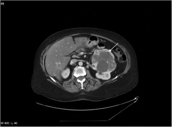
A portal venous phase enhanced CT scan was performed. In addition, the renal lesion was evaluated sonographically. The CT scan revealed an encapsulated enhancing exophytic left renal mass that measured 10 cm in maximum dimension. This heterogeneous mass contained at least three small hypoattenuating foci measuring up to 5 mm with attenuation values ranging from −19 to −32 HU, consistent with foci of macroscopic fat. No intratumoral calcium was evident on CT (Figure 1). There was no evidence of renal vein involvement or adenopathy.
Figure 1.

Axial CT image with IV contrast enhancement demonstrates a left renal mass containing a focus of macroscopic fat (arrow) without associated macroscopic calcification.
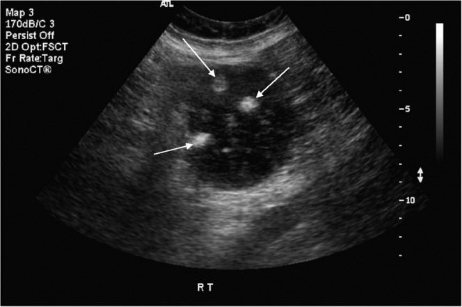
Sonographically, the mass was predominantly solid and hypoechoic, and multiple echogenic foci corresponding to the fat nodules were visualised (Figure 2).
Figure 2.

Ultrasound image of the renal mass containing echogenic foci (arrows) corresponding to the fat nodules visualised on CT.
The imaging features of the mass were highly suggestive of an angiomyolipoma (AML); nevertheless, a biopsy was performed because of the small possibility that the fat was within a renal cell carcinoma (RCC). The pathology was consistent with a conventional clear-cell RCC rather than an AML.
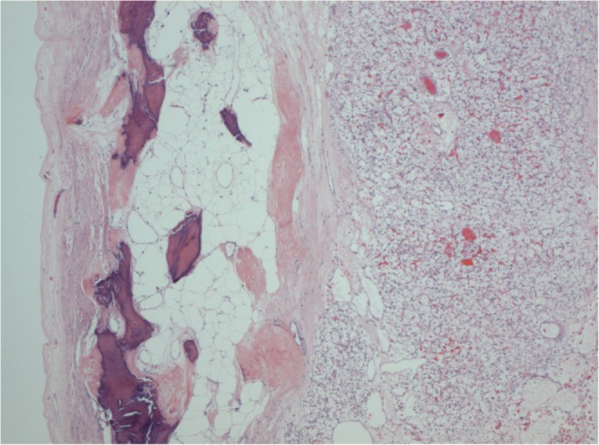
The patient underwent simultaneous left radical nephrectomy and right hemicolectomy. Pathological examination revealed a pT2 clear cell RCC with Fuhrman nuclear grade 2/4. There was no involvement of the perinephric or hilar fat. Histologically, small foci of metaplastic bone containing fat were visualised (Figure 3).
Figure 3.

Clear-cell renal cell carcinoma (right side of photo) containing focus of fat associated with osseous metaplasia (left side of photo). Original magnification 25x.
Discussion
Renal masses have traditionally been classified as AMLs if they contain macroscopic fat that is not associated with calcification or ossification and show no evidence of invasion of perinephric or renal sinus fat [1]. Fat within RCC has been pathologically accounted for by three mechanisms [1–5]. First, RCC can engulf perinephric or renal sinus fat [6]. Second, cholesterol necrosis can be misinterpreted as macroscopic fat [7, 8]. Third, osseous metaplasia of the nonepithelial stromal portion of the tumour, with growth of fatty marrow elements and trabeculae, is a rare occurrence [4]. In addition, a distinct nodule of mature, well-circumscribed adipose tissue has been identified within an encapsulated, non-calcified RCC [9]. RCCs that contain fat because of osseous metaplasia have calcification or ossification associated with that fat [1]. Engulfed renal sinus or perirenal fat is seen in masses with an invasive appearance.
If cases of engulfed fat are excluded, five cases of RCC containing macroscopic fat, but with no evidence of macroscopic calcification on CT, have been reported previously [5, 7, 9, 10], although the tumour in one of these cases contained microscopic calcification. Among the five cases, fat was found to be due to cholesterol necrosis in two cases, and the cause was unknown or not specified in three cases. To our knowledge, there are no reported cases of an RCC that contains osseous metaplasia without evidence of macroscopic calcification or ossification on CT.
However, in the present case the RCC had multiple foci of osseous metaplasia but no associated macroscopic calcification or ossification on CT. This is significant because it illustrates the potential pitfall of the current practice in which a RCC is excluded and the tumour classified as an AML on the basis of the presence of intratumoural macroscopic fat without associated macroscopic calcification or engulfed perirenal or renal hilar fat.
Angiomyolipomas are benign lesions and, traditionally, conservative treatment has been advocated unless the patient is symptomatic, there are risk factors for haemorrhage (such as pregnancy, tuberous sclerosis complex or large tumour size) or there are concerns regarding patient compliance or access to health care [11]. Given that there are now six reported cases of RCC containing macroscopic fat without macroscopic calcification evident on CT, we support Schuster et al. [9] in recommending that serial imaging is considered for suspected AMLs that are not surgically excised, and that a frozen-section biopsy should be taken if partial-nephrectomy is performed for a suspected AML.
Most lesions that contain macroscopic fat without associated calcification are in fact AMLs rather than RCCs. Therefore, we suggest that, when calcification is not identified in fat-containing renal lesions that are less than 4 cm in maximal dimension, follow-up be obtained on an annual basis by ultrasound, CT or MRI to ensure interval stability. Lesions larger than 4 cm that are not removed should initially be followed biannually and, if stable, annually thereafter.
References
- 1.Helenon O, Merran S, Paraf F, Melki P, Correas J, Chretien Y, et al. Unusual fat-containing tumors of the kidney: a diagnostic dilemma. Radiographics 1997;17:129–44 [DOI] [PubMed] [Google Scholar]
- 2.Prando A, Prando D, Prando P. Renal cell carcinoma: unusual imaging manifestations. Radiographics 2006;26:233–44 [DOI] [PubMed] [Google Scholar]
- 3.Strotzer M, Lehner K, Becker K. Detection of fat in a renal cell carcinoma mimicking angiomyolipoma. Radiology 1993;188:427–8 [DOI] [PubMed] [Google Scholar]
- 4.Helenon O, Chretien Y, Paraf F, Melki P, Denys A, Moreau J. Renal cell carcinoma containing fat: demonstration with CT. Radiology 1993;188:429–30 [DOI] [PubMed] [Google Scholar]
- 5.D’Angelo PC, Gash JR, Horn AW, Klein FA. Fat in renal cell carcinoma that lacks associated calcifications. AJR Am J Roentgenol 2002;178:931–2 [DOI] [PubMed] [Google Scholar]
- 6.Prando A. Intratumoral fat in a renal cell carcinoma. AJR Am J Roentgenol 1991;156:871a. [DOI] [PubMed] [Google Scholar]
- 7.Lesavre A, Correas J, Merran S, Grenier N, Vieillefond A, Helenon O. CT of papillary renal cell carcinomas with cholesterol necrosis mimicking angiomyolipomas. AJR Am J Roentgenol 2003;181:143–5 [DOI] [PubMed] [Google Scholar]
- 8.Castoldi M, Dellafiore L, Renne G, Schiaffino E, Casolo F. CT demonstration of liquid intratumoral fat layering in a necrotic renal cell carcinoma. Abdom Imaging 1995;20:483–5 [DOI] [PubMed] [Google Scholar]
- 9.Schuster TG, Ferguson MR, Baker DE, Schaldenbrand JD, Solomon MH. Papillary renal cell carcinoma containing fat without calcification mimicking angiomyolipoma on CT. AJR Am J Roentgenol 2004;183:1402–4 [DOI] [PubMed] [Google Scholar]
- 10.Radin D, Chandrasoma P. CT demonstration of fat density in renal cell carcinoma. Acta Radiol 1992;33:365–7 [PubMed] [Google Scholar]
- 11.Nelson CP, Sanda MG. Contemporary diagnosis and management of renal angiomyolipoma. J Urol 2002;168:1315–5 [DOI] [PubMed] [Google Scholar]


