



Abstract
Multidetector row computed tomography (MDCT) with its high spatial and temporal resolution has now become an established and complementary method for cardiac imaging. It can now be used reliably to exclude significant coronary artery disease and delineate complex coronary artery anomalies, and has become a valuable problem-solving tool. Our experience with MDCT imaging suggests that it is clinically useful for imaging the pericardium. It is important to be aware of the normal anatomy of the pericardium and not mistake normal variations for pathology. The pericardial recesses are visible in up to 44% of non-electrocardiogram (ECG)-gated MDCT images. Abnormalities of the pericardium can now be identified with increasing certainty on 64-detector row CT; they may be the key to diagnosis and therefore must not be overlooked. This educational review of the pericardium will cover different imaging techniques, with a significant emphasis on MDCT. We have a large research and clinical experience of ECG-gated cardiac CT and will demonstrate examples of pericardial recesses, their variations and a wide variety of pericardial abnormalities and systemic conditions affecting the pericardium. We give a brief relevant background of the conditions and reinforce the key imaging features. We aim to provide a pictorial demonstration of the wide variety of abnormalities of the pericardium and the pitfalls in the diagnosis of pericardial disease.
The rapid technological development of multidetector row computed tomography (MDCT) with its greatly improved spatial and temporal resolution and sophisticated ECG-gated image acquisition software has led to the more widespread use of dedicated cardiac imaging. Not only does this technology enable assessment of the coronary arteries [1, 2], but the same acquired data set also provides imaging detail of the overall cardiac morphology including the normal and diseased pericardium, features of which may also be readily appreciated on the non-ECG-gated thoracic CT [3]. It is important for the general radiologist to be familiar with both normal and variant pericardial anatomy and with that of the pericardial recesses, which can mimic some pathological processes. Several disease processes, either primary or secondary, can affect the pericardium. This review aims to illustrate normal pericardial anatomy, diagnostic pitfalls, commonly encountered abnormalities (some of which may be quite subtle) and some more unusual entities.
The pericardial recesses
The pericardium surrounds the heart and extends cranially to cover the pulmonary trunk, superior vena cava (SVC) and ascending aorta. The normal pericardial sac is made up of an inner visceral and outer parietal layer with a serosal lining; the space between them constitutes the pericardial cavity. On CT this is seen as a thin line of fibrous tissue; the visceral pericardium cannot be visualised separately. The higher-attenuation pericardium is distinguished in relation to the low-attenuation mediastinal fat anteriorly and epicardial fat posteriorly. On MRI, the pericardium is depicted by a thin rim of low signal on both T1 and T2 weighted images.
Several studies have examined the normal thickness of the pericardium on CT and MRI and at autopsy. The pericardial thickness varies over different parts of the heart; it is at its thinnest over the left ventricle. Bull et al, [4] reported that when using 1 mm high-resolution CT (HRCT) the upper limit of the thinnest part of the pericardium was 0.7 mm and when using 10 mm CT slices it was 1.2 mm. The upper limit of normal for the thickest part of the pericardium is 2 mm [5–8]. The pericardial sac contains up to 50 ml of clear fluid [9] and forms a protective layer around the heart, acting as a barrier against local inflammation and limiting its movement within the mediastinum.
As MDCT resolution has improved, the pericardial recesses can be identified as defined anatomical structures that are visible in up to 44% of thin-section scans [10]. They are cavities found between the outer fibrous and inner serous layers of the pericardium and can best be understood by subdividing them according to the part of the pericardial cavity from which they arise. The three main sites of origin are the transverse sinus, the oblique sinus and the pericardial cavity proper (Figure 1) [3].
Figure 1.

Line drawing illustrating the relative positions of the transverse and oblique sinuses; the majority of the pericardial recesses take their origin from these spaces. Note the pericardial reflection between the two spaces. The pulmonic venous recesses are also illustrated.
The transverse sinus
This space is located posterior to the ascending aorta and pulmonary trunk and cranially to the left atrium; a number of recesses take origin from this space (Figure 1). Arising superiorly from the transverse sinus is the superior aortic recess. This, in turn, has three subdivisions:
The anterior extension is seen anteriorly between the ascending aorta and pulmonary trunk, taking a characteristic triangular shape (Figure 2a).
The posterior extension, or superior pericardial recess, is seen directly behind the ascending aorta, taking a characteristic crescent shape (Figure 2c); an important variant of this recess is the high-riding superior pericardial recess, which has a more cranial extension. This can take a variety of shapes including triangular, crescent-shaped or oval and can be seen to extend up into the right paratracheal region (Figure 2b).
The right lateral extension is seen insinuating between the ascending aorta and SVC. Basile et al [11] demonstrated this recess to be present in 6.6% of patients when the chest is imaged using 16-MDCT.
Figure 2.

(a) The anterior extension of the superior aortic recess. (b) A coronal reformation demonstrating the right paratracheal position of a high-riding superior pericardial recess. (c) The posterior extension of the superior aortic recess. (d) The left pulmonic recess.
In continuity with the transverse sinus inferiorly is the inferior aortic recess. This is a caudal extension from the transverse sinus seen extending anterior to the left atrium.
Also in continuity with the transverse sinus are the right and left pulmonic recesses. These extend from the transverse sinus inferolaterally, lying caudal to the pulmonary arteries (Figure 2d).
The oblique sinus
The oblique sinus is the most posterior pericardial space (Figure 1). This space lies behind the left atrium and is inferior to the transverse sinus, from which it is separated by pericardial reflections. Arising from the oblique sinus is the posterior pericardial recess. This extends superiorly behind the right pulmonary artery and medial to the bronchus intermedius.
Pericardial effusion
The pericardium normally contains a small amount of fluid (between 15 and 50 ml) [9]. Gradual fluid build-up may be accommodated by the pericardium to the extent of volumes greater than 1 litre [12], although rapid accumulation (Figure 3) is far more problematic; tamponade can be caused by a volume of 250 ml within a previously normal pericardial sac [13]. The aetiology of a pericardial effusion is obstruction of lymphatic or venous drainage from the heart, which can be caused by a number of disease processes: the commonest are cardiac and renal failure, followed by infection, neoplasia and myocardial infarction. Any cause of acute pericarditis, although initially causing a dry and fibrinous inflammatory reaction, will lead to subsequent development of a pericardial effusion.
Figure 3.
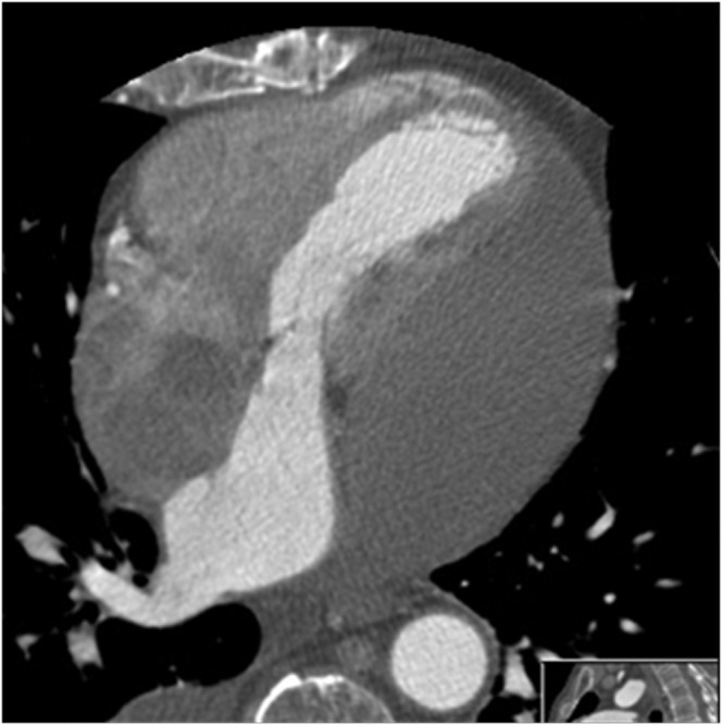
Pericardial effusion. Here, there has been a rapid accumulation of pericardial fluid, particularly on the left side, which is compressing the cardiac chambers and creating a tamponade.
Pericardial effusions can be imaged using a variety of modalities. The plain chest radiograph may suggest a pericardial effusion only in the context of a rapidly enlarging cardiac silhouette. Echocardiography is highly sensitive and specific in detecting pericardial effusions, and indeed effusions may be easily seen on subcostal views obtained during conventional abdominal ultrasound examination. CT or MRI is indicated if the effusion is suspected to be complicated by haemorrhage (Figure 4), loculations, pericardial inflammation, thickening or constriction [4, 14]. Accurate localisation of effusions can be facilitated and guide intervention and also allow further characterisation of the fluid. If the attenuation value on CT is greater than that of water, then an effusion is more likely to be due to haemopericardium, malignancy, purulent exudates or hypothyroid-associated effusion [15, 16].
Figure 4.

(a) This patient sustained a previously unsuspected iatrogenic left ventricular injury during a mitral valve annuloplasty, and presented with cardiac tamponade secondary to a rapidly accumulating haemorrhagic pericardial effusion. (b) The inferior left ventricle wall has ruptured secondary to acute myocardial infarction causing a haemopericardium and cardiovascular compromise. Note the contrast extravasation and the subendocardial myocardial perfusion defect.
On MRI, a haemorrhagic effusion is characterised by high signal on T1 weighted images and low signal on T2 weighted cine images [17]. The size and extent of the pericardial effusion may also be better assessed using CT or MRI than with transthoracic echocardiography; the smallest amount of pericardial fluid that can be detected by CT is approximately 10 ml [18]. A pericardial space greater than 5 mm anterior to the right ventricle is equated to at least a moderate effusion [19]. When the effusion is secondary to a malignancy there may also be associated pericardial irregularity and nodularity (see later).
Pericarditis without constriction
Pericarditis is essentially a fibrotic inflammatory reaction of the pericardium brought on by a large number of conditions, including radiation therapy, post cardiac surgery or post myocardial infarction (Dressler's syndrome), or by drugs, uraemia, infection (e.g. viral, bacterial tuberculosis), and hypothyroidism [16, 20]. There is thickening of the pericardium associated with a pericardial effusion, but without constriction of the myocardium (Figure 5). The pericardial fat may also be of increased, ill-defined attenuation, in keeping with inflammatory change.
Figure 5.
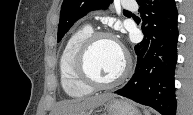
Anterosuperior pericardial thickening and fatty inflammatory change is seen in this patient who presented with acute chest pain and no clinical evidence of myocardial infarction. The patient had pericarditis.
All causes of pericarditis can lead to subsequent constriction [17]. Inflammation of the pericardium causes enhancement on post-intravenous (iv) contrast CT and also post-gadolinium MRI. In a study by Masui et al [21] there was 88% correlation between pericardial thickness at surgery and MR images. Pericarditis may affect only part of the pericardium, making it important to visualise and scrutinise the entire pericardium [12]. Ben-Horin et al [22] described the case of a patient with known systemic lupus erythematosus who presented with pleuritic chest pain; conventional methods of investigation did not detect a pericardial effusion, whereas CT demonstrated a localised area of pericardial inflammation leading to the diagnosis of lupus pericarditis.
It should be noted, however, that subtle increases in pericardial “thickness” in the absence of surrounding inflammatory change could simply result from a small amount of pericardial fluid and not from thickening. In the context of a correlative clinical history, and if there is doubt on CT, then T1/T2 MRI could be performed to confirm the presence of fluid or thickening.
Constrictive pericarditis
Constrictive pericarditits is a diagnosis requiring a combination of clinical signs and imaging. It is also of paramount importance to distinguish constrictive pericarditis from restrictive cardiomyopathy; although clinical signs, transthoracic echocardiography and cardiac catheterisation findings are very similar, the treatments are different.
Constrictive pericarditis is caused by a thickened or fibrotic pericardium, which in turn has a multifaceted aetiology (Table 1). This is contrasted with restrictive cardiomyopathy, in which there is restrictive filling or reduced diastolic filling of one or both ventricles, with a normal-thickness pericardium.
Table 1. Causes of constrictive pericarditis and restrictive cardiomyopathy.
| Constrictive pericarditis | Restrictive cardiomyopathy | |
| Causes | Post-cardiac surgery | Idiopathic endomyocardial fibrosis |
| Mediastinal radiotherapy | Amyloidosis | |
| Infection — viral, bacterial (e.g. TB) | Loeffler endomyocarditis | |
| Hydatid disease | Haemochromatosis | |
| Uraemia | Malignancy | |
| Connective tissue disease | Carcinoid | |
| Malignancy | ||
| Haematoma | ||
| Clinical signs | Heart murmur uncommon | Mitral and tricuspid regurgitation |
| Hepatomegaly | Bilateral pleural effusions | |
| Peripheral oedema | Ascites | |
| Peripheral oedema | ||
| 2-D Echocardiography | Normal ventricular size | Normal or reduced ventricular size |
| Pericardial thickening | Non-dilated left ventricle with normal contraction | |
| Pericardial effusion may be visualised | Marked dilatation of both atria | |
| Cardiac catheterisation | Square root sign of ventricular diastolic pressure | Square root sign of ventricular diastolic pressure |
| Increased right heart pressure | ||
| Treatment | Complete pericardectomy | Low dose diuretics |
| Permanent pacemaker | ||
| Cardiac transplantation |
Constriction causes small tubular-shaped ventricles, with distortion of the ventricular septum (Figure 6a). The septum becomes flattened or sigmoid in shape and can exhibit the phenomenon of septal bounce. This phenomenon is seen on echocardiography, which reveals the septum moving in an atypical manner. As the pericardium limits ventricular filling, one ventricle fills at the expense of the other with subsequent septal movement; the right atrium, inferior vena cava (IVC) and hepatic veins become dilated (Figure 6b).
Figure 6.

(a) Axial sections through the heart in a four-chamber view in a young Asian male patient with constrictive non-calcified tuberculous pericarditis (see later). The image demonstrates diffuse pericardial thickening, small-volume pericardial effusion, a flattened interventricular septum and a small right pleural effusion. (b) Axial sections through the upper abdomen in the same patient, showing ascites, hepatomegaly and dilatation of the hepatic inferior vena cava and hepatic veins with reflux of intravenous contrast.
CT and MRI can aid the diagnosis of constrictive pericarditis requiring clinical and radiological concordance. A thickened pericardium on its own does not therefore indicate constrictive pericarditis. Once the pericardium is thicker than 6 mm [19] and the patient is clinically in heart failure, constrictive pericarditis is very likely.
Masui et al [21] demonstrated that MRI has an accuracy of 93% when differentiating between constrictive pericarditis and restrictive cardiomyopathy, using pericardial thickening of greater than 4 mm as the deciding factor. MRI has also been shown to be better than CT at differentiating between pericardial fluid and thickened pericardium [11], and these modalities may therefore be seen as complementary. Pericardial constriction may just be limited to the right heart or the right atrioventicular groove [23], with pericardial thickening present only over the right atria and ventricles [19]. CT is very sensitive in demonstrating calcification of the pericardium, unlike MRI, which is also suggestive of constrictive pericarditis if found in the right clinical setting (Figure 7).
Figure 7.

Volume-rendered MDCT (c, d) demonstrates in three dimensions florid, irregular, amorphous, pericardial calcified plaques causing constrictive pericardial disease. Areas of non-calcified pericardial thickening can also be appreciated in the four-chamber (c, d) and two-chamber planes (a, b) above. In this case, CT aided in surgical resection.
About 50% of cases show some degree of calcification [21] which, if present, excludes restrictive cardiomyopathy. Enhancement of the pericardium on post-contrast CT and MRI indicates inflammation [24].
Johnson et al [25] noted that constrictive pericarditis can present with vague abdominal symptoms. If the abdominal CT shows dilated hepatic veins/IVC, ascites or liver cirrhosis then the pericardium should be carefully inspected, as in their experience the majority of patients will have an abnormal pericardium.
Tuberculous disease of the pericardium
Tuberculosis (TB) is an important cause of pericardial effusion worldwide. Cherian et al [26] looked at the features differentiating tuberculous pericardial effusion from chronic idiopathic effusion and found three important features:
Mediastinal lymphadenopathy was present in all patients with TB and absent in those with chronic idiopathic effusion.
Thinning of the pericardium was not a feature in TB.
Pericardial tamponade occurred in 64% of both groups.
TB is the commonest cause of constrictive pericarditis in developing countries, with 50% of patients developing constriction despite treatment [27, 28]. Associated findings in TB constrictive pericarditis are pericardial thickening, effusion, lymphadenopathy and calcification. The calcification tends to be thick, irregular, amorphous and in the AV grooves (Figure 6). This can be compared with the eggshell calcification seen in viral or uraemic pericarditis.
Tamponade is a frequent complication of TB constrictive pericarditis, but patients do well with appropriate therapy [29]; abscess formation is a rare complication. Gulati et al [30] demonstrated 15 abscesses in 13 patients out of a group of 120 known to have constrictive pericarditis. TB was found to be the cause in all cases; 91% of the abscesses on CT demonstrated smooth walls and 54% thin enhancing rims. The most common position was the right atriouentricular (AV) groove, in 77%, calcification was present in 27% of cases and the abscesses were shown to have a low attenuation core and may contain septations. The diagnosis was confirmed by examination of the aspirated fluid or the demonstration of other extracardiac manifestations of the disease.
Pericardial abcesses have been reported to be caused by a variety of other conditions, including infective pericarditis after blunt and penetrating trauma, thermal injuries [31, 32], endocarditis [33], sepsis [34] and oesophago-mediastinal fistula [35].
Pericardial cysts
Pericardial cysts are rare congenital defects; however, they are the most common benign pericardial mass [36]. These cysts are formed during early development when a portion of the pericardium is pinched off; they can be found anywhere in the mediastinum, but by far the commonest position is at the right cardiophrenic angle [12]. Pericardial cysts usually have thin, smooth walls without internal septations (Figure 8) and attach to the pericardium directly or by a pedicle [36]. The majority of these cysts are an incidental finding on the chest radiography.
Figure 8.
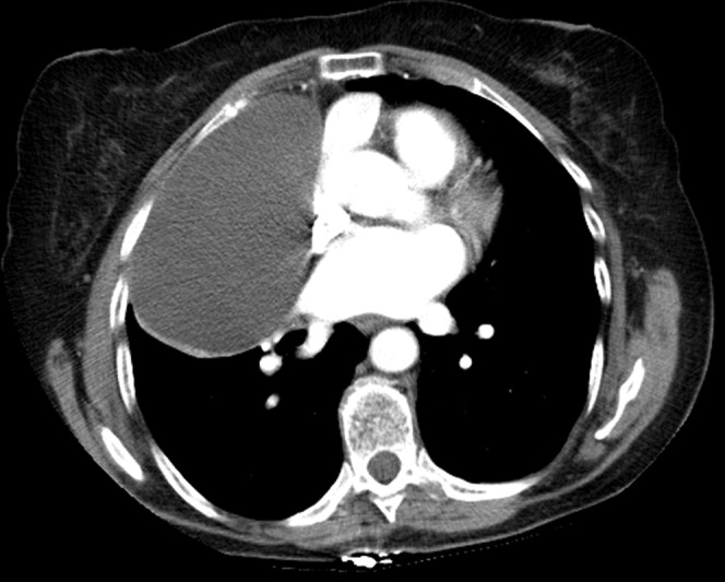
Non-gated contrast-enhanced chest CT image in the axial plane. At the right cardiophrenic angle there is an ovoid structure with attenuation value in keeping with fluid density that does not enhance with contrast. The structure proved to be a pericardial cyst, following aspiration.
Pericardial diverticula are clinically identical to cysts and can be hard to distinguish from bronchogenic or thymic cysts if in an unusual location [37]. CT will confirm the diagnosis of a pericardial cyst by clearly demonstrating the position and extent of the lesion; fluid density and characterisation of the mass; a clearly defined ovoid mass; and absent enhancement with iv contrast [38].
MRI demonstrates four key features: low signal on T1 weighted images; homogeneously high signal on T2 weighted images; no enhancement with iv gadolinium; and, occasionally, a high signal on T1 weighted images owing to the high protein content of the fluid [38].
Pericardial cysts may be aspirated under image guidance and, if the fluid is clear and watery, the diagnosis is confirmed. If there is doubt over the diagnosis, the patient is symptomatic or a complication arises, then the cyst should be resected [39]. The reported complications of pericardial cysts are cardiac compression [39], infection of the cyst with or without cardiac erosion [40] and rupture of the cyst [41]. There are no reported cases of malignant degeneration. An important identifying characteristic of pericardial cysts is their tendency to alter their size and shape with respiration or body position [37].
Cystic appearances of the pericardium may also be due to hydatid cysts. Cardiac involvement is very rare, accounting for 0.2–2% of all hydatid cyst-related cases [42], with the pericardium involved in only 10–15% of these cases [43]. CT is the main diagnostic test and illustrates the overall extent of the disease. The classical findings of a hydatid pericardial cyst are the presence of a homogeneous cystic mass that is clearly defined with thin walls, evidence of trabeculations, and the presence of daughter cysts [43, 44]. Hydatid cysts of the pericardium can cause complications, with case reports of right ventricular outflow obstruction [39] and circulatory collapse [43].
Pericardial haematoma
Pericardial haematomas are essentially the result of trauma. They may be due to blunt trauma, causing a haemopericardium that subsequently organises to create a haematoma, or can arise iatrogenically during cardiac surgery, cardiac catheterisation, paracentesis or central line insertion.
Features of pericardial haematoma on CT are not documented in the literature. The blood is of high attenuation initially, which decreases over time. As the haematoma progresses it organises and may become fibrotic and calcify. Haematomas do not enhance with iv contrast medium.
On MRI the appearances vary according to the age of the haematoma: in the acute phase they are homogeneously of high signal [45, 46], whereas between 1 and 4 weeks of maturity (the subacute phase) the signal becomes heterogeneous, with areas of high signal on both T1 and T2 weighted images [45, 47]. Chronic haematomas demonstrate low signal intensity foci that correspond to calcification or fibrosis; on T1 weighted images there is a dark peripheral rim [48, 49]. Haematomas may be confused with pseudoaneurysms of the ventricles or coronary arteries or can be mistaken for tumours. The use of iv gadolinium allows the differentiation of these diagnoses, as haematomas do not enhance [50]. Pericardial haematomas can further complicate matters by causing tamponade.
Pericardial defects
Pericardial defects are rare, with the most common cause being congenital absence. Defects can also result from surgery or trauma (Figure 9).
Figure 9.
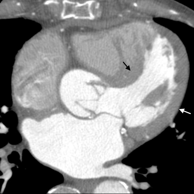
Gated contrast-enhanced axial image.This patient has undergone aortic valve replacement and grafts. There is mediastinal fibrosis (black arrow) (seen as thickened soft tissue anterior to the right ventricle and just posterior to the sternum), which could be mistaken for a thickened pericardium when the pericardium is in fact absent. The right heart is enlarged and the septum is bowed (white arrow), which are also findings in constrictive pericarditis.
With congenital absence of the pericardium there is premature atrophy of the cardinal vein leading to poor nourishment of the left pleuro-pericardial membrane. This leads to failure of the pericardium to develop [51]. These defects are three times commoner in men and are usually detected in the early 20s. The commonest defect is complete absence of the left side of the pericardium; this then allows lung tissue to be interposed between the main pulmonary artery and aorta. Bulging of the left atrial appendage can then occur through the defect. As a consequence of all these abnormalities the heart rotates to the left [38]. There may unusually be right-sided defects, diaphragmatic defects or total bilateral absence of the pericardium.
Congenital pericardial defects are associated with congenital abnormalities such as bronchogenic cysts, ventricular septal defect (VSD), patent ductus arteriosus (PDA), mitral stenosis, diaphragmatic hernia or sequestration. Most pericardial defects are asymptomatic, especially if they do not have any associated defects.
Imaging of pericardial defects usually begins with an incidental finding on the chest radiograph. There may be a focal bulge in the region of the main pulmonary artery or lung interposed between the left hemi-diaphragm and the heart. There can also be levoposition of the heart. The diagnosis can be confirmed on CT or MRI, in which the absence of the pericardium can be more clearly defined [12].
Complications of congenital pericardial defects include herniation and entrapment of the cardiac chamber, particularly the left atrial appendage, leading to ischaemic necrosis [36]; to alleviate this, surgical closure or enlargement of the defect may be necessary [38]. A further complication is that following a pneumothorax there is likely to be a pneumomediastinum.
Pericardial tumours
Metastatic disease of the pericardium is much commoner than that of primary pericardial malignancy. Metastases are present at autopsy in 10–12% of patients with known neoplasia [47, 48]. If discovered pre-autopsy, they are associated with a poor prognosis [52]. The primary tumours most likely to metastasise to the pericardium are lung, breast (Figure 10), melanoma and lymphoma [47, 48].
Figure 10.
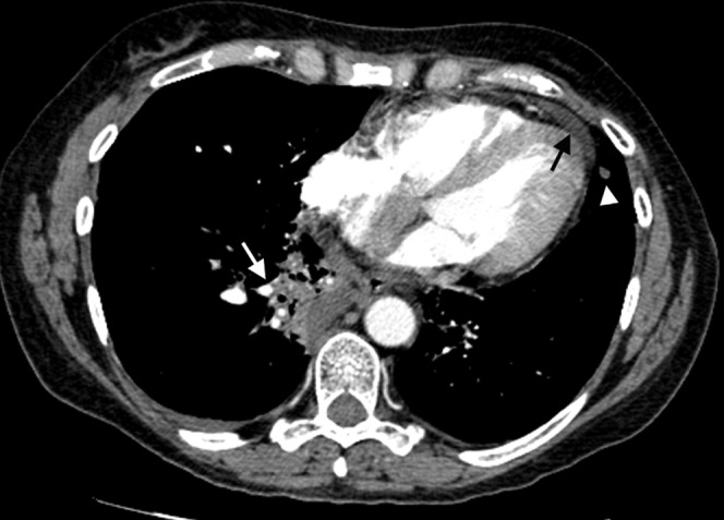
Non-gated contrast-enhanced chest CT axial image. This patient was known to have breast cancer and had undergone radiotherapy; the lung demonstrates a linear area of fibrosis consistent with this. On further imaging the pericardium has become nodular and thickened (black arrow) and enhances with contrast. There are also lung (white arrowhead) and liver deposits consistent with metastatic disease; however, without a biopsy it is impossible to tell whether the pericardial change is secondary to metastatic spread or radiation.
Metastatic spread to the pericardium happens late in the disease and is usually associated with recurrent disease. A third of cases are from carcinoma of the lung, and a third of lung cancer cases have pericardial infiltration at autopsy (Figure 11) [17].
Figure 11.
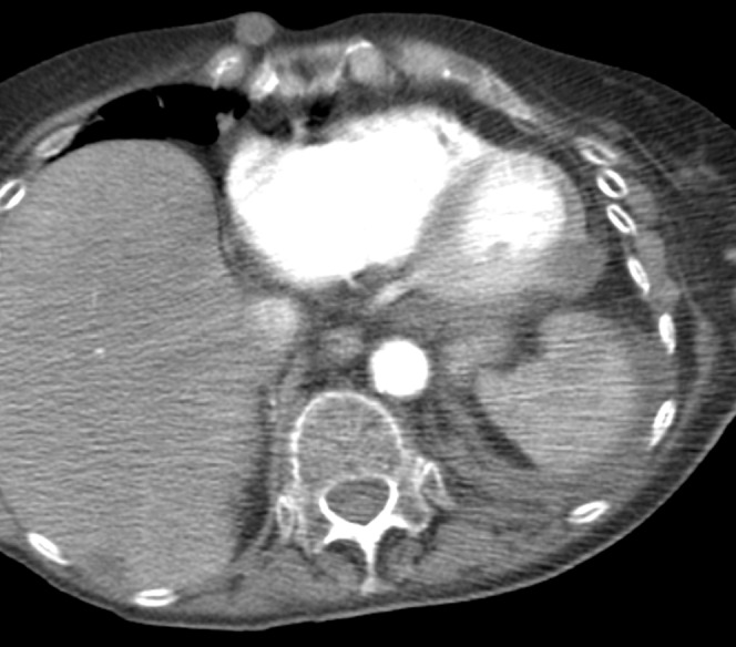
An axial section through the heart in a patient with end-stage lung cancer demonstrates a soft tissue mass infiltrating the pericardium and myocardium on the left poterolateral wall. A small volume of ascites can also be seen around the spleen.
Direct extension of primary tumours from the mediastinum or lung to the pericardium is a common mode of spread [4]. Other modes of spread are through the lymphatic system, haematogenously or transvenously [53].
Four key features of pericardial malignancy are apparent on CT: an irregular, thickened nodular pericardium (although this may also occur with an inflammatory pericardiopathy; follow-up imaging may be required); a pericardial effusion; pericardial enhancement after iv contrast administration [54]; and the possible presence of a mass in the pericardium (Figure 12).
Figure 12.

(a) ECG-gated contrast-enhanced cardiac CT images demonstrate a large, heterogeneously enhancing, pericardial mass on the antero-inferior border of the heart causing right heart compression and elevated right heart pressure. The patient initially presented with right heart failure. The tumour was entirely resected and proved to be a sarcoma on histology. (b) the same patient CT in the axial plane.
Both benign and malignant primary tumours of the pericardium are rare. Malignant primaries include mesothelioma, sarcoma, liposarcoma and lymphoma, with mesothelioma and sarcoma being the more common [17]. Mesothelioma accounts for 50% of all pericardial primary tumours [55] and may present as a pericardial effusion with pericardial nodules or plaques. Sarcoma, liposarcoma and lymphoma manifest as large pericardial masses (Figure 12) with a haemorrhagic effusion; imaging alone cannot give a definitive diagnosis, therefore biopsy becomes necessary.
Benign tumours of the pericardium include fibroma, teratoma, haemangioma and lipoma. Teratomas tend to present as a mass with either fat or calcium within them, which can readily be demonstrated on CT. Lipomas demonstrates high signal on T1 weighted images and a low attenuation (negative Hounsfield units) on CT. Fibromas have poor vascularisation and therefore have either no enhancement or irregular enhancement after iv contrast [49, 56].
Pitfalls in diagnosing pericardial disease
When diagnosing pericardial disease, there are many things to consider in order to avoid potential pitfalls. Normal superior pericardial recesses can mimic aortic dissection and intramural haematoma (Figure 13) and enlarged lymph nodes (Figure 14).
Figure 13.
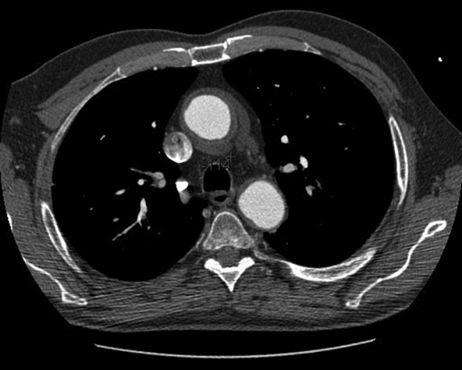
In the context of a patient presenting with acute chest pain radiating to the back, CT angiography of the aorta demonstrates a volume of fluid within normal limits within the posterior extension of the superior pericardial recess mistaken for ascending aortic intramural haematoma. Note the crescent-shaped fluid density immediately posterior to the aorta, which on multiplanar reformatting was found to be continuous with the pericardial cavity.
Figure 14.
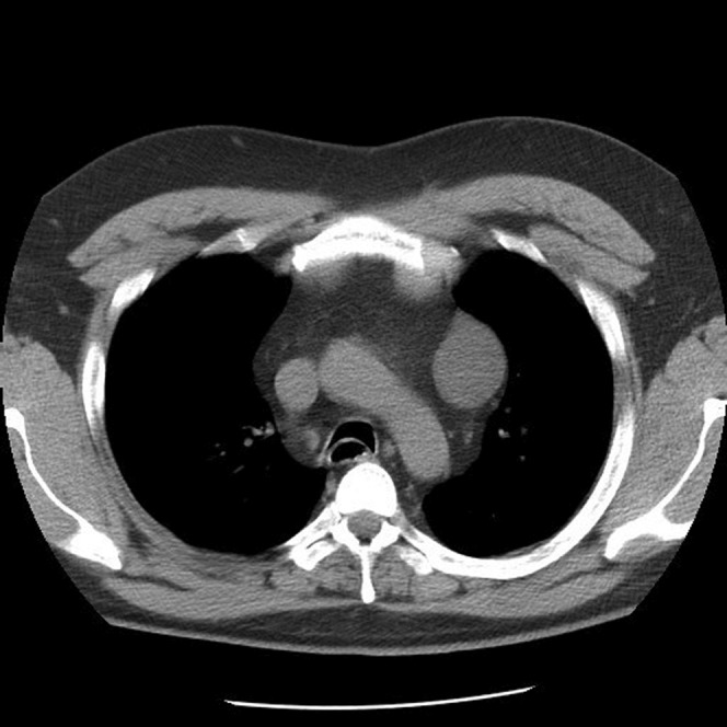
This patient with a history of thyroid malignancy underwent CT which demonstrated a well-defined fluid density within an atypical high-riding superior aortic recess. This was initially misdiagnosed as an enlarged lymph node and subsequently found at surgery to be a pericardial recess.
When considering these differential diagnoses, the Hounsfield attenuation number should be confirmed; for pericardial recesses, this number will be equivalent to that of water and will also be in continuity with the pericardial space. Small volumes of pericardial fluid may be confused with pericardial thickening even on CT, and MRI might still be needed for further characterisation. Some disease processes, such as systemic lupus erythematosus, may affect only a portion of the pericardium and therefore the entire pericardium must be scrutinised, again highlighting the importance of pericardial anatomical knowledge and normal variation.
Any process that causes thickening, nodularity or masses of the pericardium can be confused with metastatic disease, such as radiation pericarditis [13], tuberculous pericarditis (Figure 6) and severe acute pericardial inflammation following radiation therapy for the treatment of a primary malignancy.
Pericardial effusions should be assessed for size and attenuation on CT. Observed values greater than water suggest that the effusion is caused by haemorrhage, malignancy, infection or hypothyroidism.
Conclusion
MDCT has augmented our ability to identify and characterise the pericardium, allowing us to differentiate between the normal anatomy and pathology with a precise relationship to other anatomical structures. This review illustrates how ECG-gated cardiac MDCT, with submillimetre spatial resolution, should be considered as part of the diagnostic armamentarium in the context of suspected pericardial disease.
Acknowledgments
The authors would like to thank The Royal College of Radiologists Research Fellowship Award for funding Dr N Manghat to study “The clinical applications of cardiac CT” and General Electric Medical Systems for software applications support.
References
- 1.Kini S, Bis KG, Weaver L. Normal and variant coronary arterial and venous anatomy on high-resolution CT angiography. AJR Am J 2007;188:1665–74 [DOI] [PubMed] [Google Scholar]
- 2.Cury RC, Nieman K, Shapiro MD, Nasir K, Cury RC, Brady TJ. Comprehensive cardiac CT study: evaluation of coronary arteries, left ventricular function and myocardial perfusion — is it possible? J Nuc Cardiol 2007;14:229–43 [DOI] [PubMed] [Google Scholar]
- 3.Broderick LS, Brooks GN, Kuhlman JE. Anatomic pitfalls of the heart and pericardium. Radiographics 2005;25:441–53 [DOI] [PubMed] [Google Scholar]
- 4.Bull RK, Edwards PD, Dixon AK. CT dimensions of the normal pericardium. Br J Radiol 1998;71:923–5 [DOI] [PubMed] [Google Scholar]
- 5.Truong MT, Erasmus JJ, Gladish GW, Sabloff BS, Marom EM, Madewell JE, et al. Anatomy of pericardial recesses on multidetector CT: implications for oncologic imaging. AJR 2003;181:1109–13 [DOI] [PubMed] [Google Scholar]
- 6.Bogaert J, Duerinck AJ. Appearance of the normal pericardium on coronary MR angiograms. J Magn Reson Imaging 1995;5:579–87 [DOI] [PubMed] [Google Scholar]
- 7.Delile JP, Hernigou A, Sene V, Chatellier JC, Challander P, Plainjosse MC, et al. Maximal thickness of the normal human pericardium assessed by electron beam computed tomography. Eur Radiol 1999;9:1183–9 [DOI] [PubMed] [Google Scholar]
- 8.Deepak , Talreja DR, Edwards WD, Danielson GK, Schaff HV, Tajik AJ, Tazelaar HD, et al. Constrictive pericarditis in 26 patients with histologically normal pericardial thickness. Circulation 2003;108:1852. [DOI] [PubMed] [Google Scholar]
- 9.Edwards ED. Applied anatomy of the heart. In: Giulaini ER, Fuster V, editors. Cardiology: fundamentals and practice. 2nd edn. St. Louis, MO: Mosby-Year Book, 1991:47–51 [Google Scholar]
- 10.Kodama F, Fultz PJ, Wandtke JC. Comparing thin-section and thick-section CT of pericardial sinuses and recesses. AJR Am J Roentgenol 2003;181:1101–8 [DOI] [PubMed] [Google Scholar]
- 11.Basile A, Bisceglie P, Giulietti G, Calcara G, Figuera M, Mundo E, et al. Prevalence of “high riding” superior pericardial recesses on thin-section 16-MDCT scans. Eur J Radiol 2006;59:265–9 [DOI] [PubMed] [Google Scholar]
- 12.Breen JF. Imaging of the pericardium. J Thorac Imaging 2001;16:47–54 [DOI] [PubMed] [Google Scholar]
- 13.Posner MR, Cohen GI, Skarin AT. Pericardial disease in patients with cancer: the differentiation of malignant from idiopathic and radiation-induced pericarditis. Am J Med 1981;71:407–13 [DOI] [PubMed] [Google Scholar]
- 14.Isner JM, Carter BL, Bankoff MS, Konstam MA, Salem DN. Computed tomography in the diagnosis of pericardial heart disease. Ann Intern Med 1982;97:473–9 [DOI] [PubMed] [Google Scholar]
- 15.Tomoda H, Hoshiai M, Furuya H, Oeda Y, Matsumoto S, Tanabe T, et al. Evalution of pericardial effusion with computed tomography. Am Heart J 1980;99:701–6 [DOI] [PubMed] [Google Scholar]
- 16.Kamath S, Roobottom C. Hyperdense pericardial effusion in dermatomyositis and contrast induced nephropathy. Emergency Radiol 2005;11:177–9 [DOI] [PubMed] [Google Scholar]
- 17.Hancock EW. Neoplastic pericardial disease. Cardiol Clin 1990;8:673–82 [PubMed] [Google Scholar]
- 18.Ovchinnikov VI. Computerized tomography of pericardial diseases. Vestn Rentgenol Radiol 1996;1:10–5 [PubMed] [Google Scholar]
- 19.Frank H, Globits S. Magnetic resonance imaging evaluation of myocardial and pericardial disease. J Magn Reson Imaging 1990;10:617–26 [DOI] [PubMed] [Google Scholar]
- 20.Song H, Choi YW, Jang IS, Jeon SC, Park CK, Lee IS, Lee JS. Pericardium: anatomy and spectrum of disease on computed tomography. Curr Probl Diagn Radiol 2002;31:198–209 [PubMed] [Google Scholar]
- 21.Masui T, Finck S, Higgins CB. Constrictive pericarditis and restrictive cardiomyopathy: evaluation with MR imaging. Radiology 1992;182:369–73 [DOI] [PubMed] [Google Scholar]
- 22.Ben-Horin S, Portnoy O, Pauzner R, Livneh A. Localized pericardial inflammation in systemic lupus erythematosus. Clin Exp Rheumatol 2004;22:483–4 [PubMed] [Google Scholar]
- 23.Higgins CB. Acquired heart disease. In: Higgins CB, Hricak H, Helms CA, editors. Magnetic resonance imaging of the body. Philadelphia, PA: Lippincott-Raven, 1997:409–60 [Google Scholar]
- 24.Stephen WM. Imaging pericardial disease. Radiol Clin North Am 1989;27:1113. [PubMed] [Google Scholar]
- 25.Johnson KT, Julsrad PR, Johnson CD. Constrictive pericarditis at abdominal CT: a commonly overlooked diagnosis. Abdom Imaging 2008;33:349–52 [DOI] [PubMed] [Google Scholar]
- 26.Cherian G, Uthaman B, Habashy AG, Salama AL, George S. Large pericardial effusion: the differentiation of tuberculous from chronic idiopathic effusion. J Assoc Physicians India 2003;51:880–3 [PubMed] [Google Scholar]
- 27.Lin JH, Chen SJ, Wu MH, Lee PI, Chang CI. Fibrinofibrous pericarditis mimicking a pericardial tumour. J Formos Med Assoc 2000;99:59–61 [PubMed] [Google Scholar]
- 28.Kawecka-Jazcz K. Pericarditis: classification, etiology, pathogenesis. Folia Med Cracov 1991;32:15–22 [PubMed] [Google Scholar]
- 29.Cherian G, Uthaman B, Salama A, Habashy AG, Khan NA, Cherian JM. Tuberculous pericardial effusion: features, tamponade and computed tomography. Angiology 2004;55:431–40 [DOI] [PubMed] [Google Scholar]
- 30.Gulati GS, Sharma S. Pericardial abscess occurring after tuberculous pericarditis: image morphology on computed tomography and magnetic resonance imaging. Clin Radiol 2004;59:514–9 [DOI] [PubMed] [Google Scholar]
- 31.Johnson MA, Hirji MK, Hennig RC, Williams D. Pericardial abscess: diagnosis using two-dimensional echocardiography and CT. Radiology 1986;159:419–21 [DOI] [PubMed] [Google Scholar]
- 32.Sato TT, Geary RL, Ashbaugh DG, Jurkovich GJ. Diagnosis and management of pericardial abscess in trauma patients. Am J Surg 1993;165:637–41 [DOI] [PubMed] [Google Scholar]
- 33.Suzuki S, Tajimi T, Takeshita A, Nakamura M, Kinoshita K, Tokunaga K. Isolated right heart purulent pericarditis forming a large mediastinal mass. Chest 1988;93:667–8 [DOI] [PubMed] [Google Scholar]
- 34.Capov I, Wechsler J, Sumbera M, Pavlik M, Jedlicka V. Pericardial abscess — a rare complication of sepsis. Acta Chir Hung 1999;38:19–21 [PubMed] [Google Scholar]
- 35.Muto M, Ohtsu A, Boku N, Tajiri H, Yoshida S. Streptococcus milleri infection and pericardial abscess associated with oesophageal carcinoma: report of two cases. Hepatogastroenterology 1999;46:1782–4 [PubMed] [Google Scholar]
- 36.Smith WHT, Beacock DJ, Goddard A, Bloomer TN, Ridgway JP, Sivananthan UM. Magnetic resonance evaluation of the pericardium. Br J Radiol 2001;74:384–92 [DOI] [PubMed] [Google Scholar]
- 37.Pader E, Kirschner P. Pericardial diverticulum. Dis Chest 1969;55:344–6 [DOI] [PubMed] [Google Scholar]
- 38.Wang ZJ, Reddy GP, Gotway MB, Yeh BM, Hetts SW, Higgins CB. CT and MR imaging of pericardal disease. Radiographics 2003;23:S167–80 [DOI] [PubMed] [Google Scholar]
- 39.Ng AF, Olak J. Pericardial cyst causing right ventricular outflow tract obstruction. Ann Thorac Surg 1997;63:1147–8 [DOI] [PubMed] [Google Scholar]
- 40.Chopra PS, Duke DJ, Pellett JR, Rahko PS. Pericardial cyst with partial erosion of the right ventricular wall. Ann Thorac Surg 1991;51:840–1 [DOI] [PubMed] [Google Scholar]
- 41.King JF, Crosby I, Pugh D, Reed W. Rupture of pericardial cyst. Chest 1971;60:611–12 [DOI] [PubMed] [Google Scholar]
- 42.Perez-Gomez F, Duran H, Tamames S, Perrotet JL, Blanes A. Cardiac echinococcosis: clinical picture and complications. Br Heart J 1973;35:I326–I331 [DOI] [PMC free article] [PubMed] [Google Scholar]
- 43.Gossios K, Passas G, Kontogiannis D, Kakadellis J. Mediastinal and pericardial hydatid cysts: an unusual cause of circulatory collapse. AJR Am J Roentgenol 2003;181:285–6 [DOI] [PubMed] [Google Scholar]
- 44.Zidi A, Zannad-Hantous S, Mestiri I, Ghrairi H, Baccouche I, Djilani H, et al. Hydatid cyst of the mediastinum: 14 case reports. J Radiol 2006;87:1869–74 [DOI] [PubMed] [Google Scholar]
- 45.Seelos KC, Funari M, Chang JM, Higgins CB. Magnetic resonance imaging in acute and subacute mediastinal bleeding. Am Heart J 1992;123:1269–72 [DOI] [PubMed] [Google Scholar]
- 46.Vilacosta I, Gómez J, Domínguez J, Domínguez L, Baãuelos C, Ferreirós J, et al. Massive pericardiac hematoma with severe constrictive pathophysiologic complications after insertion of an epicardial pacemaker. Am Heart J 1995;130:1298–300 [DOI] [PubMed] [Google Scholar]
- 47.Meleca MJ, Hoit BD. Previously unrecognised intrapericardial hematoma leading to refractory abdominal ascites. Chest 1995;108:1747–8 [DOI] [PubMed] [Google Scholar]
- 48.Brown DL, Ivey TD. Giant organized pericardial hematoma producing constrictive pericarditis: a case report and review of the literature. J Trauma 1996;41:558–60 [DOI] [PubMed] [Google Scholar]
- 49.Funari M, Fujita N, Peck WW, Higgins CB. Cardiac tumors: assessment with Gd-DTPA enhanced MR imaging. J Comput Assist Tomogr 1991;15:953–8 [PubMed] [Google Scholar]
- 50.Higgins CB, Sakuma H. Heart disease: functional evaluation with MR imaging. Radiology 1996;199:307–15 [DOI] [PubMed] [Google Scholar]
- 51.Broadbent JC, Callahan JA, Kincaid OW, Ellis FH. Congenital deficiency of the Pericardium. Dis Chest 1966;50:237–44 [DOI] [PubMed] [Google Scholar]
- 52.Mukai K, Shinkai T, Tominaga K, Shimosato Y. The incidence of secondary tumours of the heart and pericardium: a 10 year study. Jpn J Clin Oncol 1988;18:195–201 [PubMed] [Google Scholar]
- 53.Schoen FJ, Berger BM, Guerina NG. Cardiac effects of noncardiac neoplasms. Cardiol Clin 1984;2:657–70 [PubMed] [Google Scholar]
- 54.Funari M, Fujita N, Peck WW, Higgins CB. Cardiac tumours: assessment with Gd-DTPA enhanced MR imaging. J Comput Assist Tomogr 1991;15:953–8 [PubMed] [Google Scholar]
- 55.Grebenc ML, Christenson MLR, Burke AP, Green CE, Galvin JR. Primary cardiac and pericardiac neoplasms: radiologic-pathologic correlation. Radiographics 2000;20:1073. [DOI] [PubMed] [Google Scholar]
- 56.Hoffmann U, Globits S, Frank H. Cardiac and paracardiac masses: current opinion on diagnostic evaluation by magnetic resonance imaging. Eur Heart J 1998;19:553–63 [DOI] [PubMed] [Google Scholar]