Abstract
Objectives
Digital tomosynthesis is a new digital technique based on conventional X-ray tomography. It acquires multiple low-dose projections during a single sweep of the X-ray tube, which are reassembled to provide high-resolution slices at different depths. Suggested uses include visualisation of pulmonary nodules, mammography, angiography, dental imaging and delineation of fractures. This study aims to evaluate its potential role as part of an intravenous urogram (IVU) by assessing the diagnostic quality in imaging the kidneys in clinical practice.
Methods
100 renal units from consecutive traditional IVU studies were retrospectively compared with 101 renal units imaged using digital tomosynthesis. These were scored for visualisation of the renal outline and collecting system, presence of a renal cyst or mass and overall diagnostic quality. Radiation doses were calculated.
Results
46.5% of traditional IVUs were found to be of diagnostic quality. The IVUs with digital tomosynthesis were of diagnostic quality in 95.5%. This represents a highly statistically significant difference (p<0.0001). There was also a statistically significant dose reduction, with a mean reduction of 56%, for the samples studied.
Conclusion
Digital tomosynthesis offers a significant increase in the percentage of diagnostic quality tests for assessing renal pathology, compared with traditional IVU, and significantly reduces radiation. It also offers considerable advantages in ease and speed of imaging. For these reasons, in any situation where IVU is still being used to assess the kidneys, digital tomosynthesis is likely to be of considerable benefit in improving diagnostic quality.
Digital tomosynthesis (commonly referred to as “VolumeRAD”, a trade name of General Electric (GE) Healthcare, Little Chalfont, UK) is a new digital technique based on conventional X-ray tomography. It acquires a series of up to 60 low-dose projection images during a single sweep of the X-ray tube over a limited angle. These are then assembled by computer to provide multiple high-resolution slices at different depths. This allows rapid imaging and increased resolution of deep structures, with potential for increased diagnostic accuracy with reduced radiation dose. Suggested uses include visualisation of pulmonary nodules, mammography, angiography, dental imaging and delineation of fractures [1,2]. The NHS Purchasing and Supply Agency (Centre for Evidence-based Purchasing) produced an evaluation report in February 2009 stating that “An intravenous urogram (IVU) undertaken by tomosynthesis takes less time than a standard series of radiographic and tomographic images. The change to IVU tomosynthesis may save radiographer and room time and the image quality is likely to be at least as good as tomography.” [3]. This report was based on work with phantoms. We have set out to evaluate the use of digital tomosynthesis in imaging the kidney in the context of the IVU in clinical practice.
For many years there has been a gradual decline in the use of the IVU as other imaging techniques, which can demonstrate renal tract pathology more accurately, have been developed. The CT KUB (kidneys, ureter and bladder) is now regarded as the preferred investigation for patients with flank pain owing to its unrivalled ability to detect renal and ureteric stones, speed, lack of iv contrast media and its ability to detect non-renal causes of pain. Initial concerns regarding the radiation exposure from this investigation have largely been resolved through the widespread use of low-dose protocols [4]. Limitations of IVU in detecting renal masses, even when combined with conventional tomography, have led to the adoption of ultrasound and CT for evaluating the renal parenchyma [5]. The remaining indications for the IVU are the evaluation of the upper tract urothelium in patients with haematuria, in the surveillance of the upper tracts in patients with a history of urothelial tumours, in demonstrating collecting system anatomy in a variety of congenital and acquired conditions and as a precursor to renal stone surgery. Recently, even these indications have come under threat with the emergence of the CT urogram, which can provide exquisite demonstration of the renal parenchyma and urothelium at the expense of a high-radiation dose [6]. These techniques have largely replaced the IVU in healthcare settings, to the point where Amis [7] wrote a review in 1999 entitled “Epitaph for the urogram” predicting its demise.
However, IVUs are still regularly performed in many institutions. In this study we compare the completeness of the visualisation of the renal outline, the visualisation of the renal collecting system, the ability to detect renal cysts or masses, the overall diagnostic quality and the radiation dose of the new technique to our standard IVU protocol, with and without conventional tomography.
Methods and materials
51 consecutive traditional IVU studies were compared with 52 consecutive studies performed using digital tomosynthesis to give 100 and 101 renal units in the 2 groups, respectively (the difference was due to previous nephrectomies). The patient lists were obtained by searching for studies by date, before and after installation of the new equipment, covering various indications and performed by general radiographers of mixed experience. Specific matching for age, sex, creatinine clearance and bowel loading was not performed; however, the indications, referrers and sample populations were the same and so there is no reason why these parameters should be significantly different between the two groups. Four studies were excluded because of grossly abnormal pathology or because the physiology made technically adequate assessment difficult.
The protocol used for traditional IVUs was a preliminary full-length film followed by a cross-kidney nephrogram film at 30 s and a further cross-kidney view at 5 min, after which compression was applied, if not contraindicated. Further cross-kidney films were obtained at 12/15 min, with or without tomography, plus a full-length release film and post-micturition radiograph if required. 50 ml of 300 mg ml−1 contrast was administered (Ultravist 300, Bayer Schering Pharma, Berlin, Germany).
In digital tomosynthesis, after a preliminary full-length film, an early nephrogram-phase film is unnecessary. Compression is applied at 3 min, a cross-kidney scout is obtained at 9 min and at 10 min a digital tomosynthesis is performed. Images are obtained during a single sweep of the X-ray tube with a stationary detector. 24 projections are acquired over approximately 5 s and reconstructed by filtered back projection to produce a series of slices at 5 mm intervals (compared with 1 mm used for imaging scaphoid fractures), using a sampling factor of 3 (averaging information from 3 slices at each level—one either side of, and one at the level of, each projected image). Selecting a higher sampling factor results in an improved signal-to-noise ratio, but at the expense of in-plane and depth spatial resolution. These settings have been chosen as the kidney is a relatively thick structure and they represent a compromise between detail and ease of viewing. A full-length film after release of compression (if applied) and a post-micturition film are obtained subsequently if required. Again, 50 ml of 300 mg ml−1 contrast was used.
The studies were independently reviewed by two consultant uroradiologists and scored for completeness of the renal outline, visualisation of the collecting system and presence of a renal cyst or mass. The overall diagnostic quality of the study was also assessed. The proforma used scored the visualisation of the renal outline as 4 if complete, 3 if 90–99% was visualised, 2 if 50–90% was visualised and 1 if less than 50% was visualised. Visualisation of the collecting system was scored as 3 if complete, 2 if the calyces were filled but not all the infundibula (and so probably diagnostic), and 1 if incomplete. If the renal outline was scored as 3 or 4 and the visualisation of the collecting system scored 2 or 3, the study was judged to be diagnostic overall. Blinding of the assessors to the technique used was not possible owing to the inherent differences in the images obtained. A one-tailed hypothesis test approximating binomial distribution with normal distributions was used to assess the probability of the difference between the groups being due to chance.
Individual doses for each examination were recorded on the radiology information system at the time of imaging, read from calibrated dose-area-product meters. The mean radiation dose for the two study groups was calculated.
Results
51 traditional IVUs were assessed, with a total of 101 kidneys scored for completeness of renal outline and visualisation of the collecting system (1 patient had undergone a prior nephrectomy). Of these, 25 (49%) had conventional tomography as part of their IVU. 52 digital tomosynthesis studies were assessed, with a total of 100 kidneys scored (2 patients had undergone previous nephrectomies).
After a pilot study of 10 cases to refine the scoring system, there was good agreement in scoring between the 2 consultant uroradiologists. The overall assessment of whether the study was of diagnostic quality differed in only 9 out of 101 kidneys in the traditional IVU group, and 7 out of 100 kidneys in the digital tomosynthesis group (approximately two-thirds of the disagreement was in the completeness of the renal outline and a third in the visualisation of the collecting system). This implies that the scoring system was robust.
The total percentages of studies that were deemed to be diagnostic by each observer are presented in Table 1.
Table 1. The total percentage of studies that were deemed to be diagnostic by each observer for each group.
| % of studies that were diagnostic | All traditional IVUs (%) | Traditional IVUs with conventional tomography (%) | Digital tomosynthesis (%) |
| Observer 1 | 48 | 54 | 99 |
| Observer 2 | 45 | 62 | 92 |
| Average of 2 observers | 46.5 | 58 | 95.5 |
IVU, intravenous urogram.
Combining the findings from the two observers, 46.5% of traditional IVUs were found to be of diagnostic quality. If only the conventional tomography subgroup of these are analysed, the diagnostic percentage increases to 58%. However, the IVUs with digital tomosynthesis were found to be of diagnostic quality in 95.5% of kidneys. This represents a highly statistically significant difference (p<0.0001).
In traditional IVUs, visualisation of the renal outline was greater than 90% in 52% of cases, whereas in the tomosynthesis group this figure rose to 97%. In the 101 kidneys imaged by traditional IVU, only 1 cyst or mass was identified by 1 observer, compared with 10 cysts by each observer using digital tomosynthesis (ultrasound confirmation was available in all cases).
The radiation dose findings are also impressive. The average radiation dose for traditional IVUs with and without conventional tomography was 2054 cGy cm2, with a 95% confidence interval of 1486–2622 cGy cm2. For the digital tomosynthesis group this was 896.3 cGy cm2, with confidence interval of 751–1041 cGy cm2. The confidence intervals do not overlap so it can be considered that there is a significant statistical difference between the two groups. For the sample data investigated, the dose in the digital tomosynthesis group was on average less than half that of the traditional IVUs.
Discussion
We have shown that there is a highly significant improvement in the diagnostic quality of visualisation of the kidneys using digital tomosynthesis as compared with the traditional IVU. Additionally, for our sample data we found that the mean radiation dose was reduced by 56% using digital tomosynthesis. This percentage dose reduction is inkeeping with the results from previous work using phantoms by Cole et al [3].
Planar radiography renders a three-dimensional (3D) volume onto a two-dimensional (2D) image and as a consequence over and underlying tissues and structures are superimposed on the resulting image. This results in reduced conspicuity and reduced contrast. Ziedses des Plantes introduced the first method to remove structures out of the plane of interest through tomographic imaging as early as 1921 [8]. Conventional tomography, using a linear opposing motion of the X-ray tube and film housing, can improve image quality at one defined level by proportionally blurring all other structures as a function of distance from the plane of interest owing to parallax. However, Ziedses des Plantes' theory shows it is possible to generate many tomographic scans from a single low-dose acquisition, although the technology required to perform this, specifically the requirement for fast digital storage and acquisition of multiple images, did not exist until the early 21st century. Other prerequisite developments were introduced between the 1960s and 1980s, including the shortening of acquisition time by using a fluoroscopic device, and the advent of charged coupled devices (CCD).
The switch from analogue to digital reconstruction considerably simplified the engineering required to perform geometric tomographic acquisitions. However, by the late 1970s CT had become widely accepted and removed much of the impetus for further tomographic development. It was only in the late 1990s that the cost of computing dropped low enough to allow sufficiently powerful computers to mathematically apply reconstruction and post-processing algorithms to geometric tomographic systems. In addition, the availability of fast readout rate and high image quality flat panel detectors made the work of Ziedses des Plantes feasible for the first time.
This newer technology is now termed digital tomosynthesis, where systems allow the retrospective reconstruction of an arbitrary number of coronal image planes from a series of low dose discrete projections acquired over a limited angular range using a stationary detector. This allows visualisation of pathology at any position, for example at the anterior and posterior surfaces of the kidney, which would be very difficult to identify on traditional IVU series. This might explain why 20 times more cysts and masses were picked up by digital tomosynthesis in this study compared with traditional IVUs. The studies were not carried out on the same patients so some of this difference may be due to chance, but the indications for the investigations were the same in the two groups and the population from which they were drawn is the same, so a difference this large in the number of cysts or masses identified would not be expected.
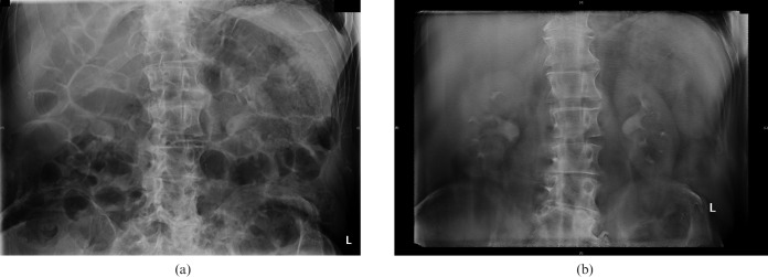
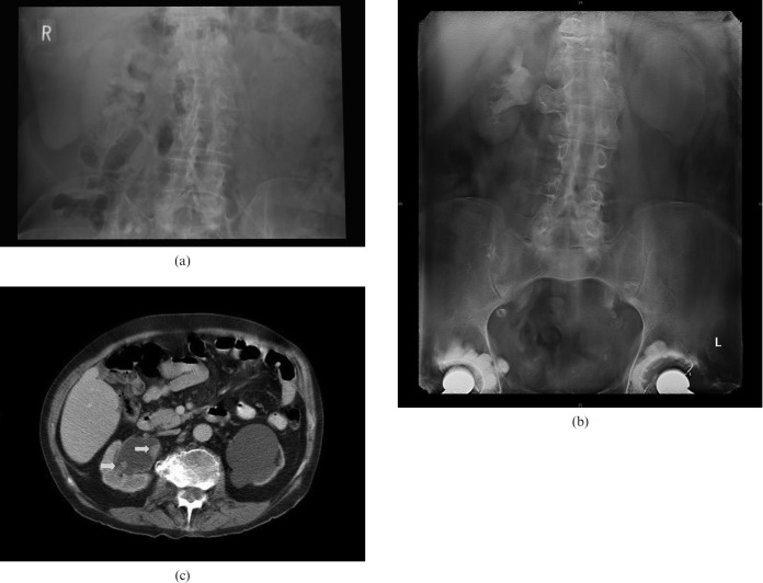
Several examples are included to illustrate the images obtained with digital tomosynthesis, and some of the pathology that can be visualised. Figure 1a is an example of a film with multiple distended bowel loops that would have made traditional IVU difficult to interpret. Figure 1b shows the quality of image obtained with digital tomosynthesis in the same patient. Figure 2a is taken from a traditional IVU in a different patient. It gives little information about the left kidney, and is not diagnostic for the right. A digital tomosynthesis obtained contemporaneously on the same patient (Figure 2b), reveals two foci of transitional cell carcinoma in the pelvis of the right kidney not seen on the traditional IVU. The hydronephrosis with thin rim of renal cortex on the left non-functioning kidney is also seen clearly; a result of a longstanding pelvi–ureteric junction obstruction. Note also the exquisite detail seen in the transverse vertebral process at the same level. Figure 2c illustrates the same features on CT in this patient for correlation.
Figure 1.
(a) Film with multiple distended bowel loops that would have made interpreting traditional intravenous urography difficult. (b) Digital tomosynthesis in the same patient.
Figure 2.
(a) Traditional intravenous urography giving little information about the left kidney and non-diagnostic for the right. (b) Digital tomosynthesis obtained contemporaneously on the same patient reveals two foci of transitional cell carcinoma in the pelvis of the right kidney, and hydronephrosis with a thin rim of renal cortex on the left owing to longstanding pelvi–ureteric junction obstruction. Note the exquisite detail seen in the transverse vertebral process at the same level. (c) The same features shown on CT in this patient, for correlation. Arrows show transitional cell carcinoma foci.
Unlike a conventional tomogram, digital tomosynthesis is technically simple. There is no need to accurately centre the radiograph or estimate the depth at which to perform the tomogram and then decide whether to go deeper or shallower for the next attempt. Instead, the whole series is obtained at the press of a button. This means that less experienced radiographers are more likely to achieve a diagnostic study. It also means that there is a much greater chance of imaging the kidney at the correct time after the administration of contrast medium as there is no delay between reviewing tomograms and performing additional cuts. Although not specifically tested, we have found that there is less requirement for patient compliance with multiple breath-holds. These benefits lead to the examination taking less time (our radiographers estimate 20 to 30 min per IVU including pre-contrast checks, vs 30 to 40 min previously), with potential associated benefits to patient throughput. In addition, if the images obtained do not cover the area of interest, it is possible to reconstruct images from the data obtained without re-irradiating the patient. We feel that the ease of performing digital tomosynthesis coupled with its imaging performance are major factors in its favour.
The standard hardware of a digital tomosynthesis system consists of an X-ray source, a 2D direct digital detector and the image reconstruction software. It also requires significant resources in computer memory to act as data buffers to store large numbers of images before, during and after reconstruction. Therefore, there is an impact on the bandwidth, networking infrastructure and storage capacity of any Picture Archiving and Communications System (PACS) server in place. The digital tomosynthesis system used is the GE VolumeRAD system, which relies on the GE Definium 8000 digital X-ray system to acquire the projection images necessary for tomosynthesis reconstruction. The complete installation cost £295 000, but if the digital X-ray system were already in place with sufficient computing power, the reconstruction algorithm software required to allow digital tomosynthesis would cost considerably less.
Conclusion
Digital tomosynthesis is a new technology that allows rapid acquisition of data and reconstruction of a series of tomographs through an organ. We have shown that it can be used for assessing the kidneys and offers a significantly increased percentage of diagnostic quality tests for assessing renal pathology compared with the traditional IVU. It also offers considerable advantages in ease of imaging and the time taken. It reduces the radiation dose by more than half compared with the traditional IVU protocol used in our institution. For these reasons we believe that in any situation where the IVU is still being used to assess the kidneys, digital tomosynthesis will be of considerable benefit in improving diagnostic quality.
References
- 1.Dobbins JT, 3rd, McAdams HP. Chest tomosynthesis: technical principles and clinical update. Eur J Radiol 2009;72:244–51 [DOI] [PMC free article] [PubMed] [Google Scholar]
- 2.Dobbins JT, 3rd, Godfrey DJ. Digital x-ray tomosynthesis: current state of the art and clinical potential. Phys Med Biol 2003;48:65–106 [DOI] [PubMed] [Google Scholar]
- 3.Cole JA, Emerton DP, Mackenzie A, Urbanczyk H, Clinch PJ, Lawinski CP, et al. Evaluation Report, Tomosynthesis for General Radiography. NHS Purchasing and Supply Agency, Feb 2009: CEP09001 [Google Scholar]
- 4.Freeman SJ, Sells H. Investigation of loin pain. Imaging 2008;20:38–56 [Google Scholar]
- 5.Warshauer DM, McCarthy SM. Detection of renal masses: sensitivities and specificities of excretion urography/linear tomography. Radiology 1988;169:363–5 [DOI] [PubMed] [Google Scholar]
- 6.Cowan NC, Turney BW, Taylor NJ, McCarthy CL, Crew JP. Multidetector computer tomography urography for diagnosing upper urinary tract urothelial tumour. BJU Int 2007;99:1363–70 [DOI] [PubMed] [Google Scholar]
- 7.Amis ES. Epitaph for the urogram. Radiology 1999;213:639–40 [DOI] [PubMed] [Google Scholar]
- 8.Ziedses DesPlantes BG. Planigraphie en Subtractie. Röntgenographische Differentiatiemethoden. Thesis, Kemink en Zoon, Utrecht, 1934: 112 [Google Scholar]


