Abstract
Myelolipomas are rare benign tumours composed of adipose tissue and haematopoietic cells that are typically found in adrenal glands but have also appeared in extra-adrenal sites. Distinguishing between extra-adrenal myelolipomas and malignant tumours, such as liposarcomas, is crucial to avoid an invasive procedure. To this end, we present a comprehensive report of the CT imaging characteristics of a pathologically proven bilateral extra-adrenal perirenal myelolipoma.
Myelolipomas are rare benign tumours typically found in adrenal glands [1]. Extra-adrenal myelolipomas are rare; bilateral extra-adrenal perirenal myelolipomas are far rarer [2]. Nonetheless, awareness of the CT findings of these lesions is important for establishing the diagnosis of extra-adrenal perirenal myelolipomas and for differentiating them from malignant tumours to avoid an invasive procedure. We herein present the CT imaging characteristics of a pathologically proven bilateral extra-adrenal perirenal myelolipoma.
Case report
A 77-year-old man with a history of Kaposi's sarcoma was admitted to the emergency department for abdominal distension and hypertension. Physical examination revealed normal findings and, apart from slightly increased leukocyte numbers (11 000 μl−1), the laboratory findings were unremarkable.
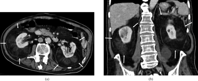
A contrast-enhanced abdominal CT scan showed large bilateral perirenal fat-containing masses (Figure 1); there was no communication between the masses. The kidneys were entirely embedded in the fat masses. There were small non-fatty soft-tissue densities interspersed within the lesions. The adrenal glands, kidneys and pararenal spaces were preserved. Bilateral renal cortical cysts were observed.
Figure 1.
An enhanced CT scan shows large well-circumscribed fat-containing masses (arrows) entirely surrounding and embedding both kidneys. (a) Transverse and (b) coronal images.
CT-guided fine-needle aspiration and core biopsy of both perirenal masses were performed. The core biopsy of both masses showed a glandular tumour composed of mature adipose tissue, fibromuscular tissue and an accumulation of myeloid tissue. A diagnosis of bilateral perirenal myelolipoma was made. CT performed at 3 months' follow-up showed stable perirenal masses with no evident change.
Discussion
Myelolipoma manifests in four distinct clinicopathological patterns: isolated adrenal myelolipoma, adrenal myelolipoma with haemorrhage, extra-adrenal myelolipoma and myelolipoma associated with other adrenal disease. Myelolipomas are most often present as isolated adrenal myelolipoma. Extra-adrenal presentation of the tumour is rare and usually in the abdominal and presacral regions, although gastric and hepatic locations have also been described. Other locations are mediastinum, lungs, pelvis, spleen, retroperitoneum, presacral region and mesentery [1]. Although sporadic case reports involving perirenal sites of myelolipomas have surfaced in the literature, only one case of bilateral extra-adrenal perirenal myelolipomas with imaging findings has been reported to date [2].
The cause of myelolipomas is not well documented; however, it is proposed that metaplasia of adrenal glands, misplacement of myeloid cells during embryogenesis, embolisation of bone marrow or a response to a trigger stimulus might be the cause [1]. The natural history of extra-adrenal myelolipoma is not well understood, but it is known that the tumour can enlarge over time and cause mass effect or haemorrhage [2]. The tumours are well encapsulated and asymptomatic when small [3]. Malignant degeneration has not been reported.
The imaging characteristics of myelolipomas vary according to the major component of the mass [2]. On sonography, fatty areas are hyperechoic, whereas cellular components are hypoechoic. Characteristically, lesions seen on CT have a negative Hounsfield unit value owing to macroscopic fat. Because of haematopoietic tissue, the attenuation values of myelolipomas are usually higher than those of the retroperitoneal fat, as was the case with our patient. High-attenuation regions may be seen as a result of haemorrhage or calcification. On T1 weighted MRI, myelolipomas are seen as high signal intensity lesions. The soft-tissue components of myelolipomas may be mildly enhanced after contrast injection [4].
Pre-operative diagnosis of extra-adrenal myelolipomas is difficult [1]. Radiologically, other fat-containing perirenal masses such as lipoma, liposarcoma, myelolipoma, myolipoma and angiomyolipoma cannot be differentiated; therefore, histological studies may be required [2, 5]. Other disorders to be considered in the differential diagnosis are extramedullary haemopoiesis, lymphoma and amyloidosis. Precise diagnosis of retroperitoneal masses with imaging techniques is challenging. Some radiological findings, such as the location, size, vascularity and local invasion, may contribute to the differential diagnosis. Percutaneous needle biopsy or core biopsy is often needed to confirm the extra-adrenal myelolipoma diagnosis. Surgical excision is thought to be unnecessary unless the diagnosis is unclear or the lesion is symptomatic.
The most important differential diagnosis for myelolipomas is retroperitoneal (perirenal) well-differentiated liposarcoma [5]. The radiological appearance of liposarcoma is dependent on its histological type. Well-differentiated liposarcomas do not invade the renal parenchyma, whereas other types tend to be poorly marginated and have infiltrative features. Histologically, well-differentiated liposarcomas are not haemorrhagic and have lipoblast zones of atypical cellular features. By contrast, extra-adrenal myelolipomas are usually well-encapsulated and have adipose tissue and haematopoietic tissue on histopathological examinations. This bilaterality and the well-encapsulated nature of the presented lesions suggest a benign nature; the diagnosis of myelolipoma was established on pathological examination [1, 5].
Myelolipoma and extramedullary haemopoiesis may have similar appearances on both imaging and histopathological studies. Extramedullary haemopoiesis is usually a multifocal, poorly circumscribed lesion; macroscopic fat is not a feature. But case reports of extramedullary haemopoiesis involving the perirenal space and having similar features as myelolipoma have been reported.
Angiomyolipoma is another diagnosis to be eliminated. It has prominent vascular structures and arises from the renal cortex, therefore producing a defect in the renal parenchyma. Myelolipomas, on the other hand, have a smooth interface between the cortex and parenchyma that might help in differentiating these lesions. In our patient, there were no prominent vascular structures in the lesion and the cortex was intact, which favoured the diagnosis of myelolipoma.
In conclusion, CT features such as location, size, vascularity and local invasion may help establish the myelolipoma diagnosis.
References
- 1.Kammen BF, Elder DE, Fraker DL, Siegelman ES. Extra-adrenal myelolipoma: MR imaging findings. AJR Am J Roentgenol 1998;171:721–3 [DOI] [PubMed] [Google Scholar]
- 2.Kumar M, Duerinckx AJ. Bilateral extra-adrenal perirenal myelolipomas: an imaging challenge. AJR Am J Roentgenol 2004;183:833–6 [DOI] [PubMed] [Google Scholar]
- 3.Meaglia JP, Schmidt JD. Natural history of an adrenal myelolipoma. J Urol 1992;147:1089–90 [DOI] [PubMed] [Google Scholar]
- 4.Cyran KM, Kenney PJ, Memel DS, Yacoub I. Adrenal myelolipoma. AJR Am J Roentgenol 1996;166:395–400 [DOI] [PubMed] [Google Scholar]
- 5.Liang EY, Cooper JE, Lam WW, Chung SC, Allen PW, Metreweli C. Case report: myolipoma or liposarcoma — a mistaken identity in the retroperitoneum. Clin Radiol 1996;51:295–7 [DOI] [PubMed] [Google Scholar]
- 6.Kocaoglu M, Bozlar U, Sanal HT, Guvenc I. Replacement lipomatosis: CT and MRI findings of a rare renal mass. Br J Radiol 2007;80:287–9 [DOI] [PubMed] [Google Scholar]
- 7.Chen KT, Felix EL, Flam MS. Extra-adrenal myelolipoma. Am J Clin Pathol 1982;78:386–9 [DOI] [PubMed] [Google Scholar]


