Abstract
Yttrium-90 (90Y) internal pair production can be imaged by positron emission tomography (PET)/CT and is superior to bremsstrahlung single-photon emission CT/CT for evaluating hepatic 90Y microsphere biodistribution. We illustrate a case of 90Y imaging using first generation PET/CT technology, producing high-quality images for qualitative diagnostic purposes.
Yttrium-90 (90Y) selective internal radiation therapy (SIRT) is an emerging treatment modality for inoperable liver tumours. 90Y has internal pair production which can be imaged by positron emission tomography with integrated CT (PET/CT) [1-3]. 90Y PET/CT is superior to bremsstrahlung single photon emission CT with integrated CT in evaluating hepatic 90Y microsphere biodistribution, which correlates with post-SIRT response [2,3].
We illustrate a case of 90Y PET/CT acquired using a first generation PET/CT scanner (Biograph WO; Siemens, Erlangen, Germany), producing high-resolution images of 90Y microsphere biodistribution (Figures 1–3). Our imaging protocol is detailed in Table 1. Total coincidences were 4.7 million over 40 min (1.2 GBq injected). No effort was made to reduce bremsstrahlung X-rays or background counts from the lutetium-based PET crystal. Background noise was visually minimised by adjusting the PET threshold. Images were analysed qualitatively for diagnostic purposes. Quantitation of 90Y activity was not performed.
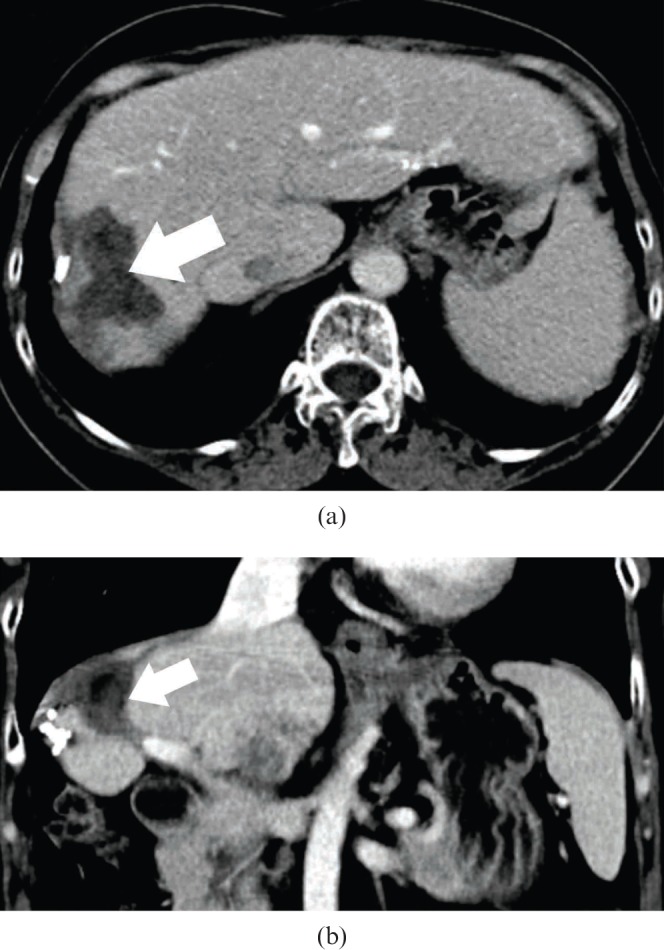
Figure 1.

56-year-old female with recurrent hepatocellular carcinoma (HCC) in the right lobe underwent yttrium-90 (90Y) selective internal radiation therapy. She was previously treated by radiofrequency ablation and liver resection. HCC recurrence was in the caudate lobe and periablation cavity region. A total of 1.2 GBq of 90Y resin microspheres was injected via the right hepatic artery. Pre-therapy triphasic CT liver (delayed phase) shows an ablation cavity (arrow) in segment VII/VIII—(a) transaxial; (b) coronal.
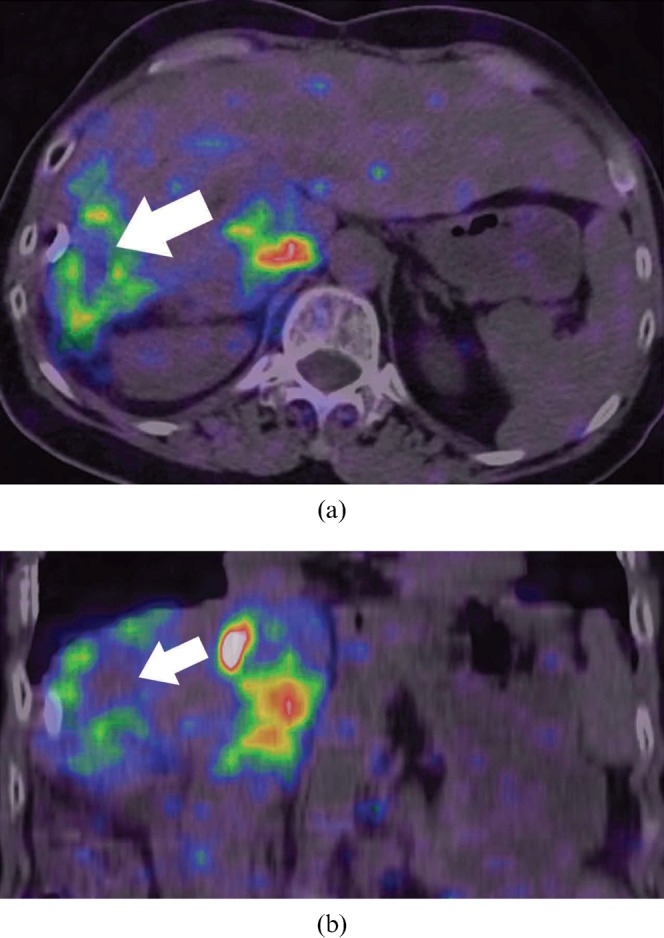
Figure 3.

Bremsstrahlung single-photon emission CT (SPECT)/CT acquired on the same day depicts bremsstrahlung activity as diffuse foci, inferior to the spatial resolution of yttrium-90 positron emission tomography/CT. Periablation cavity bremsstrahlung activity (arrow) cannot be clearly delineated. Bremsstrahlung activity in the caudate lobe is seen on SPECT/CT as a conglomerate focus with diffuse margins—(a) transaxial; (b) coronal.
Table 1. Yttrium-90 (90Y) imaging protocol using first generation positron emission tomography (PET)/CT.
| PET/CT scannerGeneral technique | Siemens Biograph LSO, Erlangen, GermanyImaging performed 6 h post-90Y injection; patient positioned supine with arms elevated; PET acquired in one bed position centred over the liver for 40 min |
| PET gantry information | Detector material: lutetium oxyorthosilicate; crystal dimension 6.45×6.45×25 mm; crystals per detector block 64; 144 detector blocks; 4 photomultiplier tubes per block; detector ring diameter 824 mm; 384 detectors per ring; 24 detector rings; total 9216 detectors |
| PET reconstruction parameters | PET matrix 128×128×47; attenuation weighted ordered subsets expectation maximisation iterative reconstruction; two iterations and eight subsets |
| CT parameters | Single-slice CT; 120 kVp; 90 mAs; field of view 50 cm; slice interval 3 mm |
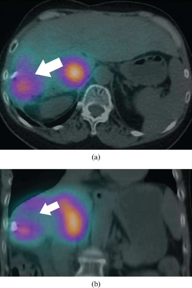
Figure 2.

Yttrium-90 (90Y) positron emission tomography/CT depicts hepatic 90Y microsphere biodistribution in high resolution. Periablation cavity 90Y activity (arrow) is well delineated and focal 90Y activity in the caudate lobe is well defined—(a) transaxial; (b) coronal.
Imaging 90Y microsphere biodistribution using first generation PET/CT technology is feasible. Its high-resolution images are useful for qualitative diagnostic purposes. Post-SIRT 90Y PET/CT permits accurate prognostication and effective planning of adjuvant modalities (e.g. radiofrequency ablation) by targeting poorly implanted tumour regions.
References
- 1.Lhommel R, van Elmbt L, Goffette P, Van denEynde M, Jamar F, Pauwels S, et al. Feasibility of 90Y TOF PET-based dosimetry in liver metastasis therapy using SIR-spheres. Eur J Nucl Med Mol Imaging 2010;37:1654–62 [DOI] [PubMed] [Google Scholar]
- 2.Lhommel R, Goffette P, Van denEynde M, Jamar F, Pauwels S, Bilbao JI, et al. Yttrium-90 TOF PET scan demonstrates high-resolution biodistribution after liver SIRT. Eur J Nucl Med Mol Imaging 2009;36:1696. [DOI] [PubMed] [Google Scholar]
- 3.Gates VL, Esmail AA, Marshall K, Spies S, Salem R. Internal pair production of 90Y permits hepatic localization of microspheres using routine PET: proof of concept. J Nucl Med 2011;52:72–6 [DOI] [PubMed] [Google Scholar]


